. 2019 Apr 16;5(1):62.
doi: 10.1186/s40792-019-0622-9.

Hepatic pleomorphic leiomyosarcoma after surgery for gastric gastrointestinal stromal tumor: a case report

Affiliations

Hepatic pleomorphic leiomyosarcoma after surgery for gastric gastrointestinal stromal tumor: a case report

Ryo Muranushi et al. Surg Case Rep. .

Abstract

Background: Pleomorphic leiomyosarcomas (PLMSs) are extremely rare tumors. We present the first case of hepatic primary PLMS after surgery for gastric gastrointestinal stromal tumor (GIST).

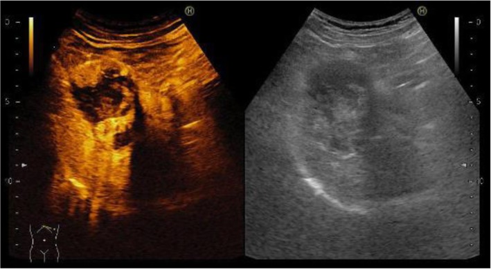
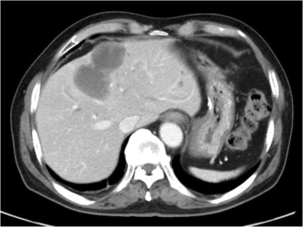
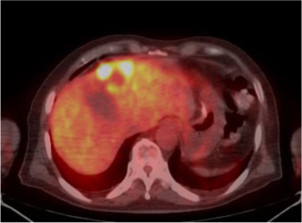
Case presentation: The patient was a 62-year-old man who was referred to our hospital for resection of a hepatic tumor arising after gastric GIST surgery that was resistant to imatinib and sunitinib. A 40-mm tumor in the left lobe of the liver and three small nodules in the right lobe were detected. We performed hepatic left lobectomy and partial resections for three lesions. According to the histopathological and immunohistochemical findings and c-kit gene mutations analysis, the main tumor was diagnosed as a PLMS.

Conclusion: It is necessary to consider the possibility that imatinib-resistant GIST recurrence lesions are a different kind of soft-tissue sarcoma. Accurate diagnosis is required to not miss the opportunity for radical excision of PLMS.

Keywords: Gastrointestinal stromal tumor; Hepatectomy; Pleomorphic hepatic leiomyosarcoma.


Conflict of interest statement

Consent for publication

Written informed consent was obtained from the patient for the publication of this case report and the accompanying images.

Competing interests

The authors declare that they have no competing interests.

Publisher’s Note

Springer Nature remains neutral with regard to jurisdictional claims in published maps and institutional affiliations.

Figures

Fig. 1
Fig. 1
Abdominal ultrasonography findings. A 51-mm-wide tumor in hepatic segment 4 with heterogeneous echo was detected and it didn't present bloodstream increase. Sonazoid enhanced ultrasonography with hypervolemic contrasting pattern revealed that the tumor was enhanced in the early phase and washed out in the late phase
Fig. 2
Fig. 2
Enhanced computed tomography findings. A 40-mm-diameter tumor in hepatic segments 3 and 4 (S3+4) with enhanced solid nodule along the wall was detected. On the right side of the tumor, there was a 50-mm tumor, which suggested hemorrhagic cyst
Fig. 3
Fig. 3
Gadolinium-enhanced magnetic resonance imaging findings. In addition to main tumor in S3+4 (a), small nodules that represented enhancement defect in the hepatocyte phase were detected in S6 (2b), S7 (2c), and S8 (2d)
Fig. 4
Fig. 4
18F‑fluorodeoxyglucose positron-emission tomography findings. The S3+4 tumor verge represented high FDG accumulation (SUVmax=12.13)
Fig. 5
Fig. 5
Photograph of resected specimen. The tumor measuring 73 mm × 65 mm × 36 mm in the resected left lobe showed a 40-mm white solid component and a 22-mm black nodule adjoining
Fig. 6
Fig. 6
Histopathological findings. The spindle‑shaped cells with nuclear atypia and eosinophilic cytoplasm proliferated diffusely. The tumor cells were full of pleomorphism. a: Hematoxylin-Eosin staining, ×50 b: Hematoxylin-Eosin staining, ×200
Fig. 7
Fig. 7
Immunohistochemical findings. Desmin was positive (a, ×200), α-smooth muscle actin (α-SMA) was slightly positive (b, ×200), and heavy-caldesmon (c, ×200) and muscle actin (HHF35) (d, ×200) was positive in 30% of the tumor tissue. Myogenic differentiation 1 (MyoD1) (e, ×200) did not present significant staining

Similar articles

Cited by

References

    1. Oda Y, Miyajima K, Kawaguchi K, Tamiya S, Oshiro Y, Hachitanda Y, et al. Pleomorphic leiomyosarcoma: clinicopathologic and immunohistochemical study with special emphasis on its distinction from ordinary leiomyosarcoma and malignant fibrous histiocytoma. Am J Surg Pathol. 2001;25(8):1030–1038. doi: 10.1097/00000478-200108000-00007. - DOI - PubMed
    1. Nazneen S, Kumari A, Choudhary V, Kumari S, Pankaj S. Prolonged survival of a young female with high grade pleomorphic leiomyosarcoma of ovary without recurrence. J Obstet Gynaecol India. 2016;66(Suppl 2):639–641. doi: 10.1007/s13224-015-0784-8. - DOI - PMC - PubMed
    1. Shamseddine A, Faraj W, Mukherji D, El Majzoub N, Khalife M, Soubra A. Unusually young age distribution of primary hepatic leiomyosarcoma: case series and review of the adult literature. World J Surg Oncol. 2010;8:56. doi: 10.1186/1477-7819-8-56. - DOI - PMC - PubMed
    1. Tsiatis AC, Atkinson JB, Wright JK, Cates JM. Primary hepatic myxoid leiomyosarcoma: a case report and review of the literature. Ultrastruct Pathol. 2008;32(1):25–28. doi: 10.1080/01913120701696122. - DOI - PubMed
    1. Shivathirthan N, Kita J, Iso Y, Hachiya H, Kyunghwa P, Sawada T, et al. Primary hepatic leiomyosarcoma: case report and literature review. World J Gastrointest Oncol. 2011;3(10):148–152. doi: 10.4251/wjgo.v3.i10.148. - DOI - PMC - PubMed

LinkOut - more resources