. 2019 Jun;23(6):4443-4453.
doi: 10.1111/jcmm.14340. Epub 2019 Apr 16.

Salidroside ameliorates Adriamycin nephropathy in mice by inhibiting β-catenin activity

Affiliations

Salidroside ameliorates Adriamycin nephropathy in mice by inhibiting β-catenin activity

Xinzhong Huang et al. J Cell Mol Med. 2019 Jun.

Abstract

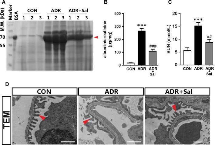
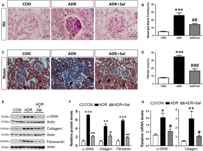
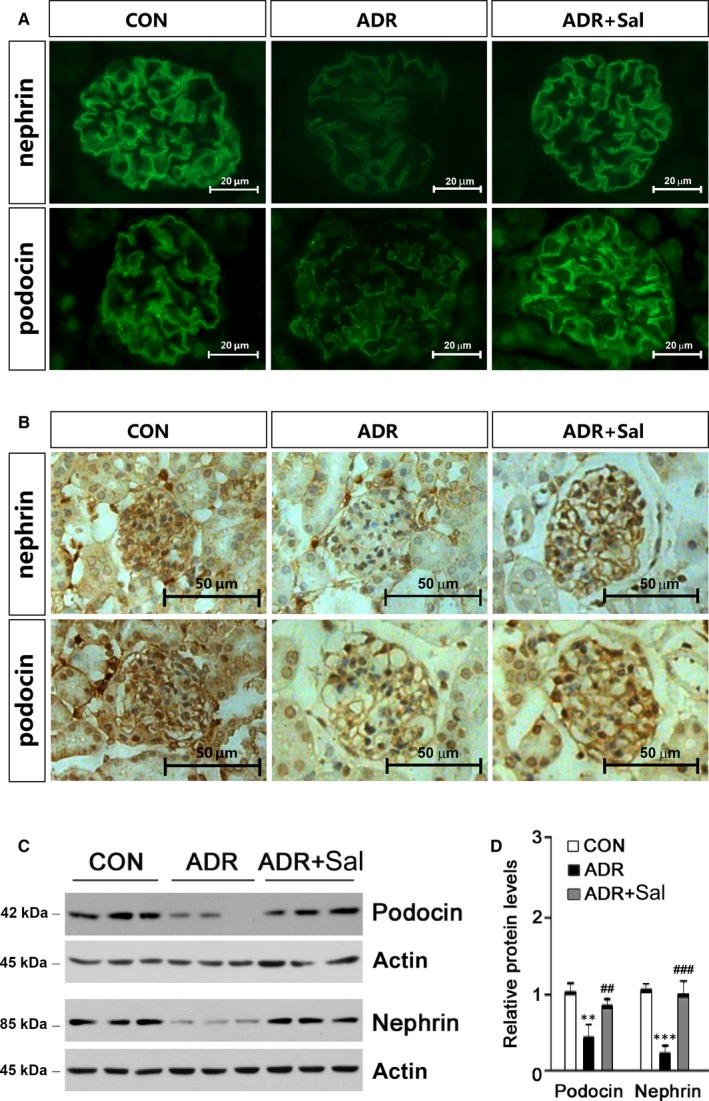
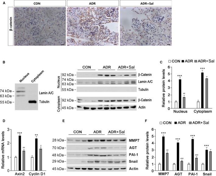
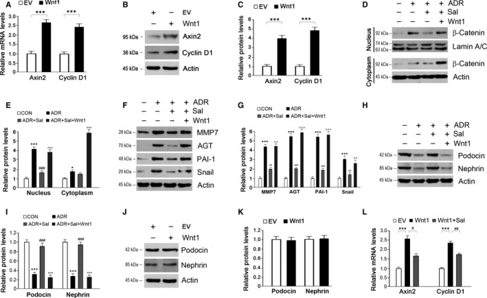
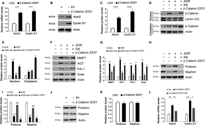
Salidroside is a major phenylethanoid glycoside in Rhodiola rosea L., a traditional Chinese medicine, with multiple biological activities. It has been shown that salidroside possesses protective effects for alleviating diabetic renal dysfunction, contrast-induced-nephropathy and other kidney diseases. However, the involved molecular mechanism was still not understood well. Herein, we examined the protective effects of salidroside in mice with Adriamycin (ADR)-induced nephropathy and the underlying molecular mechanism. The results showed that salidroside treatment ameliorates proteinuria; improves expressions of nephrin and podocin; and reduces kidney fibrosis and glomerulosclerosis induced by ADR. Mechanistically, ADR induces a robust accumulation of β-catenin in the nucleus and stimulates its downstream target gene expression. The application of salidroside largely abolishes the nuclear translocation of β-catenin and thus inhibits its activity. Furthermore, the activation of β-catenin almost completely counteracts the protective roles of salidroside in ADR-injured podocytes. Taken together, our data indicate that salidroside ameliorates proteinuria, renal fibrosis and podocyte injury in ADR nephropathy, which may rely on inhibition of β-catenin signalling pathway.

Keywords: Adriamycin nephropathy; podocytes; salidroside; β-catenin.


Conflict of interest statement

The authors declare that they have no conflict of interest.

Figures

Figure 1
Figure 1
Salidroside administration reduces proteinuria in ADR nephropathy. (A) SDS‐PAGE analysis showing the abundance of urinary proteins. Red arrow indicates the size of urinary proteins. (B) Urinary albumin levels. (C) Blood urea nitrogen (BUN) concentrations. (D) TEM analysis showing podocyte foot processes integrity. Arrowheads indicate secondary foot process and SD. Scale bar = 1 µm. 24‐hour urine samples were used for detecting urinary proteins and urinary albumin levels. The results are the means ± SEM of three independent experiments. ***P < 0.001 vs CON; ## P < 0.01 and ### P < 0.001 vs ADR alone. n = 5. ADR, adriamycin; CON, control; Sal, salidroside; TEM, transmission electron microscope
Figure 2
Figure 2
Salidroside administration ameliorates kidney injury in ADR nephropathy. (A) PAS staining. Representative micrographs were shown. Scale bar = 20 µm. (B) Morphometric analysis of PAS stained kidney sections. (C) Masson‐trichrome staining. Representative micrographs were shown. Scale bar = 20 µm. (D) Morphometric analysis of Masson‐trichrome stained kidney sections. (E) Western blot analyses showing the protein levels of α‐SMA, collagen I and fibronectin. (F) Quantitative determination for α‐SMA, collagen I and fibronectin protein levels. (G) The mRNA levels of α‐SMA and collagen I are shown. The results are the means ± SEM of three independent experiments. *P < 0.05, **P < 0.01 and ***P < 0.001 vs CON; # P < 0.05, ## P < 0.01 and ### P < 0.001 vs ADR alone. n = 5. ADR, adriamycin; CON, control; Sal, salidroside
Figure 3
Figure 3
Salidroside preserves nephrin and podocin expressions and prevents podocyte injury in ADR nephropathy. (A) Immunofluorescence analysis showing the abundance and distribution of nephrin and podocin. Representative micrographs were given. Scale bar = 20 µm. (B) Immunohistochemical analysis showing nephrin and podocin expressions. Representative micrographs were given. Scale bar = 50 µm. (C) Expressions of nephrin and podocin were analysed by Western blot. Glomerular lysates from different groups of mice were immunoblotted with specific antibodies against nephrin and podocin respectively. Actin was used as a loading control. (D) Quantitative determination of the relative abundances of nephrin and podocin. The results are the means ± SEM of three independent experiments. **P < 0.01 and ***P < 0.001 vs CON; ## P < 0.01 and ### P < 0.001 vs ADR alone. n = 5. ADR, adriamycin; CON, control; Sal, salidroside
Figure 4
Figure 4
Salidroside administration blocks renal β‐catenin activation induced by ADR. (A) Immunohistochemical analysis showing β‐catenin protein expression and localization in the kidney. Representative micrographs were shown. Scale bar = 20 µm. (B) Western blot analysis showing renal β‐catenin protein abundance in the nucleus and cytoplasm. Nucleus and cytoplasm lysates were distinguished using antibodies against Lamin A/C and Tubulin respectively (left panel). Lamin A/C and Actin were used as loading controls. (C) Quantitative analysis of β‐catenin protein levels as shown in (B). (D) Axin2 and Cyclin D1 expressions were examined. (E) Western blot analysis showing the effects of salidroside on β‐catenin downstream target gene expression. (F) Quantitative analysis of protein levels as shown in (E). Lamin A/C and Actin were used as loading controls. The results are the means ± SEM of three independent experiments. **P < 0.01; ***P < 0.001 vs CON; # P < 0.05; ## P < 0.01, ### P < 0.001 vs ADR alone. n = 5. ADR, adriamycin; AGT, angiotensinogen; CON, control; MMP7, matrix metalloproteinase 7; PAI‐1, plasminogen activator inhibitor‐1; Sal, salidroside
Figure 5
Figure 5
Activation of β‐catenin by Wnt1 abolishes the protective effects of salidroside against ADR injury in podocytes. (A‐B) The transfection of Wnt1 in cultured podocytes successfully activates β‐catenin activity. (C) Quantitative analysis of Axin2 and Cyclin D1 as shown in (B). (D) The repression of salidroside on β‐catenin activity was counteracted by Wnt1 in ADR‐treated podocytes. (E) Quantitative analysis of Western blots as shown in (D). (F) The inhibition of β‐catenin downstream target gene expressions by salidroside was re‐activated by Wnt1 in ADR‐treated podocytes. (G) Quantitative analysis of Western blots as shown in (F). (H) Activation of β‐catenin by Wnt1 reverses the effects of salidroside on podocin and nephrin expressions in ADR‐injured podocytes. (I) Quantitative analysis of Western blots as shown in (H). (J) Wnt1 has no effects on podocin and nephrin expressions. (K) Quantitative analysis of Western blots as shown in (J). (L) Effects of salidroside on Axin2 and Cyclin D1 expressions in podocytes transfected with the plasmid bearing Wnt1. Lamin A/C and Actin were used as loading controls. The results are the means ± SEM of three independent experiments. EV, empty vector; CON, control; ADR, adriamycin; Sal, salidroside; MMP7, matrix metalloproteinase 7; AGT, angiotensinogen; PAI‐1, plasminogen activator inhibitor‐1. *P < 0.05 and ***P < 0.001 vs CON; # P < 0.05; ## P < 0.01 and ### P < 0.001 vs ADR; ^^P < 0.01 and ^^^P < 0.001 vs ADR + Sal
Figure 6
Figure 6
Activation of β‐catenin by β‐catenin S33Y diminishes the protective roles of salidroside against ADR injury in podocytes. (A‐B) The transfection of β‐catenin S33Y in cultured podocytes successfully activates β‐catenin activity. (C) Quantitative analysis of Axin2 and Cyclin D1 as shown in (B). (D) The repression of salidroside on β‐catenin activity was counteracted by β‐catenin S33Y in ADR‐treated podocytes. (E) Quantitative analysis of Western blots as shown in (D). (F) The inhibition of β‐catenin downstream target gene expressions by salidroside was re‐activated by β‐catenin S33Y in ADR‐treated podocytes. (G) Quantitative analysis of Western blots as shown in (F). (H) Activation of β‐catenin by β‐catenin S33Y reverses the effects of salidroside on podocin and nephrin expressions in ADR‐injured podocytes. (I) Quantitative analysis of Western blots as shown in (H). (J) β‐catenin S33Y has no effects on podocin and nephrin expressions. (K) Quantitative analysis of Western blots as shown in (J). (L) Effects of salidroside on Axin2 and Cyclin D1 expressions in podocytes transfected with the plasmid bearing β‐catenin S33Y. Lamin A/C and Actin were used as loading controls. The results are the means ± SEM of three independent experiments. EV, empty vector; CON, control; ADR, adriamycin; Sal, salidroside; MMP7, matrix metalloproteinase 7; AGT, angiotensinogen; PAI‐1, plasminogen activator inhibitor‐1. *P < 0.05; **P < 0.01 and ***P < 0.001 vs CON; # P < 0.05; ## P < 0.01 and ### P < 0.001 vs ADR; ^P < 0.05; ^^P < 0.01 and ^^^P < 0.001 vs ADR + Sal

Similar articles

Cited by

References

    1. Agrawal S, Guess AJ, Chanley MA, Smoyer WE. Albumin‐induced podocyte injury and protection are associated with regulation of COX‐2. Kidney Int. 2014;86:1150‐1160. - PMC - PubMed
    1. He W, Kang YS, Dai C, Liu Y. Blockade of Wnt/beta‐catenin signaling by paricalcitol ameliorates proteinuria and kidney injury. J Am Soc Nephrol. 2011;22:90‐103. - PMC - PubMed
    1. Reiser J, Sever S. Podocyte biology and pathogenesis of kidney disease. Annu Rev Med. 2013;64:357‐366. - PMC - PubMed
    1. Thomas MC. Pathogenesis and progression of proteinuria. Contrib Nephrol. 2011;170:48‐56. - PubMed
    1. Dai C, Stolz DB, Kiss LP, Monga SP, Holzman LB, Liu Y. Wnt/beta‐catenin signaling promotes podocyte dysfunction and albuminuria. J Am Soc Nephrol. 2009;20:1997‐2008. - PMC - PubMed

Publication types

MeSH terms