Altered thalamo–cortical and occipital–parietal– temporal–frontal white matter connections in patients with anorexia and bulimia nervosa: a systematic review of diffusion tensor imaging studies
- PMID: 30994310
- PMCID: PMC6710091
- DOI: 10.1503/jpn.180121
Altered thalamo–cortical and occipital–parietal– temporal–frontal white matter connections in patients with anorexia and bulimia nervosa: a systematic review of diffusion tensor imaging studies
Abstract
Background: Anorexia nervosa and bulimia nervosa are complex mental disorders, and their etiology is still not fully understood. This paper reviews the literature on diffusion tensor imaging studies in patients with anorexia nervosa and bulimia nervosa to explore the usefulness of white matter microstructural analysis in understanding the pathophysiology of eating disorders.
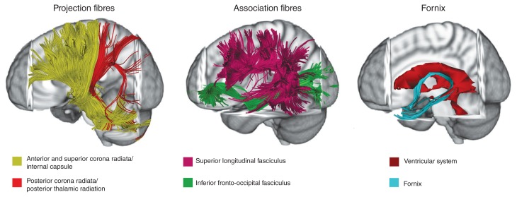
Methods: We followed the Preferred Reporting Items for Systematic Reviews and Meta-Analyses guidelines to identify diffusion tensor imaging studies that compared patients with an eating disorder to control groups. We searched relevant databases for studies published from database inception to August 2018, using combinations of select keywords. We categorized white matter tracts according to their 3 main classes: projection (i.e., thalamo–cortical), association (i.e., occipital–parietal–temporal–frontal) and commissural (e.g., corpus callosum).
Results: We included 19 papers that investigated a total of 427 participants with current or previous eating disorders and 444 controls. Overall, the studies used different diffusion tensor imaging approaches and showed widespread white matter abnormalities in patients with eating disorders. Despite differences among the studies, patients with anorexia nervosa showed mainly white matter microstructural abnormalities of thalamo–cortical tracts (i.e., corona radiata, thalamic radiations) and occipital–parietal–temporal–frontal tracts (i.e., left superior longitudinal and inferior fronto-occipital fasciculi). It was less clear whether white matter alterations persist after recovery from anorexia nervosa. Available data on bulimia nervosa were partially similar to those for anorexia nervosa.
Limitations: Study sample composition and diffusion tensor imaging analysis techniques were heterogeneous. The number of studies on bulimia nervosa was too limited to be conclusive.
Conclusion: White matter microstructure appears to be affected in anorexia nervosa, and these alterations may play a role in the pathophysiology of this eating disorder. Although we found white matter alterations in bulimia nervosa that were similar to those in anorexia nervosa, white matter changes in bulimia nervosa remain poorly investigated, and these findings were less conclusive. Further studies with longitudinal designs and multi-approach analyses are needed to better understand the role of white matter changes in eating disorders.
© 2019 Joule Inc. or its licensors
Conflict of interest statement
None declared.
Figures
References
-
- American Psychiatric Association. Diagnostic and statistical manual of mental disorders. 5th ed. Washington (DC): APA; 2013.
-
- Zipfel S, Giel KE, Bulik CM, et al. Anorexia nervosa: aetiology, assessment, and treatment. Lancet Psychiatry. 2015;2:1099–111. - PubMed
-
- Seitz J, Herpertz-Dahlmann B, Konrad K. Brain morphological changes in adolescent and adult patients with anorexia nervosa. J Neural Transm. 2016;123:949–59. - PubMed
Publication types
MeSH terms
LinkOut - more resources
Full Text Sources
Medical