. 2019 Sep 1;28(17):2811-2825.
doi: 10.1093/hmg/ddz080.

The landscape of Parkin variants reveals pathogenic mechanisms and therapeutic targets in Parkinson's disease

Affiliations

The landscape of Parkin variants reveals pathogenic mechanisms and therapeutic targets in Parkinson's disease

Wei Yi et al. Hum Mol Genet. .

Abstract

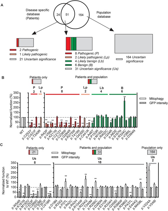
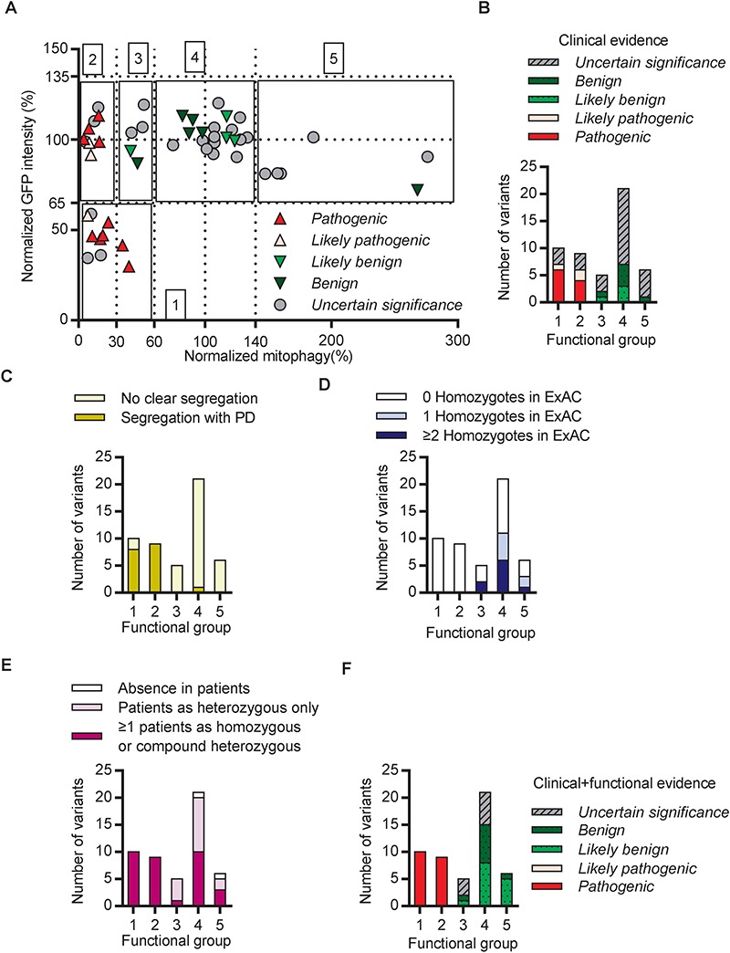
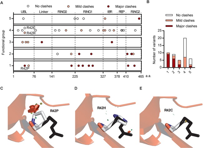
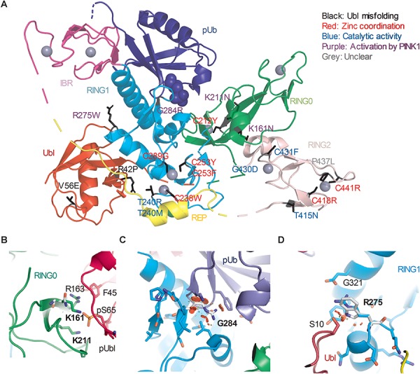
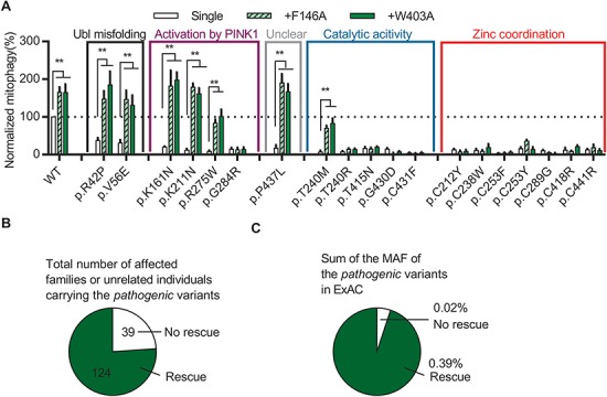
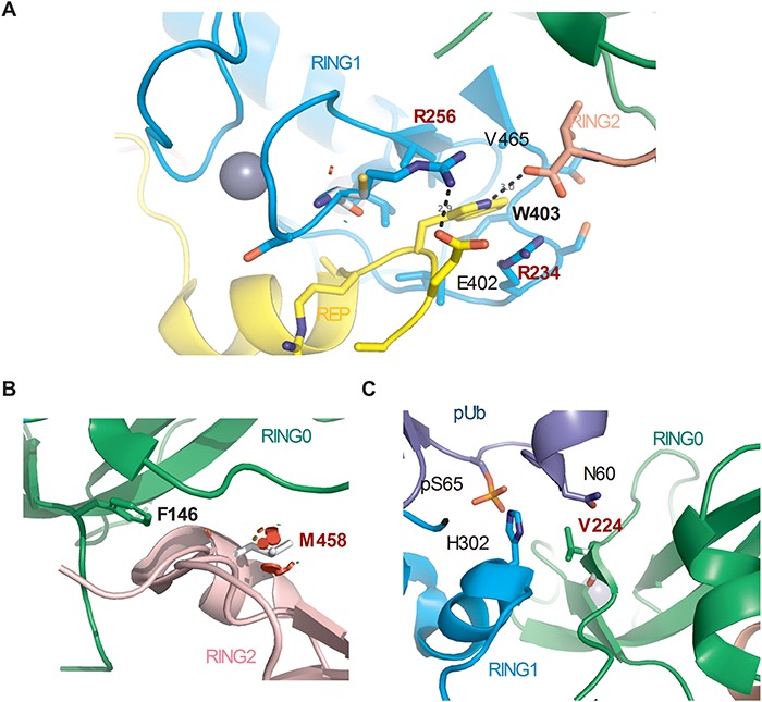
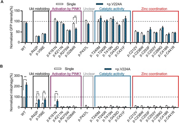
Mutations in Parkin (PARK2), which encodes an E3 ubiquitin ligase implicated in mitophagy, are the most common cause of early-onset Parkinson's disease (EOPD). Hundreds of naturally occurring Parkin variants have been reported, both in Parkinson's disease (PD) patient and population databases. However, the effects of the majority of these variants on the function of Parkin and in PD pathogenesis remain unknown. Here we develop a framework for classification of the pathogenicity of Parkin variants based on the integration of clinical and functional evidence-including measures of mitophagy and protein stability and predictive structural modeling-and assess 51 naturally occurring Parkin variants accordingly. Surprisingly, only a minority of Parkin variants, even among those previously associated with PD, disrupted Parkin function. Moreover, a few of these naturally occurring Parkin variants actually enhanced mitophagy. Interestingly, impaired mitophagy in several of the most common pathogenic Parkin variants could be rescued both by naturally occurring (p.V224A) and structure-guided designer (p.W403A; p.F146A) hyperactive Parkin variants. Together, the findings provide a coherent framework to classify Parkin variants based on pathogenicity and suggest that several pathogenic Parkin variants represent promising targets to stratify patients for genotype-specific drug design.


Figures

Figure 1
Figure 1
Parkin missense variants displayed a wide range of functional alterations. (A) 75 Parkin missense variants were reported in disease-specific databases (PDmutDB and MDSGene), 215 were reported in population databases (ExAC and dbSNP) and 51 were reported in both. Variants were assigned with one of the five standard ACMG terminologies: pathogenic (red), likely pathogenic (pink), likely benign (green), benign (olive) and uncertain significance (gray). (B and C) Quantification of the function of Parkin missense variants assigned as (B) Pathogenic (red), likely pathogenic (pink), likely benign (green), benign (olive) or (C) Uncertain significance (gray) by clinical evidence. Solid bars show mitophagy after 4 h of CCCP treatment quantified from mt-Keima signal in U2OS cells expressing GFP-Parkin variants normalized to WT Parkin. Hatched bars show GFP intensity of cells expressing GFP-Parkin variants normalized to WT Parkin. *P<0.05, **P<0.01, in one-way ANOVA with Dunnett’s post hoc test comparing the function of each variant with WT. N = 3–7.
Figure 2
Figure 2
Integration of clinical and functional evidence refined the classification of Parkin variants. (A) Functional alteration of variants assigned as pathogenic (red), likely pathogenic (pink), likely benign (green), benign (olive) and of uncertain significance (gray) based on clinical evidence were plotted for mitophagy activity on the X axis and GFP intensity on the Y axis. Functional alteration segregated into five groups, indicated by black boxes. 1. Significantly decreased mitophagy activity and GFP intensity compared with WT. 2. Severely decreased mitophagy activity with WT GFP intensity. 3. Moderately decreased mitophagy activity with WT GFP intensity. 4. WT mitophagy activity and GFP intensity. 5. Significantly increased mitophagy activity with WT GFP intensity. (B) Quantification of the variants within each of the functional groups from (A). (C) Quantification of variants within the functional groups described in (A) according to their segregation or lack of segregation with PD in families. (D) Quantification of variants within the functional groups described in (A) according to the observation of the variant as more than one homozygote (blue), one homozygote (light-blue) or no homozygotes (white) in ExAC. (E) Quantification of variants within the functional groups described in (A) according to the observation of the variant in PD patients. (F) Quantification of variants within the functional groups described in (A) according to their classification based on clinical and functional evidence.
Figure 3
Figure 3
Steric clashes in structural simulations predicted the dysfunction of Parkin variants. (A) Schematic representation of Parkin missense variants on Parkin protein 2D structure. Each circle indicates a missense variant. The location of the variant on the 2D sequence was plotted in the X axis with dotted lines separating the Parkin domains. The functional groups described in Figure 2A were plotted on the Y axis. The colors indicate the type of clash introduced by the missense variant from structural simulation. (B) Distribution of the variants from (A) within functional groups according to the type of clash they introduce. (C) Structure of human Parkin bound to pUb (PDB 5n2w) was used to illustrate the impact of the R42P mutation. Substitution of the arginine side chain (black) to proline (white) introduced major clashes (red disks), which would destabilize the β-sheet in the Ubl. (D) Substitution of the arginine side chain (black) to histidine (white) introduced mild clashes (red and green disks). (E) Substitution of the arginine side chain (black) to cysteine (white) did not introduce any clashes.
Figure 4
Figure 4
Structural analysis of Parkin variants revealed various pathogenic mechanisms. (A) Pathogenic variants were mapped onto the 3D structure of human Parkin bound to pUb (PDB 5N2W). The side chains of the amino acids substituted by the variants were highlighted in black. The blue spheres represent the phosphate of pUb. The gray spheres represent zinc. The color of the text indicates the type of disruption to Parkin caused by the variant. (B) Close-up view of pUbl-RING0 interface in the structure of fly pParkin bound to phospho-Ub (PDB 6DJX). Lys161 and Lys211 form ionic interactions with the phosphate on Ser65 of the pUbl. Mutations of these lysine residues would weaken the pUbl-RING0 interaction, preventing activation of Parkin. (C) Close-up view of the pUb:RING1 interface in human Parkin bound to pUb (PDB 5N2W). The G284R variant in Parkin RING1 would introduce major clashes with pUb, disrupting the interaction. (D) Close-up view of Arg275 in human Parkin bound to pUb (PDB 5N2W). Arg275 interacts with Glu321 in the helix that interacts with pUb. Mutation to a tryptophan (white) would introduce clashes with this helix as well as Ser10 in the Ubl domain.
Figure 5
Figure 5
Structure-guided designer hyperactive Parkin mutants can rescue mitophagy in pathogenic variants. (A) Quantification of induced mitophagy after 4 h of CCCP treatment in U2OS cells expressing WT GFP-Parkin, pathogenic missense variants or W403A or F146A in cis with WT or pathogenic variants. Mitophagy mediated by each Parkin missense variant was normalized to that of WT Parkin in each replicate. *P<0.05, **P<0.01, in two-way ANOVA with Dunnett’s post hoc test comparing the function of each variant with the variant in cis with W403A or F146A. N = 3–7. (B) The number of families or individuals with PD carrying the pathogenic missense variants for which mitophagy was or was not rescued by the designer mutations are shown. (C) The sum of the MAF in ExAC of the pathogenic missense variants for which mitophagy was or was not rescued by the designer mutations are shown.
Figure 6
Figure 6
Structural basis for the effects of the naturally occurring hyperactive Parkin variants. (A) Close-up view of R234Q and R256C variant sites in the structure of human Parkin bound to pUb (PDB 5N2W). Arg256 forms a hydrogen bond with Glu402, and its mutation to a cysteine would destabilize the REP:RING1 interaction, similar to W403A. The side-chain of Arg234 also stacks with the indole ring of Trp403. (B) Close-up view of M458L variant site in the RING0:RING2 interface (PDB 5N2W). M458L introduced major clashes that would destabilize the RING0:RING2 interaction, similarly to F146A. (C) Close-up view of V224A variant site (PDB 5N2W). Val224 interacts with pUb and forms van der Waals force interactions with Asn60. Mutation to alanine could modulate the affinity for pUb.
Figure 7
Figure 7
The naturally occurring Parkin p.V224A hyperactive variant rescued mitophagy in several pathogenic variants. (A) Quantification of GFP intensity from GFP signal by FACS in untreated cells expressing WT GFP-Parkin, pathogenic missense variants or p.V224A in cis with WT or pathogenic variants. The GFP intensity for each Parkin missense variant was normalized to that for WT Parkin in each replicate. (B) Quantification of induced mitophagy after 4 h of CCCP treatment in cells expressing WT GFP-Parkin, pathogenic missense variants or p.V224A in cis with WT or pathogenic variants. Mitophagy mediated by each Parkin missense variant was normalized to that of WT Parkin in each replicate. *P<0.05; **P<0.01, in two-way ANOVA with Dunnett’s post hoc test comparing the function of each variant with variant in cis with p.V224A. N = 3–7.

References

    1. Poewe W., Seppi K., Tanner C.M., Halliday G.M., Brundin P., Volkmann J., Schrag A.E. and Lang A.E. (2017) Parkinson disease. Nat. Rev. Dis. Primers, 3, 17013. - PubMed
    1. Koros C., Simitsi A. and Stefanis L. (2017) Genetics of Parkinson's disease: genotype–phenotype correlations. Int. Rev. Neurobiol., 132, 197–231. - PubMed
    1. Nuytemans K., Theuns J., Cruts M. and Van Broeckhoven C. (2010) Genetic etiology of Parkinson disease associated with mutations in the SNCA, PARK2, PINK1, PARK7, and LRRK2 genes: a mutation update. Hum. Mutat., 31, 763–780. - PMC - PubMed
    1. Cruts M., Theuns J. and Van Broeckhoven C. (2012) Locus-specific mutation databases for neurodegenerative brain diseases. Hum. Mutat., 33, 1340–1344. - PMC - PubMed
    1. Kasten M., Hartmann C., Hampf J., Schaake S., Westenberger A., Vollstedt E.J., Balck A., Domingo A., Vulinovic F., Dulovic M. et al. (2018) Genotype–phenotype relations for the Parkinson's disease genes Parkin, PINK1, DJ1: MDSGene systematic review. Mov. Disord., 33, 730–741. - PubMed

Publication types

MeSH terms