. 2019 May;47(6):1385-1395.
doi: 10.1177/0363546519832864. Epub 2019 Apr 17.

Anterior Cruciate Ligament Tear Promotes Skeletal Muscle Myostatin Expression, Fibrogenic Cell Expansion, and a Decline in Muscle Quality

Affiliations

Anterior Cruciate Ligament Tear Promotes Skeletal Muscle Myostatin Expression, Fibrogenic Cell Expansion, and a Decline in Muscle Quality

Bailey D Peck et al. Am J Sports Med. 2019 May.

Abstract

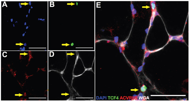
Background: Anterior cruciate ligament (ACL) tears result in significant quadriceps muscle atrophy that is resistant to recovery despite extensive rehabilitation. Recent work suggests an elevated fibrotic burden in the quadriceps muscle after the injury, which may limit recovery. Elucidating the mechanisms and cell types involved in the progression of fibrosis is critical for developing new treatment strategies.

Purpose: To identify factors contributing to the elevated fibrotic burden found after the injury.

Study design: Descriptive laboratory study.

Methods: After an ACL injury, muscle biopsy specimens were obtained from the injured and noninjured vastus lateralis of young adults (n = 14, mean ± SD: 23 ± 4 years). The expression of myostatin, transforming growth factor β, and other regulatory factors was measured, and immunohistochemical analyses were performed to assess turnover of extracellular matrix components.

Results: Injured limb skeletal muscle demonstrated elevated myostatin gene ( P < .005) and protein ( P < .0005) expression, which correlated ( R2 = 0.38, P < .05) with fibroblast cell abundance. Immunohistochemical analysis showed that human fibroblasts express the activin type IIB receptor and that isolated primary human muscle-derived fibroblasts increased proliferation after myostatin treatment in vitro ( P < .05). Collagen 1 and fibronectin, primary components of the muscle extracellular matrix, were significantly higher in the injured limb ( P < .05). The abundance of procollagen 1-expressing cells as well as a novel index of collagen remodeling was also elevated in the injured limb ( P < .05).

Conclusion: These findings support a role for myostatin in promoting fibrogenic alterations within skeletal muscle after an ACL injury.

Clinical relevance: The current work shows that the cause of muscle quality decline after ACL injury likely involves elevated myostatin expression, and future studies should explore therapeutic inhibition of myostatin to facilitate improvements in muscle recovery and return to sport.

Keywords: collagen; extracellular matrix; fibroblast; quadriceps.


Figures

Figure 1.
Figure 1.
Increased myostatin (MSTN) mRNA expression is observed following anterior cruciate ligament tear in quadriceps skeletal muscle. Gene expression, measured by quantitative polymerase chain reaction, from injured (I) and noninjured (NI) vastus lateralis muscle. Target genes (A) MSTN, (B) TGFβ1, (C) activin A, and (D) GDF11 were normalized to the geomean of 4 stable housekeeping genes and further normalized to the relative expression in NI control muscle. Values are presented as mean ± SD relative quotient (RQ), N = 14. **P < .005 vs NI limb.
Figure 2.
Figure 2.
Elevated myostatin (MSTN) protein expression and phosphorylation of SMAD3 in anterior cruciate ligament–injured quadriceps muscle. Whole muscle protein lysates were probed with (A) anti-TGFβ1, (B) anti-GDF8/MSTN, and (C) anti-p-SMAD3 and anti-SMAD3 antibodies, in addition to stable housekeeping proteins β-actin and β-tubulin. Band intensities were collected from the noninjured (NI) and injured (I) muscle samples, and differences were represented as a fold change in arbitrary units (AUs) across limbs. Values are presented as mean ± SD, n = 11. **P < .005 vs NI limb. ***P < .0005 vs NI limb. SMAD3, Small mothers against decapentaplegic 3.
Figure 3.
Figure 3.
Myostatin (MSTN) protein expression correlates with fibrogenic cell abundance in quadriceps muscle following anterior cruciate ligament injury. Correlation of (A) Tcf4+ fibroblasts and (B) PDGFRα+ FAPs relative to myofiber number with myostatin protein expression in the injured limb. Values are presented as mean ± SD, n = 10. AU, arbitrary unit.
Figure 4.
Figure 4.
Elevated inflammatory marker mRNA and protein expression is observed following anterior cruciate ligament tear in quadriceps skeletal muscle. Gene expression is measured by quantitative polymerase chain reaction from injured (I) and noninjured (NI) vastus lateralis muscle. Target genes (A) IL-6 and (B) TNFα were normalized to the geomean of 4 stable housekeeping genes and then further normalized to the relative expression in NI control muscle. Values are presented as mean ± SD relative quotient (RQ). Whole muscle protein lysates were probed with (C) anti-TNFα in addition to a stable housekeeping protein β-tubulin. Band intensities were represented as a fold change in arbitrary units (AUs) across limbs, N = 14. *P < .05 vs noninjured limb. ***P < .0005 vs NI limb.
Figure 5.
Figure 5.
Macrophage abundance is not different in the quadriceps muscle following anterior cruciate ligament tear. Representative images of (A) DAPI, (B) CD11b, (C) CD206, and (D) merged immunohistochemistry demonstrating an M1 macrophage (CD11b+/CD206−, yellow arrowhead) and an M2 macrophage (CD11b+/CD206+, white arrowhead). Abundance of (E) M1 and (F) M2 macrophages in the injured (I) and noninjured (NI) limbs. Values are represented as mean macrophages per fiber ± SD, n = 10. Scale bar = 50 μm.
Figure 6.
Figure 6.
Activin type IIB receptor (ACVR2B) is expressed by human skeletal muscle fibroblasts. Representative images of (A) DAPI, (B) Tcf4, (C) ACVR2B, and (D) wheat germ agglutinin staining in human skeletal muscle. (E) Merged immunohistochemical image demonstrating ACVR2B+/Tcf4+ cells (yellow arrows). Scale bar = 50 μm.
Figure 7.
Figure 7.
Myostatin treatment induces proliferation of human skeletal muscle–derived fibroblasts. (A-D) Representative images of EdU-labeled fibroblasts following 12-hour VEH or MSTN (300 ng/mL) treatment. (E) Myostatin treatment results in a 70% increase in EdU+ fibroblast frequency. Values are presented as percentage EdU+/DAPI+, mean ± SD, n = 3, with isolates assessed in duplicate. *P < .05 vs VEH-treated group. Scale bar = 200 μm. DAPI, 4#,6-diamidino-2-phenylindole; EdU, 5-ethynyl-2′-deoxyuridine; VEH, vehicle.
Figure 8.
Figure 8.
Elevated abundance of collagen 1, procollagen 1–positive cells, and CHP in the injured limb quadriceps muscle. (A) Representative images of collagen 1 (green) and procollagen 1 (red) staining in injured (I) and noninjured (NI) limbs. (B) Percentage area of collagen 1 staining in the I and NI limbs. (C) Number of procollagen 1+ cells relative to muscle area in the I and NI limbs. (D) Representative images of collagen 4 (red) and CHP (green) in I and NI limbs. Percentage area of (E) collagen 4 staining and (F) CHP staining in the I and NI limbs. Values are represented as mean ± SD, n = 10. *P < .05 vs from NI limb. **P < .005 vs noninjured limb. Scale bar = 100 μm.
Figure 9.
Figure 9.
Fibronectin and tenascin C accumulation in the injured limb quadriceps muscle following an anterior cruciate ligament tear. Representative images of fibronectin staining (red) in the (A) noninjured and (B) injured limbs. (C) Percentage area of fibronectin staining in the noninjured and injured limbs. Representative images of tenascin C staining (red), collagen 4 (green), and DAPI (blue) in the (D) noninjured and (E) injured limbs. (F) Percentage area of tenascin C staining in the noninjured and injured limbs. Values are presented as mean ± SD, n = 10. *P < .05 vs noninjured limb. Scale bar = 100 μm. I, injured; NI, noninjured. DAPI, 4#,6-diamidino-2-phenylindole.

References

    1. Allen DL, Unterman TG. Regulation of myostatin expression and myoblast differentiation by FoxO and SMAD transcription factors. Am J Physiol Cell Physiol. 2007;292(1):C188–C199. - PubMed
    1. Angelozzi M, Madama M, Corsica C, et al. Rate of force development as an adjunctive outcome measure for return-to-sport decisions after anterior cruciate ligament reconstruction. J Orthop Sports Phys Ther. 2012;42(9):772–780. - PubMed
    1. Ardern CL, Taylor NF, Feller JA, Webster KE. Fifty-five per cent return to competitive sport following anterior cruciate ligament reconstruction surgery: an updated systematic review and meta-analysis including aspects of physical functioning and contextual factors. Br J Sports Med. 2014;48(21):1543–1552. - PubMed
    1. Ardern CL, Taylor NF, Feller JA, Webster KE. Return-to-sport outcomes at 2 to 7 years after anterior cruciate ligament reconstruction surgery. Am J Sports Med. 2012;40(1):41–48. - PubMed
    1. Ardern CL, Webster KE, Taylor NF, Feller JA. Return to sport following anterior cruciate ligament reconstruction surgery: a systematic review and meta-analysis of the state of play. Br J Sports Med. 2011;45(7):596–606. - PubMed

Publication types