. 2019 Apr 17;14(4):e0214516.
doi: 10.1371/journal.pone.0214516. eCollection 2019.

TLR2/4 signaling pathway mediates sperm-induced inflammation in bovine endometrial epithelial cells in vitro

Affiliations

TLR2/4 signaling pathway mediates sperm-induced inflammation in bovine endometrial epithelial cells in vitro

Mohamed Aboul Ezz et al. PLoS One. .

Abstract

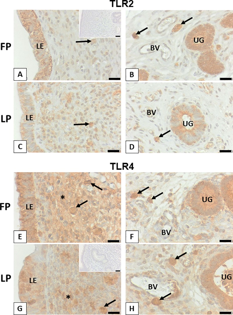
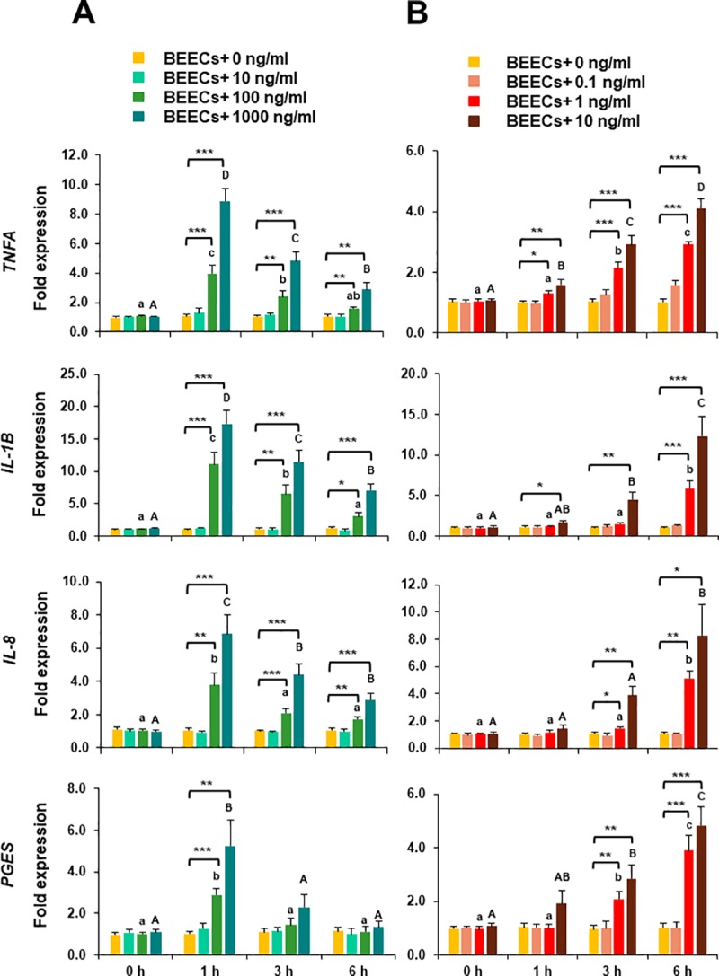
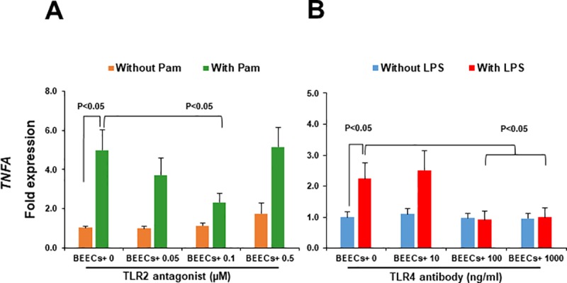
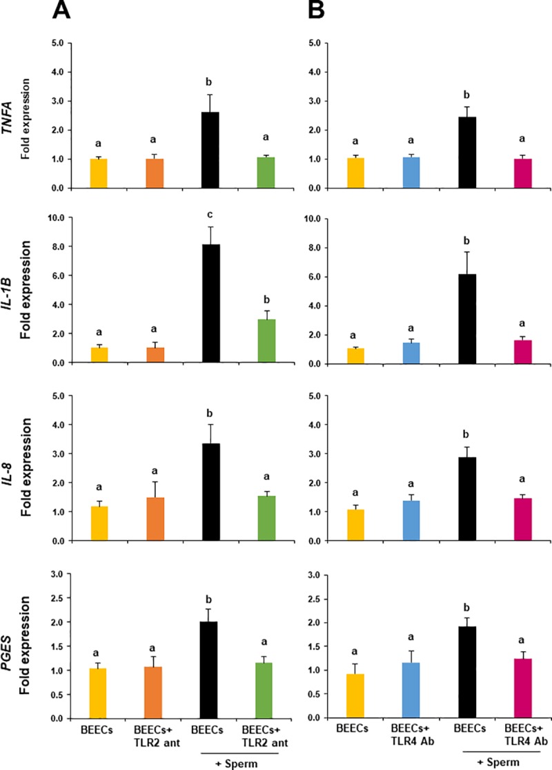
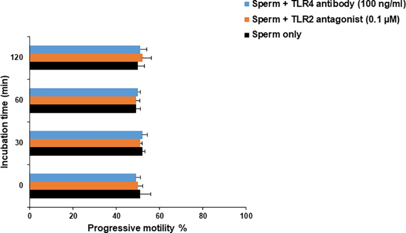
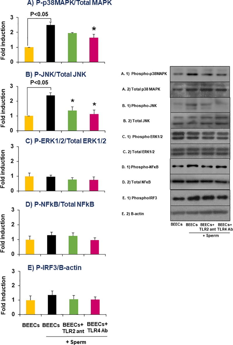
We have recently shown that sperm attachment to bovine endometrial epithelial cells (BEECs) triggers uterine local innate immunity with induction of a pro-inflammatory response in vitro, however details of the mechanism remain unknown. Here, we investigated the involvement of Toll-like receptor 2/4 (TLR2/4) pathway in mediating sperm-BEECs inflammatory process. Immunohistochemistry of the uterine tissue revealed that TLR2 and TLR4 proteins were present in the luminal and glandular epithelia of bovine endometrium. Moreover, BEECs monolayers were treated with TLR2 agonist (Pam; 0, 10, 100, and 1000 ng/ml) or TLR4 agonist (LPS; 0, 0.1, 1, and 10 ng/ml) for 0, 1, 3, or 6 h, followed by evaluating mRNA expression of the pro-inflammatory genes (TNFA, IL-1B, IL-8, and PGES) in BEECs using a real-time PCR. Both Pam and LPS treatments showed a dose-dependent stimulation of mRNA expression of the pro-inflammatory genes. To elucidate the functional role of TLR2/4 in sperm-BEECs interaction, BEECs monolayers were incubated with either TLR2 antagonist or TLR4 antibody for 2 h prior to the co-culture with sperm for 3 h. Importantly, pre-incubation of BEECs with TLR2 antagonist or TLR4 antibody prevented the stimulatory effect of sperm on the transcription of pro-inflammatory genes in BEECs. Furthermore, sperm increased the phosphorylation levels of TLR2/4 downstream targets (p38MAPK and JNK) in BEECs within 1 h of the co-culture. Treatment of BEECs with TLR2 antagonist prior to sperm addition inhibited JNK phosphorylation, while TLR4 antibody inhibited the phosphorylation of both p38MAPK and JNK. In conclusion, the present in vitro findings strongly suggest that bovine endometrial epithelial cells respond to sperm via TLR2/4 signal transduction.


Conflict of interest statement

The authors declare that there is no conflict of interest that could be preserved as prejudicing the impartiality of the research reported.

Figures

Fig 1
Fig 1. Bovine endometrium expressed TLR2 and TLR4 proteins.
Representative sections for luminal epithelium and stratum compactum (A, C, E, G), as well as stratum reticulare with endometrial glands (B, D, F, H) and blood vessels (BV) are shown. TLR2 is localized in luminal (LE) and glandular epithelium (UG) as well as a few immune cells (arrows) during follicular phase (FP) and luteal phase (LP). TLR4 is found in the same cells and additionally in stromal cells of the stratum compactum (asterisks) in some areas. Scale bars, 20 μm from A to H. Insets to A and G show representative controls for TLR2 and TLR4, respectively with scale bars: 40 μm.
Fig 2
Fig 2. TLR2/4 activation induced transcription of the pro-inflammatory genes in BEECs.
Sub-confluent bovine endometrial epithelial cells (BEECs) monolayers were exposed to A) TLR2 agonist (Pam; 0, 10, 100, and 1000 ng/ml) or B) TLR4 agonist (LPS; 0, 0.1, 1, and 10 ng/ml) for 0, 1, 3, or 6 h. At each time point, mRNA expressions of TNFA, IL-1B, IL-8, and PGES in BEECs were quantified by a real-time PCR. Data are presented as mean ± SEM of 4 independent experiments using epithelial cells from 4 different uteri (3 wells per treatment per experiment). Asterisks denote a significant variance [* (P < 0.05), ** (P < 0.01), *** (P < 0.001)] between the different doses of Pam/LPS when compared to the control group (0 ng/ml) at each time point. Different small letters denote a significant variance (P < 0.05) between the different time points of 100 ng/ml Pam (A) or 1 ng/ml LPS (B). Different capital letters denote a significant difference (P < 0.05) between the different time points of 1000 ng/ml Pam (A) or 10 ng/ml LPS (B).
Fig 3
Fig 3. TLR2/4 blocker inhibited the inflammatory response of BEECs towards TLR2/4 agonist.
Sub-confluent BEECs monolayers were 2 h incubated with A) TLR2 antagonist (0 μM, 0.05 μM = 18.12 ng/ml, and 0.1 μM = 36.24 ng/ml, and 0.5 μM = 181.20 ng/ml) prior to stimulation with Pam (100 ng/ml) for 1 h or B) TLR4 antibody (0, 10, 100, and 1000 ng/ml) prior to stimulation with LPS (1 ng/ml) for 3 h. TNFA mRNA expression was then quantified in BEECs. Data are presented as mean ± SEM of 3 independent experiments using epithelial cells from 3 different uteri (3 wells per treatment per experiment).
Fig 4
Fig 4. TLR2/4 blocker prevented the stimulatory effect of sperm on the transcription of pro-inflammatory genes in BEECs.
Sub-confluent BEECs monolayers were incubated for 2 h with A) 0.1 μM TLR2 antagonist or B) 100 ng/ml TLR4 antibody, then stimulated with 106/ml sperm for 3 h. mRNA expressions of TNFA, IL-1B, IL-8, and PGES were then quantified in BEECs. Data are presented as mean ± SEM of 4 independent experiments using epithelial cells from 4 different uteri (3 wells per treatment per experiment). Different letters denote a significant variance (P< 0.05) between the different groups.
Fig 5
Fig 5. Direct effect of TLR2/4 blocker on sperm motility.
Sperm cells at 106/ml Sp-TALP were exposed to either 0.1 μM TLR2 antagonist or 100 ng/ml TLR4 antibody for 0, 30, 60, and 120 min. At each time point, progressive motility of recovered sperm from both groups was assessed and compared with those of the control group (without either TLR2 antagonist or TLR4 antibody). Data are presented as mean ± SEM of 3 independent experiments using epithelial cells from 3 different uteri (3 wells per treatment per experiment).
Fig 6
Fig 6. TLR2/4 blocker inhibited TLR signal transduction pathways in BEECs in response to sperm.
Sub-confluent BEECs monolayers were incubated for 2 h with 0.1 μM TLR2 antagonist or 100 ng/ml TLR4 antibody, then stimulated with 106/ml sperm for 1 h. Western blotting was then carried out to estimate the phosphorylation levels of A) p38MAPK, B) JNK, C) ERK1/2, D) NFkB, and E) IRF3 in the different groups. Data are presented as mean ± SEM of 3 independent experiments using epithelial cells from 3 different uteri (3 wells per treatment per experiment). Asterisk (*) denotes a significant variance (P< 0.05) between TLR2/4 blocking groups when compared with sperm group.
Fig 7
Fig 7. A diagrammatic illustration of the mechanism by which sperm activate TLR2/4 signaling pathway in BEECs in vitro.
The proposed working model showing that stimulation of BEECs with sperm activates MAPKs components (p38MAPK and JNK; solid lines) without any effect on other downstream targets of TLR2/4 signaling pathways (ERK1/2, NFKB or IRF3; dotted line) down to the nuclear translocation of AP-1 protein which in turn regulates the transcription of pro-inflammatory genes in BEECs. “?” refers to unknown endogenous ligand(s) which may link sperm to TLR2/4 signaling pathway in BEECs.

Similar articles

Cited by

References

    1. Rath D, Schuberth HJ, Coy P, Taylor U. Sperm interactions from insemination to fertilization. Reprod Domest Anim. 2008;43 Suppl 5:2–11. 10.1111/j.1439-0531.2008.01250.x . - DOI - PubMed
    1. Katila T. Post-mating inflammatory responses of the uterus. Reprod Domest Anim. 2012;47 Suppl 5:31–41. 10.1111/j.1439-0531.2012.02120.x . - DOI - PubMed
    1. Bazer FW, Wu G, Spencer TE, Johnson GA, Burghardt RC, Bayless K. Novel pathways for implantation and establishment and maintenance of pregnancy in mammals. Molecular human reproduction. 2010;16(3):135–52. 10.1093/molehr/gap095 - DOI - PMC - PubMed
    1. Robertson SA. Control of the immunological environment of the uterus. Rev Reprod. 2000;5(3):164–74. . - PubMed
    1. Quayle AJ. The innate and early immune response to pathogen challenge in the female genital tract and the pivotal role of epithelial cells. J Reprod Immunol. 2002;57(1–2):61–79. . - PubMed

Publication types

MeSH terms