. 2019 Apr 16;27(3):708-718.e10.
doi: 10.1016/j.celrep.2019.03.076.

Single-Cell RNA-Sequencing-Based CRISPRi Screening Resolves Molecular Drivers of Early Human Endoderm Development

Affiliations

Single-Cell RNA-Sequencing-Based CRISPRi Screening Resolves Molecular Drivers of Early Human Endoderm Development

Ryan M J Genga et al. Cell Rep. .

Abstract

Studies in vertebrates have outlined conserved molecular control of definitive endoderm (END) development. However, recent work also shows that key molecular aspects of human END regulation differ even from rodents. Differentiation of human embryonic stem cells (ESCs) to END offers a tractable system to study the molecular basis of normal and defective human-specific END development. Here, we interrogated dynamics in chromatin accessibility during differentiation of ESCs to END, predicting DNA-binding proteins that may drive this cell fate transition. We then combined single-cell RNA-seq with parallel CRISPR perturbations to comprehensively define the loss-of-function phenotype of those factors in END development. Following a few candidates, we revealed distinct impairments in the differentiation trajectories for mediators of TGFβ signaling and expose a role for the FOXA2 transcription factor in priming human END competence for human foregut and hepatic END specification. Together, this single-cell functional genomics study provides high-resolution insight on human END development.

Keywords: CRISPRi; chromatin accessibility; dCas9-KRAB; endoderm; hepatic endoderm; human development; perturbation screen; pluripotent stem cells; single-cell RNA-seq; stem cell differentiation.


Conflict of interest statement

DECLARATION OF INTERESTS

The authors declare no competing interests.

Figures
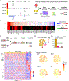
Figure 1.
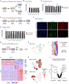
Figure 1.. scRNA-Seq CRISPRi Screen Identifies Molecular Drivers of Human END-Diff
(A) Representative integrative genomics viewer (IGV) tracks at the OCT4 and SOX17 loci. RNA-seq and ATAC-seq datasets for H1 ESC or END highlight dynamic transcriptome and chromatin changes. (B) Schematic of the atacTFAP analysis demonstrating how H1 ESC and END ATAC-seq and RNA-seq data (n = 2 biological replicates) are integrated to predict TF candidates during differentiation. Criteria for ATAC-seq peak analysis are FDR < 0.05 and log fold change ≥ 1.0. (C) 50 TF candidates ordered by atacTFAP score (top) and differential transcript expression (RNAdiff) between ESC and END (bottom). (D) Schematic of the scRNA-seq CRISPRi screening experiment during END-Diff. Expression of dCas9-KRAB is induced (via the addition of doxycycline) only after cells are pooled. (E) tSNE and cluster assignments resulting from scRNA-seq CRISPRi experiment (n = 2 biological replicates). (F) For each cluster, proportion of cells assigned to scramble gRNAs (p < 2.2E-16 versus random allocation; hypergeometric test). (G) Heatmap of all 16,110 cells passing screen quality control. Genes shown are a subset of cluster markers with q < 0.05, FC > 1.5 in either direction, and detection in at least 10% of cells in some cluster. (H) Feature plots selected from among top marker transcripts in each cluster. See also Figures S1 and S2, and Tables S1, S2, and S3.
Figure 2.
Figure 2.. Dissecting the Role of TGFβ Mediators during END-Diff at the Single-Cell Level
(A) Cluster enrichment of gRNAs for each TF target. Heatmap shows Pr(cluster | gRNA) minus Pr(cluster | scramble). (B) tSNE of cluster assignments and feature plots of gRNAs targeting specific TGFβ mediator genes. Each dot represents one cell. (C) Staging of END-Diff via Quant-seq (top; n = 2 biological replicates) compared with scRNA-seq CRISPRi cluster characteristics (bottom). (D) Model of the effects of TGFβ mediator perturbations on human END-Diff. See also Tables S1 and S3.
Figure 3.
Figure 3.. Loss of FOXA2 Expression Results in Distinct Transcriptomic and Chromatin Changes within END
(A) A gene module affected across multiple perturbations. Within the main cluster only, log fold changes (color intensity) were estimated via MIMOSCA. Genes were selected by running sparse PCA and thresholding the top component. Bars to the right show the number of differentially expressed genes by target (MAST q < 0.05; cluster 0 only). (B) Schematic of FOXA2 perturbation in conjunction with scramble-gRNA control conditions during differentiation. (C) Immunofluorescence analysis for SOX17, OCT4, and FOXA2 of scramble and FOXA2 perturbed END. Nuclei are counterstained with Hoechst. Scale bar: 100 μm. (D) Summary of dynamic changes (FDR < 0.05, log fold change ≥ 1.0) in H3K27ac (left) and chromatin accessibility as measured by ATAC-seq (right) between ESC, scramble-gRNA control END, and FOXA2 perturbed END (n = 2 biological replicates). (E) ATAC-seq, histone ChIP-seq, and TF ChIP-seq data at the TTR and HHEX loci showing decreased gene activity in FOXA2-gRNA containing END at foregut-associated genes that are potentially regulated by FOXA2. See also Tables S1 and S4.
Figure 4.
Figure 4.. Loss of FOXA2 Impairs Differentiation to Foregut END and Subsequent Hepatic END, while Mid-Hindgut END Differentiation Is Unaffected
(A) Schematic of perturbation experiments for assessment of effect of FOXA2 repression on competency toward foregut and mid-hindgut END. (B and C) qPCR analysis of transcripts in scramble or FOXA2 perturbed foregut END (B) and mid-hindgut END (C). Relative transcript expression was calculated using the ΔΔCT method; all transcripts were normalized to ACTB. Error bars correspond to SD; n = 3 biological replicates. (D) Schematic of perturbation experiments during human hepatic END differentiation. (E) qPCR analysis of transcripts in scramble or FOXA2 perturbed hepatic END. Relative transcript expression was calculated using the ΔΔCT method; all transcripts were normalized to ACTB. Error bars correspond to SD; n = 3 biological replicates. (F) Immunofluorescence analysis for HNF4a and FOXA2 of scramble and FOXA2 perturbed hepatic END cells. Nuclei are counterstained with Hoechst. Scale bar: 100 μm. (G) Schematic of the scRNA-seq CRISPRi experiment during hepatic END differentiation. Scramble- and FOXA2-gRNA cells were co-cultured throughout differentiation. (H) Unsupervised cluster assignments visualized via tSNE. Each dot represents one cell (n = 2 biological replicates). (I) Heatmap of cluster average expression of selected hepatic END-associated transcripts. Asterisk denotes MAST q < 0.05; ln FC > 0.25. (J) Cellwise heatmap (n = 903 cells) containing all cluster markers with MAST q < 0.05, FC > 1.5 in either direction, and detection rate > 10% in at least one cluster. Each transcript is standardized to have mean 0 and variance 1. (K) Feature plots of scramble- and FOXA2-gRNA. (L) Expression differences from single-cell data between FOXA2-gRNA versus scramble-gRNA containing hepatic END cells. All transcripts are displayed. See also Figure S3 and Table S5.

References

    1. Adamson B, Norman TM, Jost M, Cho MY, Nuñez JK, Chen Y, Villalta JE, Gilbert LA, Horlbeck MA, Hein MY, et al. (2016). A multiplexed single-cell CRISPR screening platform enables systematic dissection of the unfolded protein response. Cell 167, 1867–1882.e21. - PMC - PubMed
    1. Alexander J, and Stainier DY (1999). A molecular pathway leading to endoderm formation in zebrafish. Curr. Biol 9, 1147–1157. - PubMed
    1. Allison TF, Smith AJH, Anastassiadis K, Sloane-Stanley J, Biga V, Stavish D, Hackland J, Sabri S, Langerman J, Jones M, et al. (2018). Identification and single-cell functional characterization of an endodermally biased pluripotent substate in human embryonic stem cells. Stem Cell Reports 10, 1895–1907. - PMC - PubMed
    1. Ang SL, and Rossant J (1994). HNF-3 beta is essential for node and noto-chord formation in mouse development. Cell 78, 561–574. - PubMed
    1. Avery S, Zafarana G, Gokhale PJ, and Andrews PW (2010). The role of SMAD4 in human embryonic stem cell self-renewal and stem cell fate. Stem Cells 28, 863–873. - PubMed

Publication types

MeSH terms