Review
. 2019 Apr 16;50(4):955-974.
doi: 10.1016/j.immuni.2019.03.016.

Immune Signaling in Neurodegeneration

Affiliations
Review

Immune Signaling in Neurodegeneration

Timothy R Hammond et al. Immunity. .

Abstract

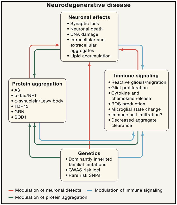
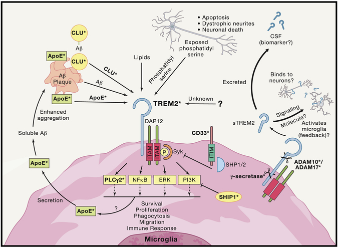
Neurodegenerative diseases of the central nervous system progressively rob patients of their memory, motor function, and ability to perform daily tasks. Advances in genetics and animal models are beginning to unearth an unexpected role of the immune system in disease onset and pathogenesis; however, the role of cytokines, growth factors, and other immune signaling pathways in disease pathogenesis is still being examined. Here we review recent genetic risk and genome-wide association studies and emerging mechanisms for three key immune pathways implicated in disease, the growth factor TGF-β, the complement cascade, and the extracellular receptor TREM2. These immune signaling pathways are important under both healthy and neurodegenerative conditions, and recent work has highlighted new functional aspects of their signaling. Finally, we assess future directions for immune-related research in neurodegeneration and potential avenues for immune-related therapies.

Keywords: Alzheimer’s disease; Complement; Cytokine; GWAS; Inflammation; Innate Immunity; Microglia; Neuro-immune; Neurodegeneration; Neuroinflammation; Signaling; TGF-β; TREM2.


Conflict of interest statement

Conflicts of Interest:

No conflicts of interest to declare.

B.S. is a member of the Scientific Advisory Board and minor shareholder in Annexon Biosciences. B.S. is an inventor on multiple patents: “Modulation of Synaptic Maintenance” (US8148330B2, US9149444B2) and is inventor on pending patent “Biomarkers for dementia and dementia related neurological disorders” (WO2015103594A1/US20160327572A1). T.R.H. & S.E.M. have no interests to declare.

Figures

Figure 1:
Figure 1:. Dramatic Increase in Immune Related Publications in Alzheimer’s Disease:
Analysis of publications via PubMed reveals dramatic increase in immune-focused Alzheimer’s research since the year 2000 that significantly exceeds overall increase in AD-focused research. Each data point represents the percentage increase in publications normalized to number of publications in year 2000 for their respective publication focus. The most precipitous increases in AD immune-related research have occurred in the last 10 years, that corresponds with the publication of GWASs, which have implicated genes enriched in immune cell expression and immune signaling, and the publication of rare high-risk variants in microglial-specific gene TREM2. Milestone citations: Reactive Microglia: (McGeer et al., 1987; Rogers et al., 1988); fAD APPV717I: (Goate et al., 1991); ApoE4 Risk: (Corder et al., 1993; Strittmatter et al., 1993); PDAPP & Tg2576: (Games et al., 1995; Hsiao et al., 1996); Aβ immunotherapoy: (Bard et al., 2000); AD GWAS: (Hollingworth et al., 2011; Lambert et al., 2009; Lambert et al., 2013; Naj et al., 2011); Trem2 R47H: (Guerreiro et al., 2013b; Jonsson et al., 2013).
Figure 2:
Figure 2:. Immune Signaling Is a Complex Multi-Dimensional Process in Neurodegenerative Disease
Neurodegenerative diseases involve a complex interplay between immune signaling, genetics, and neural damage that result in debilitating cognitive phenotypes. The neuronal and synaptic dysfunction and loss (top) in neurodegenerative disease can be mediated directly by protein accumulation/aggregation (both intracellular and extracellular), by genetic polymorphisms that modulate neuronal function, or by immune cell signaling (red arrows). Immune signaling in neurodegenerative disease can occur, in response to aggregated toxic proteins, in response to neuronal damage, and/or as result of genetic polymorphisms altering immune cell function (blue arrows). Protein aggregation can in turn also be altered by neuronal dysfunction, altered immune function, and/or genetic polymorphisms (black arrows). All of these interconnected effects lead to the memory loss, cognitive decline, motor dysfunction, and death that are the clinical manifestations of these debilitating diseases. Arrow color is indicative of the component being modulated.
Figure 3:
Figure 3:. TGF-β Signaling and the Complement Cascade in Neurodegeneration:
A. TGF-β is a secreted signal that binds to TGFBRI and TGFBRII complexes on the cell surface of neurons and microglia. TGF-β levels are elevated in the brain and CSF of AD, PD, and FTD patients. In mouse models of AD, PD, and FTD, TGF-β is neuroprotective and decreases amyloid plaques in AD models, acting in part through interleukin-like epithelial-mesenchymal transition inducer (ILEI) in neurons to reduce Aβ production. TGF-β is also necessary for normal microglia maturation and for maintaining their unique brain profile. by Microglia in TGF-β receptor knockout mice or mice lacking the TGF-β associated milieu molecule LRRC33 exhibit increased lysosomal content, decreased ramification, and altered transcriptional profile. Altered TGF-β levels could modulate microglial state and function in neurodegenerative disease. B. The complement pathway is upregulated in AD, PD, and FTD patients. In AD and FTD mouse models, heterotrimeric C1q molecules (made up of C1qa, C1qb, C1qc) bind to vulnerable synapses, leading to opsonization by C3 and phagocytosis of the structure by microglia which express the C3 receptor (CR3/CD11b). This process could contribute to synapse loss found in early stages of neurodegenerative disease. Blocking complement activation limits neuroimmune alterations and ameliorates neurocognitive deficits in mouse models of AD and FTD. C1q molecules (produced by microglia) also bind to amyloid plaques to trigger Aβ phagocytosis. ApoE can limit complement pathway activation by binding to C1q, and an association found in AD plaques and is reduced in the presence of the APOE-ε4 risk allele.
Figure 4:
Figure 4:. Convergence of Multiple AD Risk Genes/Loci on TREM2 Signaling
TREM2 signaling pathways involve potential complex signaling interactions with other AD genetic risk factors with a diverse array of functional consequences. Studies of TREM2 have begun to uncover a number of possible ligands for TREM2 that are highly expressed in the CNS and increased in AD brain, and more ligands potentially await discovery. Among the proposed ligands for TREM2 are ApoE, Clu/ApoJ, various lipids, phosphatidyl serine, and even potentially Aβ itself. TREM2 lacks its own signal transduction component and must interact with ITAM adaptor protein DAP12 in order to mediate downstream signaling. DAP12 activation and Syk phosphorylation led to a number of potential intracellular signals including NF-kB, PI3K, ERK, and newly identified AD risk gene PLCG2. Recent evidence also indicates that activation of TREM2 increases microglial production of ApoE which may then act in reciprocal cycle binding to amyloid and then binding to TREM2. The natural inhibitor of ITAM signaling comes from ITIM signaling. Interestingly, another AD risk gene CD33 contains an ITIM signaling domain, creating a potential link between TREM2 and CD33. Additionally, the gene which encodes for the protein SHIP1 has been identified as potential risk allele in AD and studies have shown that SHIP1 inhibition of PI3K activation exerts an inhibitory effect on TREM2 signaling. Ultimately, activation of TREM2 signaling results in several potential phenotypic alterations including altered migration, survival, proliferation, cytokine release, and other functions. Which intracellular pathways are directly responsible for the different functional consequences are still being worked out and may be ligand specific. TREM2 is also cleaved into a soluble fragment (sTREM2) by α-secretase (ADAM10/17, which are also implicated in AD risk and play key roles in APP processing). The function of sTREM2 is still not fully understood but sTREM2 can be detected in CSF and is being examined as potential biomarker of disease state in AD and other CNS disorders. sTREM2 has also been recently proposed to potentially bind to neurons, as well as potentially binding to an unknown receptor reciprocally on microglial. Finally, after α-secretase cleavage the remaining membrane fragment of TREM2 is cleaved by γ-secretase (components of which, PSEN1, and PSEN2, are known to cause early onset fAD). Inhibition of this cleavage results in inability of DAP12 to release from non-functional TREM2 and therefore sequestration of DAP12. Bold terms with asterisks indicate proteins coded for by genes that alter risk of AD.

References

    1. Abud E, Ramirez R, Martinez E, Healy L, Nguyen C, Newman S, Yeromin A, Scarfone V, Marsh S, Fimbres C, et al. (2017). iPSC-Derived Human Microglia-like Cells to Study Neurological Diseases. Neuron 94, 278–293.e279. - PMC - PubMed
    1. Afagh A, Cummings BJ, Cribbs DH, Cotman CW, and Tenner AJ (1996). Localization and cell association of C1q in Alzheimer's disease brain. Exp Neurol 138, 22–32. - PubMed
    1. Alzheimer A (1907). Über eine eigenartige Erkrankung der Hirnrinde. Allgemeine Zeitschrift für Psychiatrie und psychisch-gerichtliche Medizin 64.
    1. Alzheimer A (1911). Über eigenartige Krankheitsfälle des späteren Alters.
    1. Atagi Y, Liu C, Painter M, Chen X, Verbeeck C, Zheng H, Li X, Rademakers R, Kang S, Xu H, et al. (2015). Apolipoprotein E Is a Ligand for Triggering Receptor Expressed on Myeloid Cells 2 (TREM2). J Biol Chem 290, 26043–26050. - PMC - PubMed

Publication types

MeSH terms

Substances