. 2019 Apr 16;12(2):58.
doi: 10.3390/ph12020058.

Insights into the Molecular Mechanisms of Eg5 Inhibition by (+)-Morelloflavone

Affiliations

Insights into the Molecular Mechanisms of Eg5 Inhibition by (+)-Morelloflavone

Tomisin Happy Ogunwa et al. Pharmaceuticals (Basel). .

Abstract

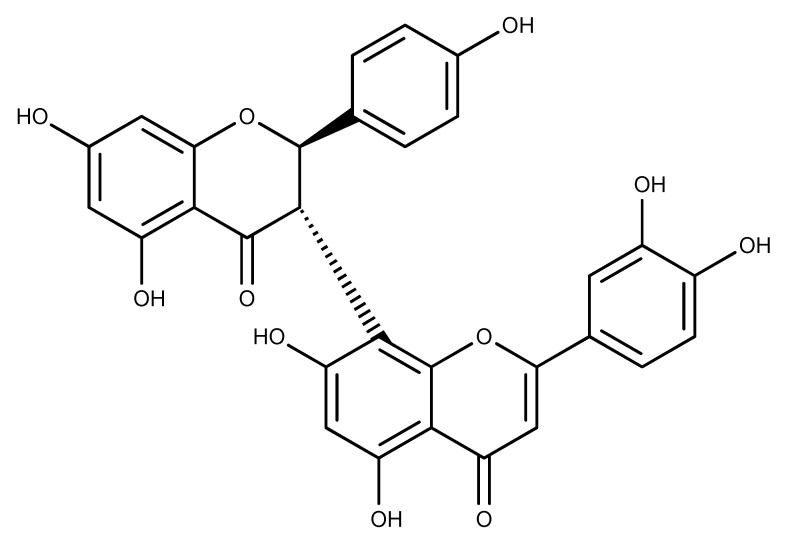
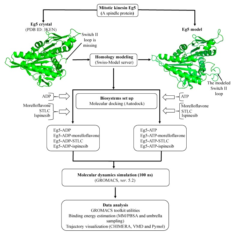
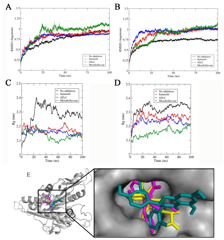
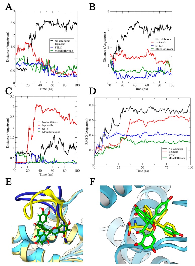
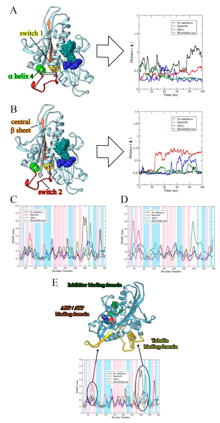
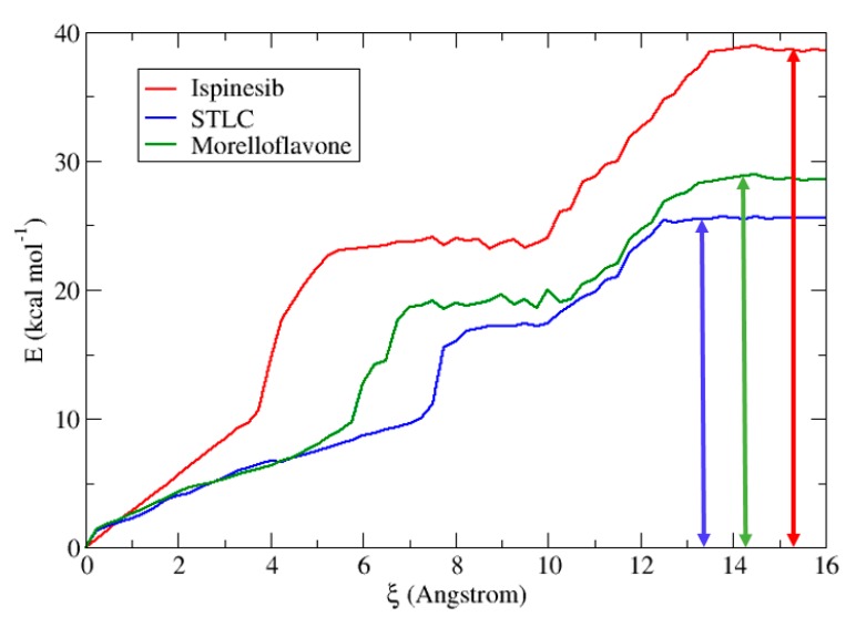
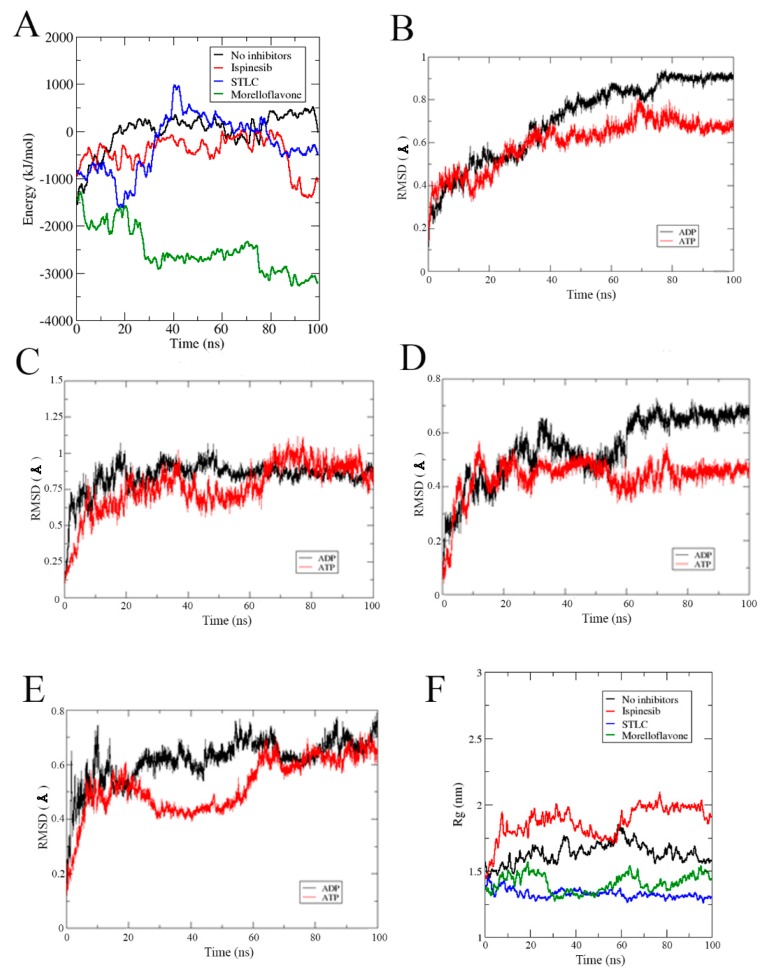
(+)-Morelloflavone (MF) is an antitumor biflavonoid that is found in the Garcinia species. Recently, we reported MF as a novel inhibitor of ATPase and microtubules-gliding activities of the kinesin spindle protein (Eg5) in vitro. Herein, we provide dynamical insights into the inhibitory mechanisms of MF against Eg5, which involves binding of the inhibitor to the loop5/α2/α3 allosteric pocket. Molecular dynamics simulations were carried out for 100 ns on eight complexes: Eg5-Adenosine diphosphate (Eg5-ADP), Eg5-ADP-S-trityl-l-cysteine (Eg5-ADP-STLC), Eg5-ADP-ispinesib, Eg5-ADP-MF, Eg5-Adenosine triphosphate (Eg5-ATP), Eg5-ATP-STLC, Eg5-ATP-ispinesib, and Eg5-ATP-MF complexes. Structural and energetic analyses were done using Umbrella sampling, Molecular Mechanics Poisson-Boltzmann Surface Area (MM/PBSA) method, GROMACS analysis toolkit, and virtual molecular dynamics (VMD) utilities. The results were compared with those of the known Eg5 inhibitors; ispinesib, and STLC. Our data strongly support a stable Eg5-MF complex, with significantly low binding energy and reduced flexibility of Eg5 in some regions, including loop5 and switch I. Furthermore, the loop5 Trp127 was trapped in a downward position to keep the allosteric pocket of Eg5 in the so-called "closed conformation", comparable to observations for STLC. Altered structural conformations were also visible within various regions of Eg5, including switch I, switch II, α2/α3 helices, and the tubulin-binding region, indicating that MF might induce modifications in the Eg5 structure to compromise its ATP/ADP binding and conversion process as well as its interaction with microtubules. The described mechanisms are crucial for understanding Eg5 inhibition by MF.

Keywords: (+)-morelloflavone; biflavonoid; inhibitors; kinesin Eg5; molecular dynamics; molecular interaction.


Conflict of interest statement

The authors declare no conflict of interest.

Figures

Figure 1
Figure 1
Chemical structure of (+)-morelloflavone [30].
Figure 2
Figure 2
Flow-chart for the computational strategy used in this study.
Figure 3
Figure 3
Stability and compactness of Eg5 complexes. RMSD of complexes in the presence of (A) ADP, and (B) ATP. Rg values of complexes in the presence of (C) ADP and (D) ATP. (E) MF (green stick), STLC (yellow), and ispinesib (magenta) are stably resident within the loop5/α2/α3 binding pocket.
Figure 4
Figure 4
Distances (in Å) between Eg5 Trp127 and Glu215 residues in the presence of (A) ADP and (B) ATP. (C) Estimated distances (in Å) between Trp127 and Tyr211 on Eg5 in the presence of ADP and inhibitors. (D) RMSD of loop5 in Eg5-ADP-inhibitor complexes. (E) Loop5, yellow cartoon, trapped in the close conformation by inhibitor (MF, reported in green) versus without inhibitor (blue cartoon). (F) Comparison between final conformations of STLC (in yellow sticks) and MF (in green sticks) in complex with Eg5 (in blue ribbons) and ADP. The movement of the two inhibitors toward loop5 is comparable.
Figure 5
Figure 5
RMSD profiles of the allosteric pocket in Eg5 in the presence of (A) ADP and (B) ATP. Rg values of the Eg5 allosteric pocket in the presence of (C) ADP and (D) ATP.
Figure 6
Figure 6
Structural behavior of Eg5 in the presence and absence of inhibitors. Distance of (A) switch I from α2-helix and (B) switch II from the central β-sheet. Graphs are shown as Eg5 without inhibitors (black), with ispinesib (red), with STLC (blue), and with MF (green). Inhibitor (sphere, cyan) is bound to the allosteric pocket whereas the nucleoside-phosphate (sphere, blue) complexed with Eg5 at the ATP/ADP-binding site. The estimated distance between selected Eg5 segments plotted for STLC and MF are similar. RMSF profiles of (C) ADP-bound Eg5 complexes and (D) ATP-bound Eg5 complexes. (E) Fluctuations in Eg5 regions induced by MF. The β-sheets, α-helices, and loops of secondary structure are highlighted in blue, pink, and white, respectively.
Figure 7
Figure 7
Potential of mean force along the reaction coordinate for the dissociation of inhibitors in Eg5 protein. We show the behavior of ispinesib (red), STLC (blue), and MF (green). The vertical solid double-headed arrows indicate the free energy barriers for the inhibitor dissociations.
Figure 8
Figure 8
(A) Binding affinity of ADP with Eg5 during MD trajectories calculated using the MM/PBSA method on the 100 ns trajectories. RMSD profiles of the active site in (B) Eg5-ADP versus Eg5-ATP, (C) Eg5-ADP-ispinesib versus Eg5-ATP-ispinesib, (D) Eg5-ADP-STLC versus Eg5-ATP-STLC, and (E) Eg5-ADP-MF versus Eg5-ATP-MF. (F) Rg of Eg5-ATP complexes.

Similar articles

Cited by

References

    1. Mandelkow E., Mandelkow E.M. Kinesin motors and disease. Trends Cell Biol. 2002;12:585–591. doi: 10.1016/S0962-8924(02)02400-5. - DOI - PubMed
    1. Hirokawa N. Kinesin and dynein superfamily proteins and the mechanism of organelle transport. Science. 1998;279:519–526. doi: 10.1126/science.279.5350.519. - DOI - PubMed
    1. Endow S.A. Microtubule motors in spindle and chromosome motility. Eur. J. Biochem. 1999;262:12–18. doi: 10.1046/j.1432-1327.1999.00339.x. - DOI - PubMed
    1. Hirokawa N., Noda Y., Tanaka Y., Niwa S. Kinesin superfamily motor proteins and intracellular transport. Nat. Rev. Mol. Cell Biol. 2009;10:682–696. doi: 10.1038/nrm2774. - DOI - PubMed
    1. Rath O., Kozielski F. Kinesins and cancer. Nat. Rev. Cancer. 2012;12:527–539. doi: 10.1038/nrc3310. - DOI - PubMed

LinkOut - more resources