. 2019 Apr 16;20(8):1881.
doi: 10.3390/ijms20081881.

Clinical and Biological Significance of ESR1 Gene Alteration and Estrogen Receptors Isoforms Expression in Breast Cancer Patients

Affiliations

Clinical and Biological Significance of ESR1 Gene Alteration and Estrogen Receptors Isoforms Expression in Breast Cancer Patients

Anna Nagel et al. Int J Mol Sci. .

Abstract

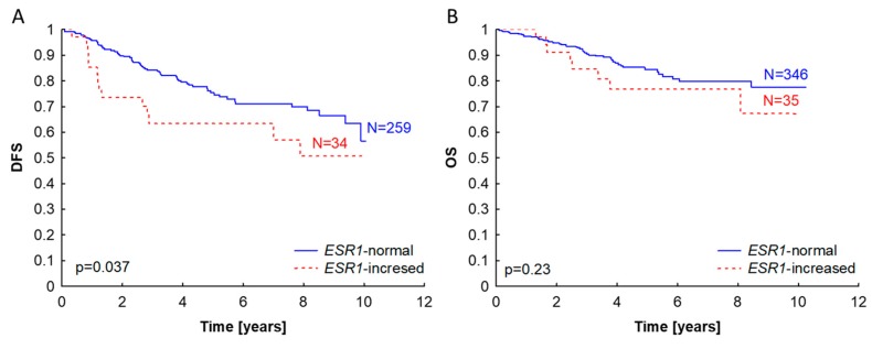
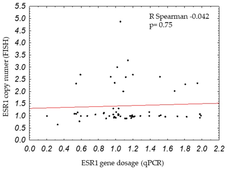
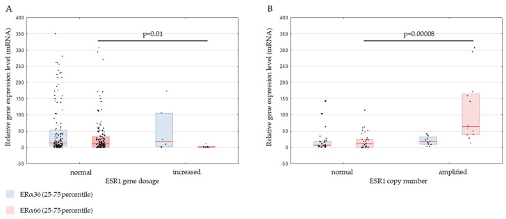
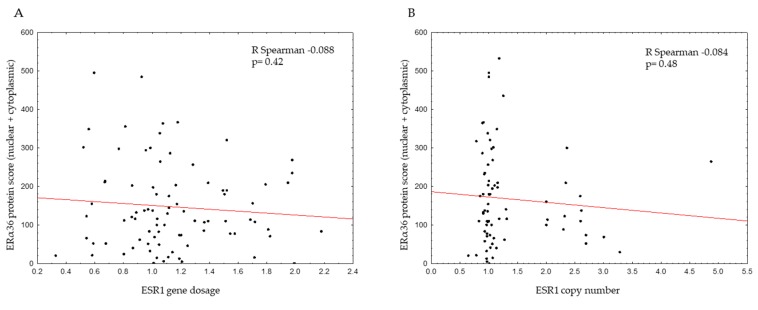
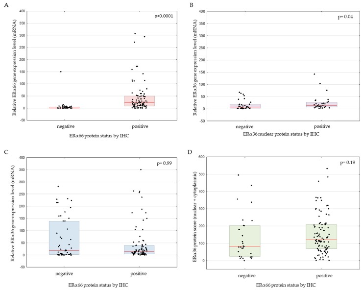
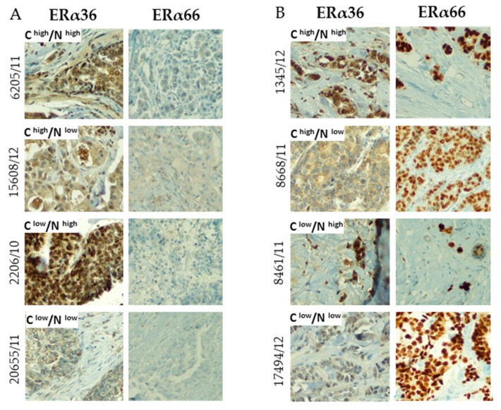
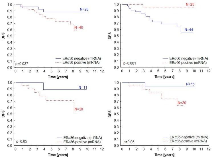
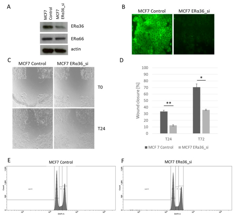
The amplification of estrogen receptor alpha (ERα) encoded by the ESR1 gene has been described as having a prognostic role in breast cancer patients. However, increased dosage of the ESR1 gene (tested by real-time PCR) is also observed in ER-negative breast cancers, which might suggest the expression of alternative isoforms of ERα (other than classical ERα of 66 kDa). In the current work, we have investigated the ESR1 gene dosage in 402 primary breast cancer patients as well as the expression of ERα isoforms-ERα66 and ERα36-on mRNA and protein levels. The obtained results were correlated with clinicopathological data of the patients. Results showed that increased ESR1 gene dosage is not related to ESR1 gene amplification measured by fluorescent in situ hybridization (FISH), but it correlates with the decreased expression of ERα66 isoform (p = 0.01). Interestingly, the short ER isoform ERα36 was expressed in samples with increased ESR1 gene dosage, suggesting that genomic aberration might influence the expression of that particular isoform. Similarly to ESR1 increased gene dosage, high ERα36 expression was linked with the decreased disease-free survival of the patients (p = 0.05), which was independent of the status of the classical ERα66 level in breast tumors.

Keywords: ERα36, ERα66, gene amplification; breast cancer; estrogen receptor; prognostic factor.


Conflict of interest statement

The authors declare no conflict of interest.

Figures

Figure 1
Figure 1
Kaplan–Meier survival curves according to ESR1 gene dosage status (measured by qPCR) in primary breast tumors. ESR1-normal status was described as ESR1/APP ratio <2, ESR1-increased as ESR1/APP ratio ≥2. The probability of disease-free survival (A) and overall survival (B) are shown.
Figure 2
Figure 2
Correlation between ESR1 copy number (measured by fluorescent in situ hybridization, or FISH) and ESR1 gene dosage (measured by qPCR).
Figure 3
Figure 3
ERα36 and ERα66 relative gene expression levels in primary breast cancers classified according to ESR1 gene dosage status measured by qPCR (A) or ESR1 gene copy number measured by FISH (B). The vertical red line represents the median gene expression level. * p < 0.05, ** p < 0.0001. ERα: estrogen receptor alpha.
Figure 4
Figure 4
Correlation between ERα36 protein score (total nuclear and cytoplasmic score and ESR1 gene dosage (measured by qPCR) (A) or ESR1 copy number (measured by FISH) (B).
Figure 5
Figure 5
ERα66 relative expression according to ERα66 immunohistochemical (IHC) protein status (A), ERα36 relative expression according to ERα36 nuclear protein (B), ERα36 expression according to ERα66 protein status (C), and ERα36 protein immunohistochemical score according to ERα66 protein status (D).
Figure 6
Figure 6
Exemplary photos of immunohistochemical staining of breast cancer samples with different ERα66 status (panel A – negative, panel B – positive according to Allred score) and ERα36 status. The ERa36 staining pattern has additionally been divided into positive and negative in cytoplasmic (C) and nuclear (N) localization based on the immunohistochemical score of the samples. All photos were taken under 20× magnification.
Figure 7
Figure 7
Kaplan–Meier survival curves presenting disease-free survival (DFS) according to ERα36 (A) and ERα66 (B) mRNA levels (measured by qPCR) in primary breast tumors. (C,D) Subgroup analysis for ERα36 expression in ERα66 protein-negative (p = 0.05) (C) and positive (D) patients (p = 0.05). Median relative gene expression was a cut-off value for the classification of samples into negative and positive.
Figure 8
Figure 8
Immunofluorescent staining of two breast cancer cell lines: MCF7 (AD) and BT474 (EH). Staining for: ERα66 (red), ERα36 (green), and nucleus (blue).
Figure 9
Figure 9
Silencing the ERα36 by siRNA confirmed by Western blotting (A) and immunofluorescence (B). Decrease in cell migration measured by wound healing assay in cells with silenced ERα36 (C,D, * p = 0.005, ** p = 0.002). Cell cycle analysis by flow cytometry in the MCF7 wild-type cell line (MCF7 Control) and with ERα36 silenced (MCF7 ERα36_si) showed no differences in the number of cells in a given cell cycle phase (E,F).

Similar articles

Cited by

References

    1. Orlando L., Schiavone P., Fedele P., Calvani N., Nacci A., Rizzo P., Marino A., D’Amico M., Sponziello F., Mazzoni E., et al. Molecularly targeted endocrine therapies for breast cancer. Cancer Treat Rev. 2010;36:67–71. doi: 10.1016/S0305-7372(10)70023-2. - DOI - PubMed
    1. Hammond M.E., Hayes D.F., Dowsett M., Allred D.C., Hagerty K.L., Badve S., Fitzgibbons P.L., Francis G., Goldstein N.S., Hayes M., et al. American Society of Clinical Oncology/College of American Pathologists guideline recommendations for immunohistochemical testing of estrogen and progesterone receptors in breast cancer. Arch. Pathol. Lab. Med. 2010;134:907–922. doi: 10.1043/1543-2165-134.6.907. - DOI - PMC - PubMed
    1. Goldhirsch A., Winer E.P., Coates A.S., Gelber R.D., Piccart-Gebhart M., Thürlimann B., Senn H.J., Panel members Personalizing the treatment of women with early breast cancer: Highlights of the St Gallen International Expert Consensus on the Primary Therapy of Early Breast Cancer 2013. Ann. Oncol. 2013;24:2206–2223. doi: 10.1093/annonc/mdt303. - DOI - PMC - PubMed
    1. Holst F., Stahl P.R., Ruiz C., Hellwinkel O., Jehan Z., Wendland M., Lebeau A., Terracciano L., Al-Kuraya K., Jänicke F., et al. Estrogen receptor alpha (ESR1) gene amplification is frequent in breast cancer. Nat. Genet. 2007;39:655–660. doi: 10.1038/ng2006. - DOI - PubMed
    1. Moelans C.B., Holst F., Hellwinkel O., Simon R., van Diest P.J. ESR1 amplification in breast cancer by optimized RNase FISH: Frequent but low-level and heterogeneous. PLoS ONE. 2013;8:e84189. doi: 10.1371/journal.pone.0084189. - DOI - PMC - PubMed