. 2019 Apr 16;20(8):1884.
doi: 10.3390/ijms20081884.

Oligomeric Architecture of Mouse Activating Nkrp1 Receptors on Living Cells

Affiliations

Oligomeric Architecture of Mouse Activating Nkrp1 Receptors on Living Cells

Ljubina Adámková et al. Int J Mol Sci. .

Abstract

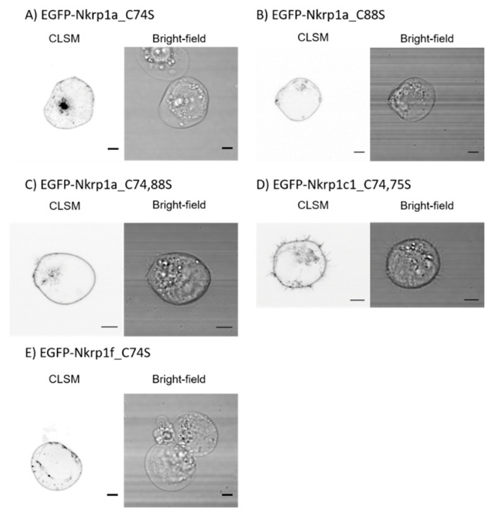
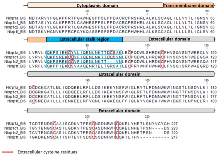
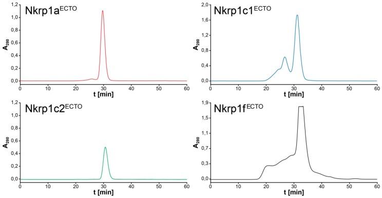
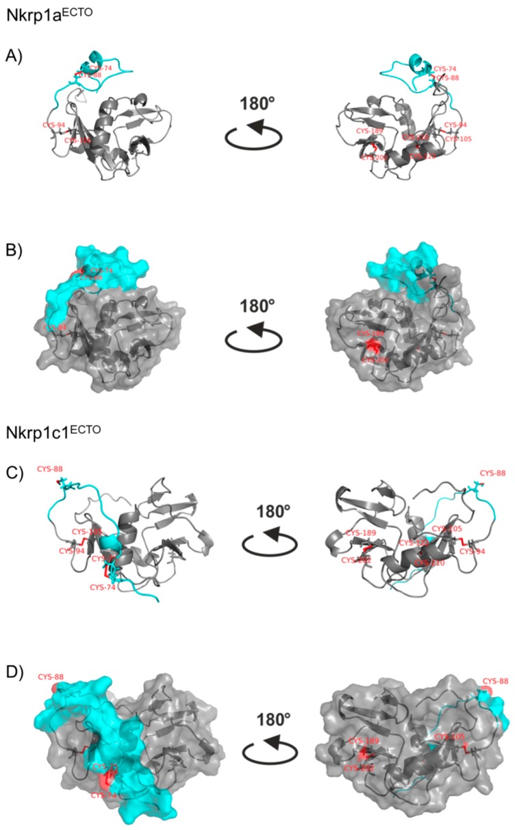
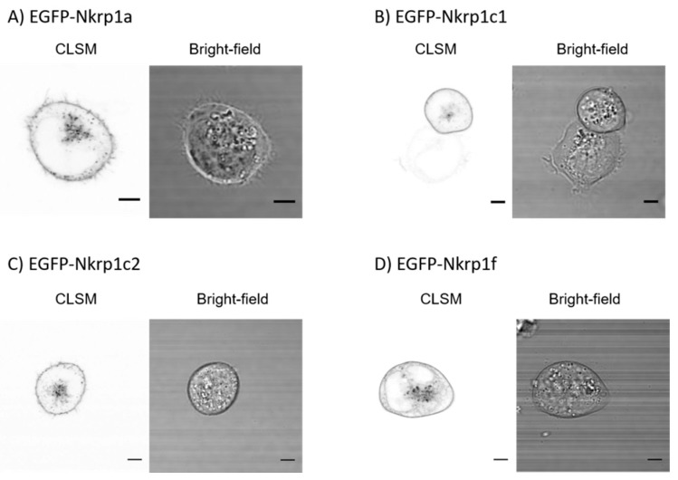
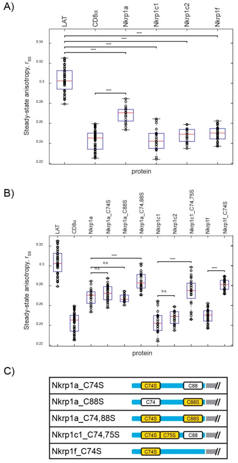
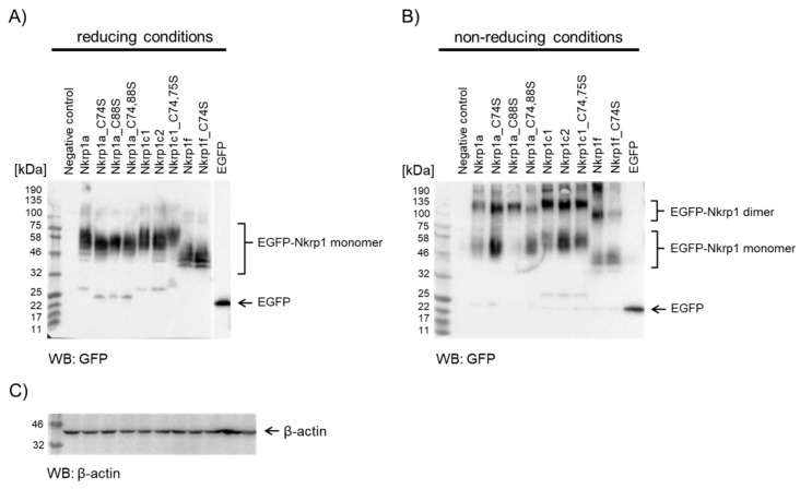
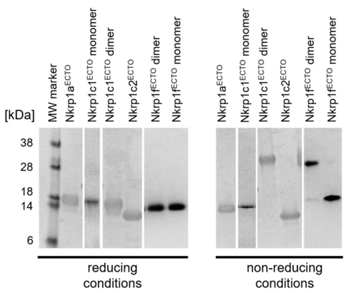
Mouse activating Nkrp1 proteins are commonly described as type II transmembrane receptors with disulfide-linked homodimeric structure. Their function and the manner in which Nkrp1 proteins of mouse strain (C57BL/6) oligomerize are still poorly understood. To assess the oligomerization state of Nkrp1 proteins, mouse activating EGFP-Nkrp1s were expressed in mammalian lymphoid cells and their oligomerization evaluated by Förster resonance energy transfer (FRET). Alternatively, Nkrp1s oligomers were detected by Western blotting to specify the ratio between monomeric and dimeric forms. We also performed structural characterization of recombinant ectodomains of activating Nkrp1 receptors. Nkrp1 isoforms c1, c2 and f were expressed prevalently as homodimers, whereas the Nkrp1a displays larger proportion of monomers on the cell surface. Cysteine-to-serine mutants revealed the importance of all stalk cysteines for protein dimerization in living cells with a major influence of cysteine at position 74 in two Nkrp1 protein isoforms. Our results represent a new insight into the oligomerization of Nkrp1 receptors on lymphoid cells, which will help to determine their function.

Keywords: Förster resonance energy transfer; Nkrp1; cysteine; dimerization; disulfide bond arrangement.


Conflict of interest statement

The authors declare no conflict of interest.

Figures

Figure A1
Figure A1
Subcellular localization of cysteine-to-serine mutant forms of EGFP-Nkrp1 fusion proteins. Confocal laser scanning microscopy (CLSM) images of live Jurkat cells transfected with (A) EGFP-Nkrp1a_C74S, (B) EGFP-Nkrp1a_C88S, (C) EGFP-Nkrp1a_C74,88S, (D) EGFP-Nkrp1c1_C74,75S fusion protein. The left picture of each panel shows the EGFP channel scan, and the right half perform the corresponding bright-field image. All EGFP-Nkrp1 cysteine-to-serine mutants were expressed mainly on the plasma membrane of live Jurkat cells. Scale bars in each panel: 5 µm.
Figure A2
Figure A2
Protein sequence alignment of activating Nkrp1 proteins from C57BL/6 mouse. Extracellular cysteine residues are in red boxes and the stalk region is indicated in a blue box. The most different isoform by protein sequence is the Nkrp1f. The Nkrp1a and the Nkrp1c have 87% simillarity.
Figure A3
Figure A3
Nkrp1 purification. Size-exclusion chromatography profile of refolded Nkrp1aECTO, Nkrp1c1ECTO, Nkrp1c2ECTO and Nkrp1fECTO. Proteins were purified on calibrated Superdex 75 column with flow rate 0.4 mL/min. The Nkrp1aECTO was eluted in 150 mM NaCl, 15 mM Tris-HCl (pH 7.5) and 1 mM NaN3. The elution buffer for Nkrp1c1ECTO, Nkrp1c2ECTO and Nkrp1fECTO consisted of 10 mM HEPES (pH 7.4), 100 mM NaCl and 1 mM NaN3.
Figure A4
Figure A4
Structural models of Nkrp1aECTO and Nkrp1c1ECTO proteins. Ribbon diagrams of Nkrp1aECTO (A) and Nkrp1c1ECTO (C) with the stalk regions indicated in cyan. The molecular surfaces of Nkrp1aECTO (B) and Nkrp1c1ECTO (D) are represented in faint gray, and the stalk regions in cyan. Cysteine residues are shown in red.
Figure 1
Figure 1
Subcellular localization of EGFP-Nkrp1 fusion proteins in living Jurkat T cells. Confocal laser scanning microscopy (CLSM) images of Jurkat cells transfected with (A) EGFP-Nkrp1a, (B) EGFP-Nkrp1c1, (C) EGFP-Nkrp1c2, and (D) EGFP-Nkrp1f. Left part of each panel shows the image from EGFP channel and right part the corresponding bright-field image. Scale bars: 5 µm.
Figure 2
Figure 2
Homo-FRET measurements of EGFP-Nkrp1 proteins in living Jurkat cells. (A) Dot plot of steady-state anisotropy, rss, of Nkrp1a, c1, c2 and f isoforms as compared to the monomeric (LAT) and dimeric (CD8α) controls. (B) Assessment of the steady-state anisotropy values of mutants Nkrp1/a_C74S,/a_C88S,/a_C74,88S, Nkrp1c1_C74,75S and Nrkp1f_C74S with their native counterparts. The red line marks median of the whole set and the bottom and top edges of the box indicate the 25th and 75th percentiles, respectively. The whiskers extend to the most extreme data points not considered outliers. Statistical significance of differences is marked with asterisks: p < 0.00 ***, p < 0.0001 ****, ‘n.s.’ not significant. All median steady-state anisotropy and p-values are in the Appendix A Table A2 and Table A3, respectively. (C) Scheme showing cysteine residues in the stalk region and their serine mutants (yellow).
Figure 3
Figure 3
Analysis of EGFP-Nkrp1 fusion proteins by immunoblotting of reducing and non-reducing SDS-PAGE. (A) Immunoblotting of EGFP-Nkrp1 proteins transiently expressed in COS-7 cells, lysed and separated by SDS-PAGE under reducing conditions (100 mM DTT). All EGFP-Nkrp1 fusion proteins appear as monomers (apparent at 40-60 kDa). (B) Samples as in A were separated by SDS-PAGE under non-reducing conditions. EGFP-Nkrp1 fusion proteins appear as monomers, dimers and higher order oligomers with different ratio between these populations. Molecular markers are to the left of the panels. Regions corresponding to the Nkrp1 monomers and dimers, as well as EGFP only are indicated to the right. The negative control corresponds to untransfected COS-7 cells treated as all other samples. For positive control, EGFP transiently expressed in COS-7 cells was used. (C) Loading control: immunoblotting of β-actin detected by specific antibody at 1:1500 dilution.
Figure 4
Figure 4
Analysis of purified recombinant Nkrp1ECTO proteins. Fractions from size-exclusion chromatography of the Nkrp1ECTO proteins were analyzed by 15% SDS-PAGE. Proteins appeared homogenous under reducing and non-reducing conditions. Nkrp1c1ECTO and Nkrp1fECTO isoforms have formed a dimer under non-reducing conditions (≈ 35 kDa).
Figure 5
Figure 5
Cysteine disulfide bonds determined for the recombinant Nkrp1ECTO proteins. Schematic illustration of intramolecular disulfide bonds of murine recombinant Nkrp1ECTO proteins studied by LC-MS. A stalk region is indicated in blue, the extracellular C-Type Lectin-like Domain in gray (predicted by InterProScan tool) and cysteine residues involved in intermolecular bonding are in red boxes.

Similar articles

Cited by

References

    1. Vivier E., Tomasello E., Baratin M., Walzer T., Ugolini S. Functions of natural killer cells. Nat. Immunol. 2008;9:503–510. doi: 10.1038/ni1582. - DOI - PubMed
    1. Vivier E., Raulet D.H., Moretta A., Caligiuri M.A., Zitvogel L., Lanier L.L., Yokoyama W.M., Ugolini S. Innate or adaptive immunity? The example of natural killer cells. Science. 2011;331:44–49. doi: 10.1126/science.1198687. - DOI - PMC - PubMed
    1. Lanier L.L. NK cell receptors. Annu. Rev. Immunol. 1998;16:359–393. doi: 10.1146/annurev.immunol.16.1.359. - DOI - PubMed
    1. Giorda R., Trucco M. Mouse NKR-P1. A family of genes selectively coexpressed in adherent lymphokine-activated killer cells. J. Immunol. 1991;147:1701–1708. - PubMed
    1. Giorda R., Rudert W.A., Vavassori C., Chambers W.H., Hiserodt J.C., Trucco M. NKR-P1, a signal transduction molecule on natural killer cells. Science. 1990;249:1298–1300. doi: 10.1126/science.2399464. - DOI - PubMed

LinkOut - more resources