Review
. 2019 Apr 16;8(4):522.
doi: 10.3390/jcm8040522.

Personalized Three-Dimensional Printed Models in Congenital Heart Disease

Affiliations
Review

Personalized Three-Dimensional Printed Models in Congenital Heart Disease

Zhonghua Sun et al. J Clin Med. .

Abstract

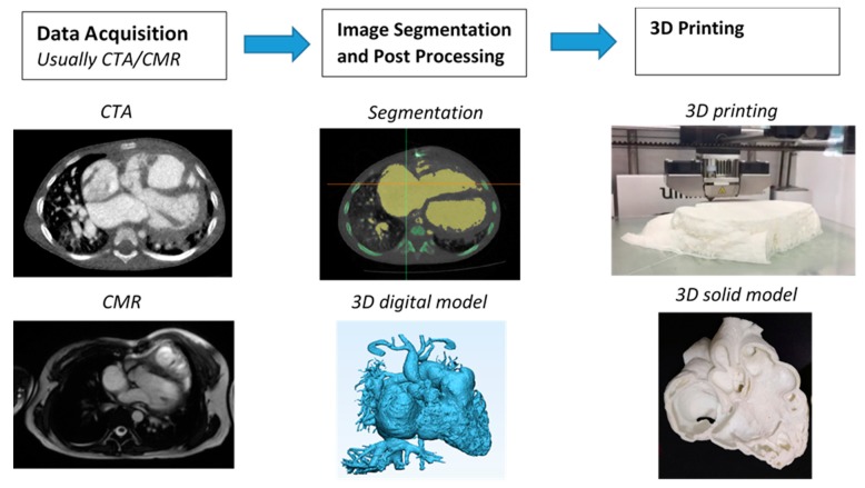
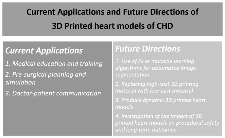
Patient-specific three-dimensional (3D) printed models have been increasingly used in cardiology and cardiac surgery, in particular, showing great value in the domain of congenital heart disease (CHD). CHD is characterized by complex cardiac anomalies with disease variations between individuals; thus, it is difficult to obtain comprehensive spatial conceptualization of the cardiac structures based on the current imaging visualizations. 3D printed models derived from patient's cardiac imaging data overcome this limitation by creating personalized 3D heart models, which not only improve spatial visualization, but also assist preoperative planning and simulation of cardiac procedures, serve as a useful tool in medical education and training, and improve doctor-patient communication. This review article provides an overall view of the clinical applications and usefulness of 3D printed models in CHD. Current limitations and future research directions of 3D printed heart models are highlighted.

Keywords: congenital heart disease; heart models; medical education; pre-operative planning; simulation; three-dimensional printing.


Conflict of interest statement

Authors declare no conflict of interest.

Figures

Figure 1
Figure 1
Steps involved in fabrication of 3D printed heart models. CTA—computed tomography angiography; CMR—cardiac magnetic resonance; 3D—three-dimensional.
Figure 2
Figure 2
Comparison of virtual 3D reconstruction model (left) and 3D printed heart model (right). Reprinted with permission under the open access from Lau et al. [16].
Figure 3
Figure 3
Scatterplot showing measurements of 3D printed model in comparison with those from cardiac computed tomography (CT) images at 10 anatomical locations. CCTA—cardiac computed tomography angiography. Reprinted with permission under the open access from Lau et al. [16].
Figure 4
Figure 4
3D printed heart models showing normal anatomy and pathology. (a) Normal heart model created from cardiac CT and is partitioned into three pieces allowing visualization of interventricular septum. (b) Repaired Tetralogy of Fallot (ToF) from an adult patient. The model was created from cardiac magnetic resonance imaging (MRI) and separated into two pieces allowing for clear visualization of overriding aorta and pulmonary infundibular stenosis. (c) Unrepaired ToF heart model from an infant. The model was created from 3D echocardiographic images and partitioned into two pieces showing the ventricular septal defect (VSD). (d) Unrepaired ToF heart model from an infant with superior and inferior portions showing VSD and the aortic overriding in relation to the VSD. Reprinted with permission under the open access from Loke et al. [22]. Ao—Aorta; MPA—Main Pulmonary Artery; LV—Left Ventricle; RV—Right Ventricle; RVOT—Right Ventricular Outflow Tract; VSD—Ventricular Septal Defect; ToF—Tetralogy of Fallot.
Figure 5
Figure 5
Impact of 3D printed heart models on medical education. Improvement was found in residents’ knowledge on congenital heart disease with use of 3D printed models when compared to 2D images. A statistically significant difference was noticed in satisfaction ratings in the group having 3D printed heart models when compared to the control group (p = 0.03). While residents in the 3D printed model group had higher self-efficacy scores, this did not reach significant difference compared to the control group using 2D images/drawings (p = 0.39). Reprinted with permission under the open access from Loke et al. [22].
Figure 6
Figure 6
Statistically significant differences were noted in confidence (A), knowledge (B), and satisfaction (C) amongst participants comparing responses before (“Pre”) and after (“Post”) their medical consultation. (A) 1 refers to not at all confident and 5 very confident. (B) Each point represents a point in knowledge, as marked based on the correct name of primary diagnosis, correctly identified keywords, and correct use of diagrams. (C) 1 indicates very dissatisfied and 5 very satisfied. The red lines indicate average score. Reprinted with permission under the open access from Biglino et al. [36].
Figure 7
Figure 7
Participants’ response to different statements on the usefulness of 3D printed models. Reprinted with permission under the open access from Biglino et al. [36].
Figure 8
Figure 8
CT scan of 3D printed heart models created using different printing materials. (A) 3D volume rendering showing the 3D printed models without contrast medium (top: Tango Plus material, bottom: TPU material). (B,C) Coronal multiplanar reformatted contrast-enhanced CT images showing 3D printed models with Tango Plus (left) and TPU (right) materials. Air bubbles are noticed in the model with TPU material. TPU—thermoplastic polyurethane. Reprinted with permission under the open access from Lau et al. [45].
Figure 9
Figure 9
Comparison of low-cost (left image) with high-cost (right image) 3D printed heart model with similar accuracy in delineating cardiac anatomy and ventricular septal defect.
Figure 10
Figure 10
Example of double outlet right ventricle with aorta and pulmonary artery arising from the right ventricle and perimembranous ventricular septal defect from computed tomography images (AC). Anterior view of the 3D printed heart model, aorta, and pulmonary artery are side-by-side with both arising from the right ventricle (D). Perimembranous VSD remoted from the arteries. Position of potential intracardiac tunnel from the left ventricle to the aorta is shown as the solid lines (E). AO—ascending aorta; LA—left atrium; LV—left ventricle; PA—pulmonary artery; RA—right atrium; RV—right ventricle; VSD—ventricular septal defect. Reprinted with permission from Zhao et al. [32].
Figure 11
Figure 11
Summary of current applications and future research directions of 3D printing in congenital heart disease. 3D—three-dimensional; CHD—congenital heart disease; AI—artificial intelligence.

References

    1. Yao R., Xu G., Mao S.S., Yang H.Y., Sang X.T., Sun W., Mao Y.L. Three-dimensional printing: Review of application in medicine and hepatic surgery. Cancer. Biol. Med. 2016;13:443–451. - PMC - PubMed
    1. Chepelev L., Souza C., Althobaity W., Miguel O., Krishna S., Akyuz E., Hodgdon T., Torres C., Wake N., Alexander A., et al. Preoperative planning and tracheal stent design in thoracic surgery: A primer for the 2017 Radiological Society of North America (RSNA) hands-on course in 3D printing. 3D Print Med. 2017;3:14. doi: 10.1186/s41205-017-0022-3. - DOI - PMC - PubMed
    1. Matsumoto J.S., Morris J.M., Foley T.A., Williamson E.E., Leng S., McGee K.P., Kuhlmann J.L., Nesberg L.E., Vrtiska T.J. Three-dimensional physical modeling: applications and experience at Mayo Clinic. Radiographics. 2015;35:1989–2006. doi: 10.1148/rg.2015140260. - DOI - PubMed
    1. Mitsouras D., Liacouras P., Imanzadeh A., Giannopolous A.A., Cai T., Kumamaru K.K., George E., Wake N., Caterson E.J., Pomahac B., et al. Medical 3D printing for the radiologist. Radiographics. 2015;35:1965–1988. doi: 10.1148/rg.2015140320. - DOI - PMC - PubMed
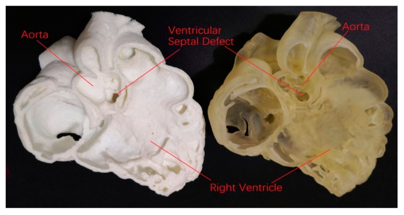
    1. Costello J., Olivieri L., Krieger A., Thabit O., Marshall M.B., Yoo S.J., Kim P.C., Jonas R.A., Nath D.S. Utilizing three-dimensional printing technology to assess the feasibility of high-fidelity synthetic ventricular septal defect models for simulation in medical education. World J. Pediatr. Congenit. Heart Surg. 2014;5:421–426. doi: 10.1177/2150135114528721. - DOI - PubMed

LinkOut - more resources