. 2019 Apr 16;24(8):1496.
doi: 10.3390/molecules24081496.

Anticancer Effects of Five Biflavonoids from Ginkgo Biloba L. Male Flowers In Vitro

Affiliations

Anticancer Effects of Five Biflavonoids from Ginkgo Biloba L. Male Flowers In Vitro

Min Li et al. Molecules. .

Abstract

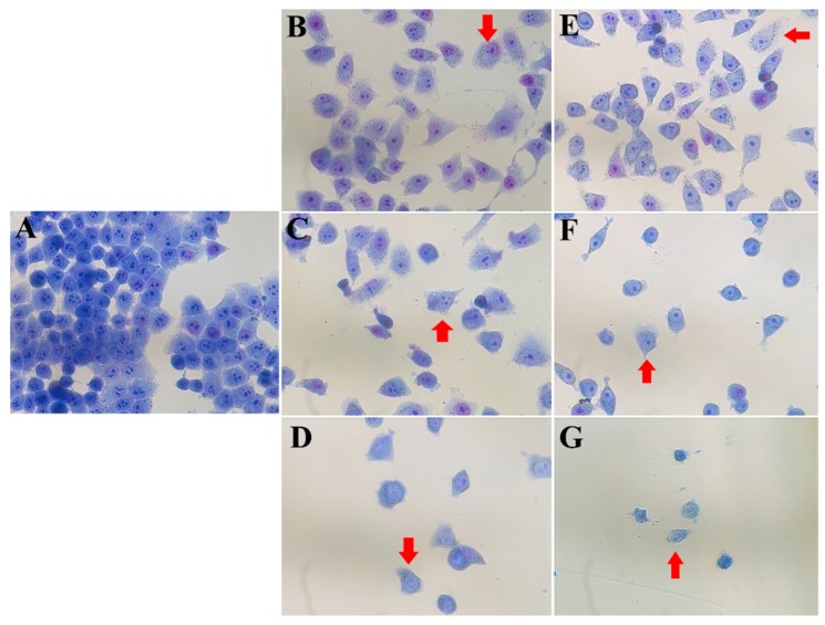
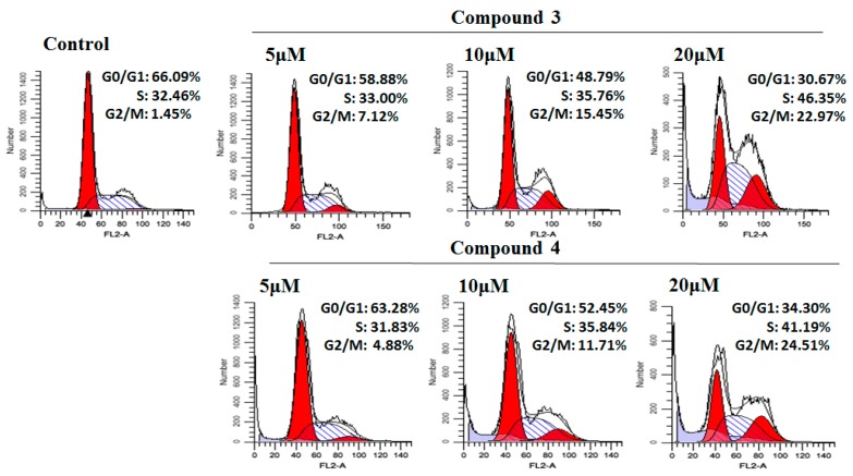
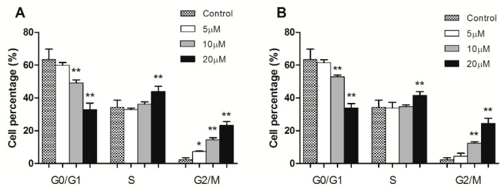
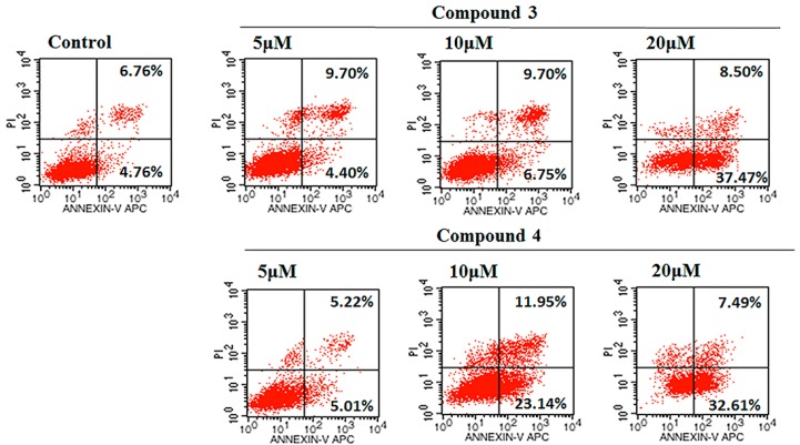
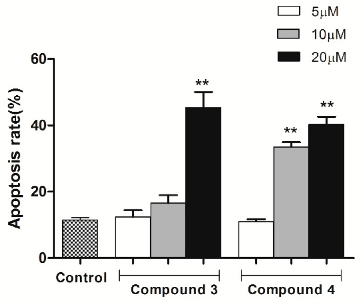
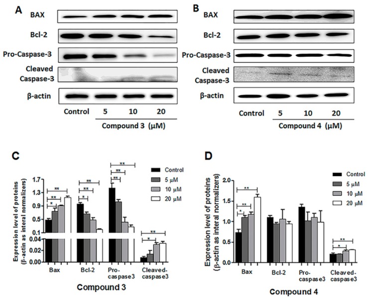
Ginkgo biloba L., an ancient dioecious gymnosperm, is now cultivated worldwide for landscaping and medical purposes. A novel biflavonoid-amentoflavone 7''-O-β-D-glucopyranoside (1)-and four known biflavonoids were isolated and identified from the male flowers of Ginkgo. The anti-proliferative activities of five biflavonoids were evaluated on different cancer lines. Bilobetin (3) and isoginkgetin (4) exhibited better anti-proliferative activities on different cancer lines. Their effects were found to be cell-specific and in a dose and time dependent manner for the most sensitive HeLa cells. The significant morphological changes validated their anticancer effects in a dose-dependent manner. They were capable of arresting the G2/M phase of the cell cycle, inducing the apoptosis of HeLa cells dose-dependently and activating the proapoptotic protein Bax and the executor caspase-3. Bilobetin (3) could also inhibit the antiapoptotic protein Bcl-2. These might be the mechanism underlying their anti-proliferation. In short, bilobetin (3) and isoginkgetin (4) might be the early lead compounds for new anticancer agents.

Keywords: Ginkgo biloba flowers; anticancer; biflavonoids; bilobetin; isoginkgetin.


Conflict of interest statement

The authors declare no conflict of interest.

Figures

Figure 1
Figure 1
Structures of compounds 15 (A). Key heteronuclear multiple bond correlation (HMBC) correlations (formula image
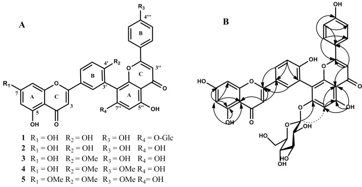
) and nuclear overhauser enhancement spectroscopy (NOESY)correlations (formula image) of compound 1 (B).
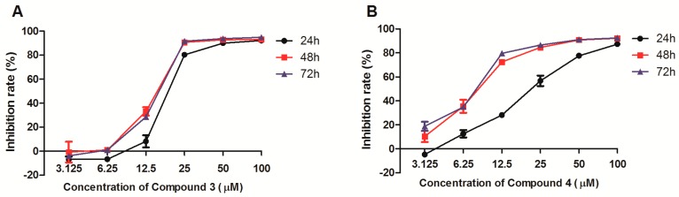
Figure 2
Figure 2
Effects of compounds 3 (A) and 4 (B) on viability of HeLa cell at different concentrations for different incubation time. HeLa cells were incubated with compound 3 or 4 at the concentration of 3.1 μM to 100 μM, and then the cytotoxic activity was evaluated using the 3-(4,5-dimethylthiazol-2-yl)-2,5-diphenyltetrazolium bromide (MTT) assay at 24 h, 48 h, and 72 h, respectively. The values are expressed as mean ± SD of triplicate experiments.
Figure 3
Figure 3
Morphological structure of HeLa cancer cells through Giemsa staining after 48 h of treatment with different concentration of compounds 3 or 4. (A) control (untreated), (B) compound 3 (15 μM), (C) compound 3 (20 μM), (D) compound 3 (25 μM), (E) compound 4 (10 μM), (F) compound 4 (15 μM), and (G) compound 4 (20 μM). The arrows show the obviously changed cells.
Figure 4
Figure 4
Cell cycle analysis of HeLa cells treated with compounds 3 and 4 (5 μM, 10 μM, and 20 μM) for 24 h by flow cytometry.
Figure 5
Figure 5
Histograms (A,B) showed the percentage of compounds 3 and 4 treated cells in different phases of the cell cycle, respectively. The values are expressed as mean ± SD of triplicate experiments. * p < 0.05, ** p < 0.01 compared with the control group.
Figure 6
Figure 6
Apoptosis was assessed using Annexin V- fluorescein isothiocyanate (FITC) and propidium iodide (PI) staining after HeLa cells were treated with compounds 3 and 4 (5 μM, 10 μM, and 20 μM) for 24 h, measured by flow cytometry.
Figure 7
Figure 7
Histograms showed the apoptosis rate (early and late apoptosis) of HeLa cells treated with compounds 3 and 4. The values are expressed as mean ± SD of triplicate experiments. ** p < 0.01 compared with the control group.
Figure 8
Figure 8
Western blots were performed to analyze relative protein expression levels involved in the cell apoptosis including Bax, Bcl-2, pro-caspase-3, and cleaved caspase-3(A,B). β -actin was used as a control. HeLa cells were treated with different concentrations (5, 10, 20 μM) of compound 3 or 4 for 24 h. The obvious change could be seen in the histogram in a concentration-dependent manner (C,D), * p < 0.05, ** p < 0.01, compared with the control group.

References

    1. Ni L., Zhao H., Tao L., Li X., Zhou Z., Sun Y., Chen C., Wei D., Liu Y., Diao G. Synthesis, in vitro cytotoxicity, and structure-activity relationships (SAR) of multidentate oxidovanadium(iv) complexes as anticancer agents. Dalton Trans. 2018;47:10035–10045. doi: 10.1039/C8DT01778F. - DOI - PubMed
    1. Du H., Huang Y., Hou X., Quan X., Jiang J., Wei X., Liu Y., Li H., Wang P., Zhan M., et al. Two novel camptothecin derivatives inhibit colorectal cancer proliferation via induction of cell cycle arrest and apoptosis in vitro and in vivo. Eur J Pharm Sci. 2018;123:546–559. doi: 10.1016/j.ejps.2018.08.018. - DOI - PubMed
    1. Ahmad K., Hafeez Z.B., Bhat A.R., Rizvi M.A., Thakura S.C., Azam A., Athara F. Antioxidant and apoptotic effects of Callistemon lanceolatus leaves and their compounds against human cancer cells. Biomed. Pharmacother. 2018;106:1195–1209. doi: 10.1016/j.biopha.2018.07.016. - DOI - PubMed
    1. Mignani S., Rodrigues J., Tomas H., Zablocka M., Shi X.Y., Caminade A., Majoral J.P. Dendrimers in combination with natural products and analogues as anti-cancer agents. Chem. Soc. Rev. 2018;47:514. doi: 10.1039/C7CS00550D. - DOI - PubMed
    1. Zhang B., Huang R.Z., Hua J., Liang H., Pan Y.M., Dai L.M., Liang D., Wang H.S. Antitumor lignanamides from the aerial parts of Corydalis saxicola. Phytomedicine. 2016;23:1599–1609. doi: 10.1016/j.phymed.2016.09.006. - DOI - PubMed

MeSH terms