. 2019 Apr 16;24(8):1498.
doi: 10.3390/molecules24081498.

Melittin Exerts Beneficial Effects on Paraquat-Induced Lung Injuries In Mice by Modifying Oxidative Stress and Apoptosis

Affiliations

Melittin Exerts Beneficial Effects on Paraquat-Induced Lung Injuries In Mice by Modifying Oxidative Stress and Apoptosis

Bishoy El-Aarag et al. Molecules. .

Abstract

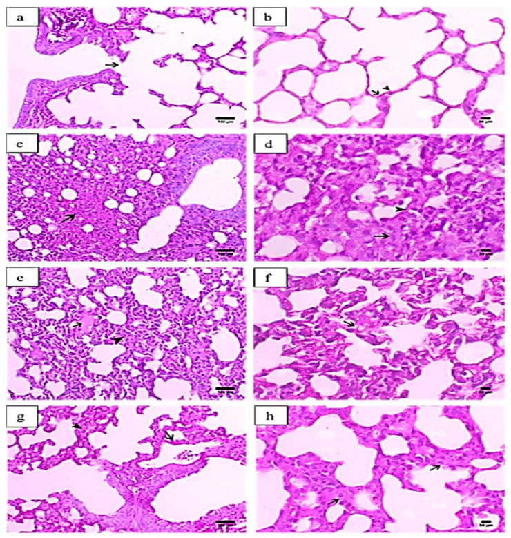
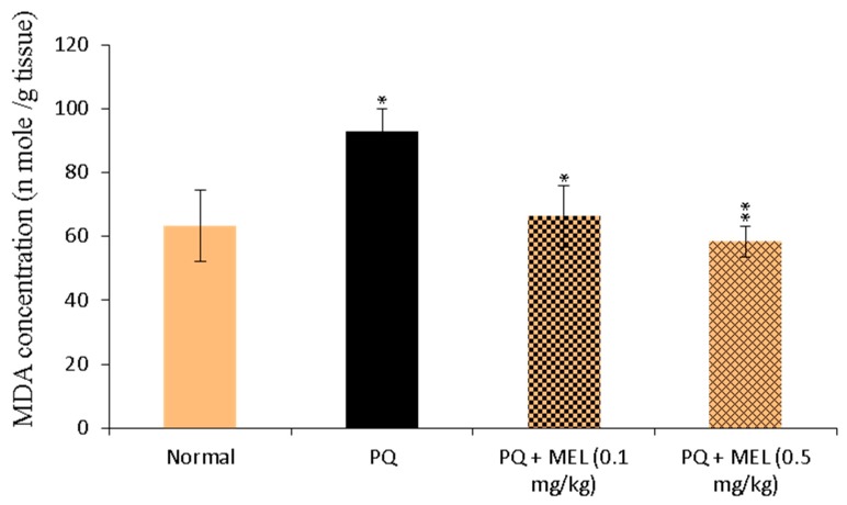
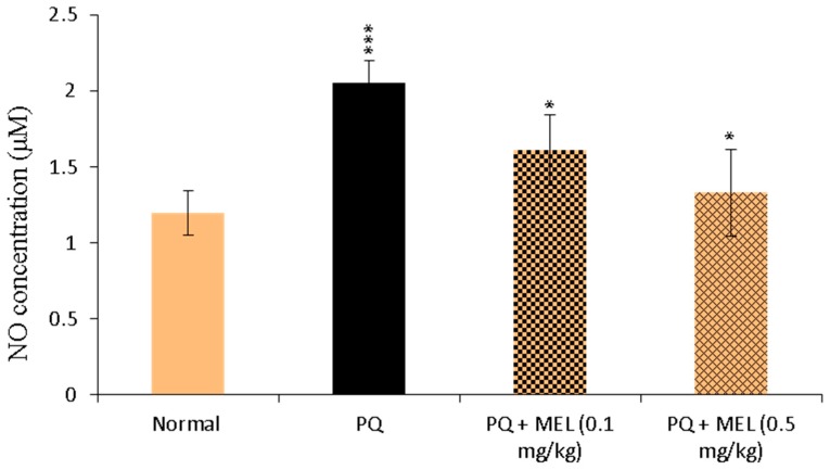
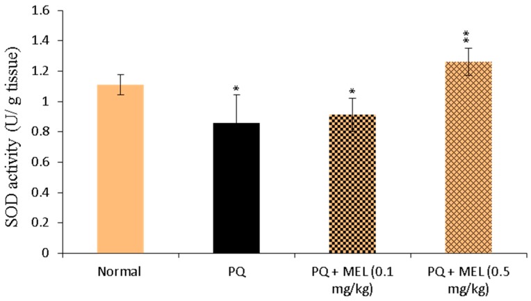
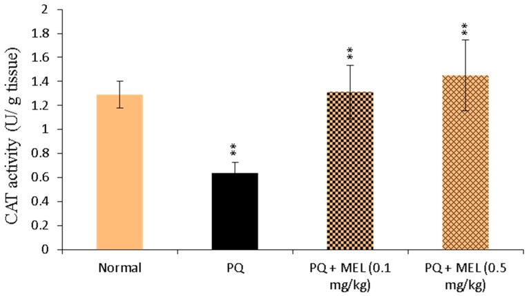
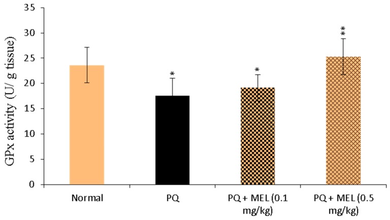
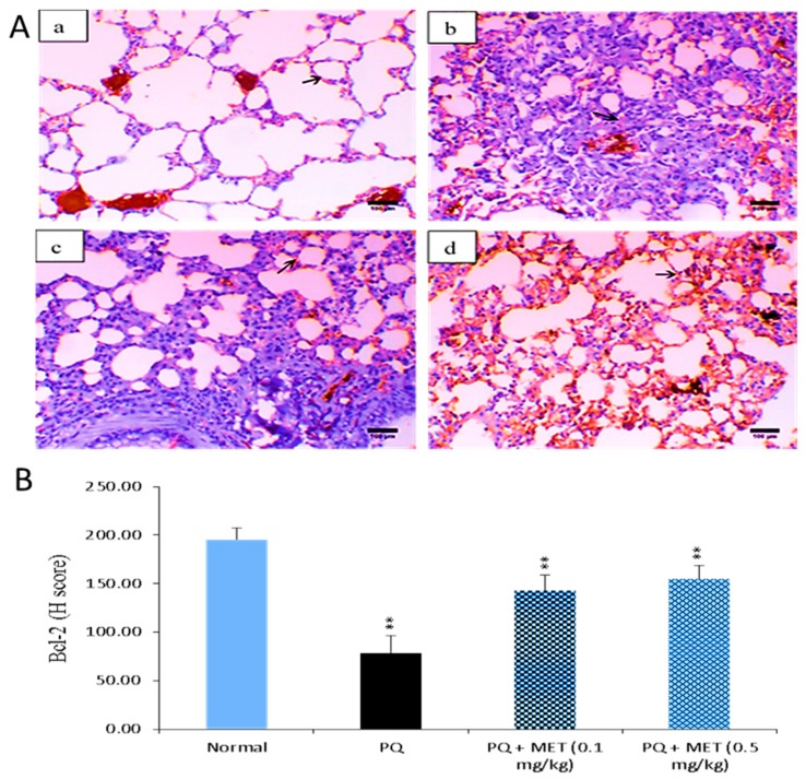
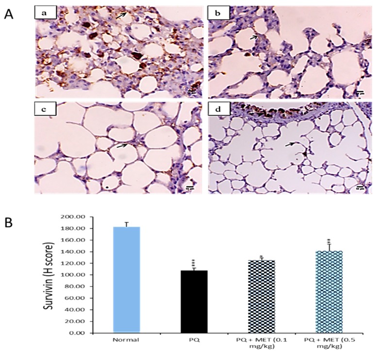
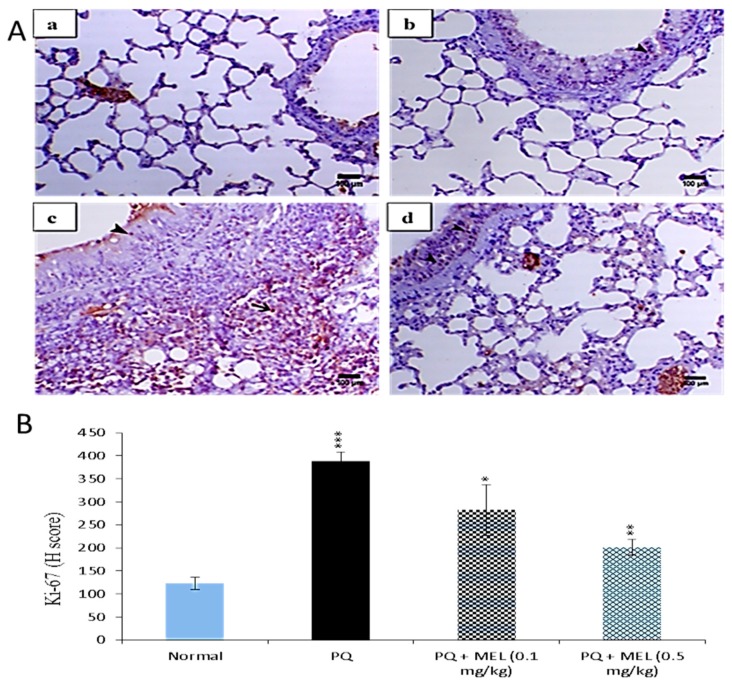
Melittin (MEL) is a 26-amino acid peptide with numerous biological activities. Paraquat (PQ) is one of the most widely used herbicides, although it is extremely toxic to humans. To date, PQ poisoning has no effective treatment, and therefore the current study aimed to assess for the first time the possible effects of MEL on PQ-induced lung injuries in mice. Mice received a single intraperitoneal (IP) injection of PQ (30 mg/kg), followed by IP treatment with MEL (0.1 and 0.5 mg/kg) twice per week for four consecutive weeks. Histological alterations, oxidative stress, and apoptosis in the lungs were studied. Hematoxylin and eosin (H&E) staining indicated that MEL markedly reduced lung injuries induced by PQ. Furthermore, treatment with MEL increased superoxide dismutase (SOD), catalase (CAT), and glutathione peroxidase (GPx) activity, and decreased malonaldehyde (MDA) and nitric oxide (NO) levels in lung tissue homogenates. Moreover, immunohistochemical staining showed that B-cell lymphoma-2 (Bcl-2) and survivin expressions were upregulated after MEL treatment, while Ki-67 expression was downregulated. The high dose of MEL was more effective than the low dose in all experiments. In summary, MEL efficiently reduced PQ-induced lung injuries in mice. Specific pharmacological examinations are required to determine the effectiveness of MEL in cases of human PQ poisoning.

Keywords: apoptosis; lung injury; melittin; oxidative stress; paraquat.


Conflict of interest statement

The authors declare no conflict of interest.

Figures

Figure 1
Figure 1
Histopathological graphs of lung sections from all groups. The lung sections were analyzed by hematoxylin and eosin (H&E) staining. Control group (a,b): (a) indicated normal terminal bronchiole ended with respiratory portion (arrow) (X200, scale bar = 100 µm), (b) showed alveoli lined with alveolar epithelial cells pnemocyte type I (arrow) and type II (arrowhead) (X400, scale bar = 50 µm). Paraquat (PQ) group (c,d): (c) presented necrosis in the alveolar tissue (arrow) (X200, scale bar = 100 µm), (d) showed excessive interstitial tissue thickening associated with septal cell proliferation (arrow) and alveolar hyaline membrane formation (arrowhead) (X400, scale bar = 50 µm). PQ + MEL (melittin) (0.1 mg/kg) group (e,f): (e) illustrated mild focal serous exudate within the alveoli (arrow) accompanied with a moderate degree of inter-alveolar interstitial tissue thickening (arrowhead) (X200, scale bar = 100 µm) (f) presented a moderate degree of thickening in the inter-alveolar septa and marked hyperplasia of pneumocyte type II in the lining epithelium (arrow) (X400, scale bar = 50 µm). PQ + MEL (0.5 mg/kg) group (g,h): (g) displayed decrease in the inter-alveolar thickening (arrowhead) with marked hyperplasia of pneumocyte type II (arrow) (X200, scale bar = 100 µm) (h) presented a decrease in the inter-alveolar septa (arrowhead) with an increase in the alveolar spaces associated with hyperplasia of pneumocyte type II (arrow) (X400, scale bar = 50 µm).
Figure 2
Figure 2
Effect of treatment with melittin (MEL) on the levels of malonaldehyde (MDA) in lung tissue homogenate. MDA (lipid peroxidation index) levels were determined in the lung tissue homogenate using MDA colorimetric assay. The absorbance of the formed product was measured at 534 nm. The results were expressed as nmol MDA per g tissue. Data are presented as mean ±SD. Significantly (* p < 0.05, ** p < 0.01) different from PQ group.
Figure 3
Figure 3
Effect of treatment with melittin (MEL) on the levels of nitric oxide (NO) level in lung tissue homogenate. The level of NO in the lung homogenate was measured according to the Griess method, using NO colorimetric assay. The formed azo dye was measured at 540 nm. NO levels were measured and expressed as µM. Data are presented as mean ±SD. Significantly (* p < 0.05, *** p < 0.001) different from PQ group.
Figure 4
Figure 4
Effect of treatment with melittin (MEL) on superoxide dismutase (SOD) activity in lung tissue homogenate. SOD activity was measured according to Beyer method, using SOD colorimetric assay. SOD activity was measured and expressed as U/g tissue. Data are presented as mean ±SD. Significantly (* p < 0.05, ** p < 0.01) different from PQ group.
Figure 5
Figure 5
Effect of treatment with melittin (MEL) on catalase (CAT) activity in lung tissue homogenate. CAT was determined in the lung tissue homogenate through the Aebi method, using CAT colorimetric assay. The color intensity was measured, and CAT activity was determined and expressed as U/g tissue. Data are presented as mean ±SD. Results are significantly (** p < 0.01) different from the PQ group.
Figure 6
Figure 6
Effect of treatment with melittin (MEL) on glutathione Peroxidase (GPx) activity in lung tissue homogenate. GPx was measured in the lung tissue homogenate by UV spectroscopic method. The decrease in the absorbance was measured at 340 nm. The level of GPx was measured and expressed as U/g tissue. Data are presented as mean ±SD. Results are significantly (* p < 0.05, ** p < 0.01) different from PQ group.
Figure 7
Figure 7
Effect of treatment with melittin (MEL) on the B-cell lymphoma-2 (Bcl-2) protein level in lung tissues. A: Representative lung immunohistochemical graphs (a: control group, b: paraquat (PQ) group, c: PQ + MEL (0.1 mg/kg) group, d: PQ + MEL (0.5 mg/kg) group); (a) showed a noticeable expression of bcl-2 immunostaining in the alveolar lining epithelium (arrow). (b) presented marked decrease in bcl-2 expression within the alveolar lining epithelium (arrow). (c) displayed mild to moderate increase of bcl-2 expression in the alveolar epithelium (arrow). (d) illustrated an obvious increase in bcl-2 expression within the alveolar epithelium (arrow). Magnification = X200 and scale bar = 100 µm. B: Statistical analysis of Bcl-2 H-score. Data are expressed as mean ±SD. Significantly (** p < 0.01) different to the PQ group.
Figure 8
Figure 8
Effect of treatment with melittin (MEL) on the survivin protein level in lung tissues. A: Representative lung immunohistochemical graphs (a: control group, b: paraquat (PQ) group, c: PQ + MEL (0.1 mg/kg) group, d: PQ + MEL (0.5 mg/kg) group); (a) showed a strong survivin expression within the alveolar epithelium (arrow) (X400, scale bar = 50 µm). (b) presented a low to mild survivin expression within the alveolar epithelium (arrow) (X400, scale bar = 50 µm). (c) indicated a moderate survivin expression in the alveolar epithelium (arrow) as well as in the bronchial epithelium (arrowhead) (X400, scale bar = 50 µm). (d) illustrated an increase in the survivin expression in the alveolar epithelium (arrow) and in the bronchial epithelium (arrowhead) (X200, scale bar = 100 µm). B: Statistical analysis of survivin H-score. Data are expressed as mean ±SD. Significantly (* p < 0.05, ** p < 0.01, *** p < 0.001) different from PQ group.
Figure 9
Figure 9
Effect of treatment with melittin (MEL) on Ki-67 protein level in lung tissues. A: Representative lung immunohistochemical graphs (a: control group, b: paraquat (PQ) group, c: PQ + MEL (0.1 mg/kg) group, d: PQ + MEL (0.5 mg/kg) group); (a) showed mild expression of Ki-67 immunostaining within the alveolar and bronchial lining epithelium (arrow). (b) presented an obvious increase in the inter-alveolar septa (arrow) as well as in the necrotic epithelial bronchial lining (arrowhead). (c) illustrated a marked decrease in Ki-67 expression within the alveolar and bronchial epithelium (arrowhead). (d) indicated a noticeable decline in Ki-67 expression within the alveolar and bronchial epithelium (arrowhead). Magnification = X200 and scale bar = 100 µm. B: Statistical analysis of Ki-67 H-score. Data are expressed as mean ±SD. Significantly (* p < 0.05, ** p < 0.01, *** p < 0.001) different from the PQ group.

References

    1. Tortorelli M.C., Hernandez D.A., Rey Vazquez G., Salibian A. Effects of paraquat on mortality and cardiorespiratory function of catfish fry Plecostomus commersoni. Arch. Environ. Contam. Toxicol. 1990;19:523–529. doi: 10.1007/BF01059071. - DOI - PubMed
    1. Baltazar T., Dinis-Oliveira R.J., Duarte J.A., de Lourdes Bastos M., Carvalho F. Paraquat research: Do recent advances in limiting its toxicity make its use safer? Br. J. Pharmacol. 2013;168:44–45. doi: 10.1111/j.1476-5381.2012.02017.x. - DOI - PMC - PubMed
    1. Novaes R.D., Goncalves R.V., Cupertino M.C., Marques D.C., Rosa D.D., Peluzio Mdo C., Neves C.A., Leite J.P. Bark extract of Bathysa cuspidata attenuates extra-pulmonary acute lung injury induced by paraquat and reduces mortality in rats. Int. J. Exp. Pathol. 2012;93:225–233. doi: 10.1111/j.1365-2613.2012.00808.x. - DOI - PMC - PubMed
    1. Xu S., Hu H., Jiang Z., Tang S., Zhou Y., Sheng J., Chen J., Cao Y. APACHE score, Severity Index of Paraquat Poisoning, and serum lactic acid concentration in the prognosis of paraquat poisoning of Chinese patients. Pediatr. Emerg. Care. 2015;31:117–121. doi: 10.1097/PEC.0000000000000351. - DOI - PubMed
    1. Sabzghabaee A.M., Eizadi-Mood N., Montazeri K., Yaraghi A., Golabi M. Fatality in paraquat poisoning. Singap. Med. J. 2010;51:496–500. - PubMed

MeSH terms

LinkOut - more resources