. 2019 Apr 17;17(1):34.
doi: 10.1186/s12915-019-0651-7.

In vivo assembly and trafficking of olfactory Ionotropic Receptors

Affiliations

In vivo assembly and trafficking of olfactory Ionotropic Receptors

Liliane Abuin et al. BMC Biol. .

Abstract

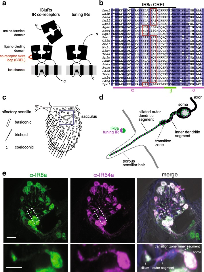
Background: Ionotropic receptors (IRs) are a large, divergent subfamily of ionotropic glutamate receptors (iGluRs) that are expressed in diverse peripheral sensory neurons and function in olfaction, taste, hygrosensation and thermosensation. Analogous to the cell biological properties of their synaptic iGluR ancestors, IRs are thought to form heteromeric complexes that localise to the ciliated dendrites of sensory neurons. IR complexes are composed of selectively expressed 'tuning' receptors and one of two broadly expressed co-receptors (IR8a or IR25a). While the extracellular ligand-binding domain (LBD) of tuning IRs is likely to define the stimulus specificity of the complex, the role of this domain in co-receptors is unclear.

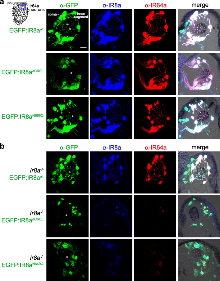
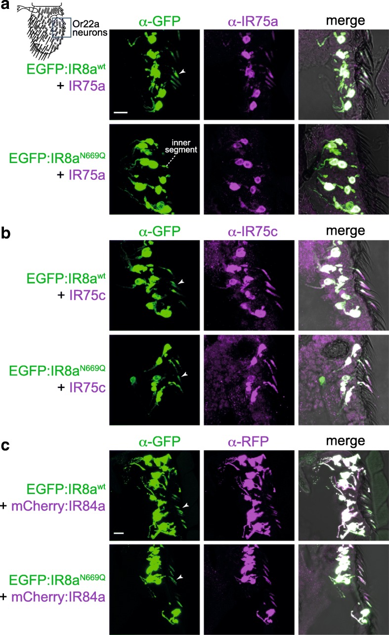
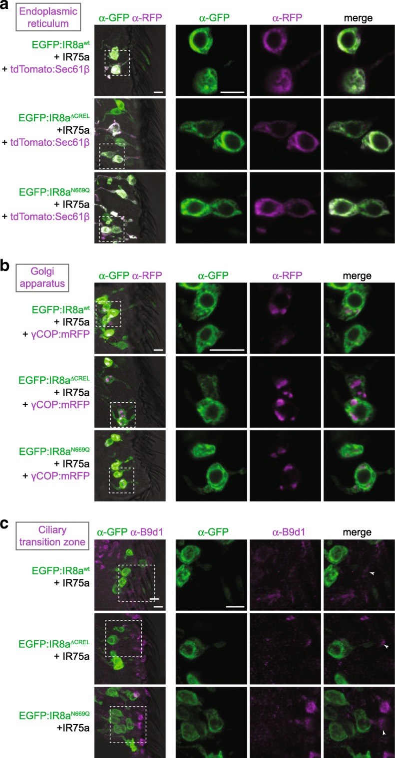
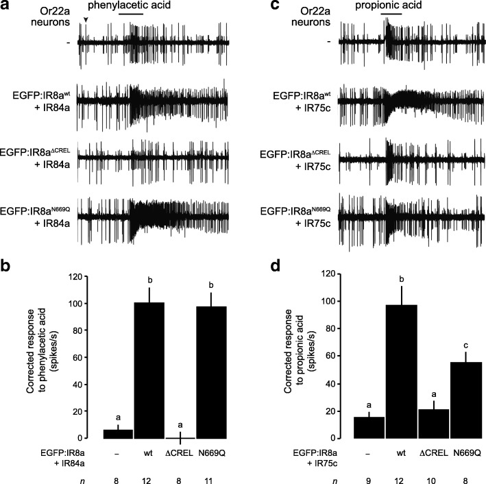
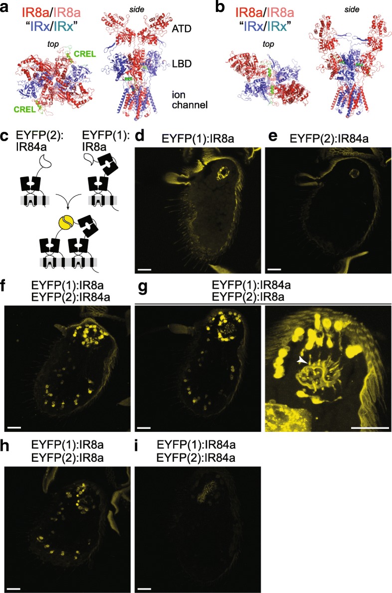
Results: We identify a sequence in the co-receptor LBD, the 'co-receptor extra loop' (CREL), which is conserved across IR8a and IR25a orthologues but not present in either tuning IRs or iGluRs. The CREL contains a single predicted N-glycosylation site, which we show bears a sugar modification in recombinantly expressed IR8a. Using the Drosophila olfactory system as an in vivo model, we find that a transgenically encoded IR8a mutant in which the CREL cannot be N-glycosylated is impaired in localisation to cilia in some, though not all, populations of sensory neurons expressing different tuning IRs. This defect can be complemented by the presence of endogenous wild-type IR8a, indicating that IR complexes contain at least two IR8a subunits and that this post-translational modification is dispensable for protein folding or complex assembly. Analysis of the subcellular distribution of the mutant protein suggests that its absence from sensory cilia is due to a failure in exit from the endoplasmic reticulum. Protein modelling and in vivo analysis of tuning IR and co-receptor subunit interactions by a fluorescent protein fragment complementation assay reveal that the CREL N-glycosylation site is likely to be located on the external face of a heterotetrameric IR complex.

Conclusions: Our data reveal an important role for the IR co-receptor LBD in control of intracellular transport, provide novel insights into the stoichiometry and assembly of IR complexes and uncover an unexpected heterogeneity in the trafficking regulation of this sensory receptor family.


Conflict of interest statement

Ethics approval and consent to participate

Not applicable.

Consent for publication

Not applicable.

Competing interests

The authors declare that they have no competing interests.

Publisher’s Note

Springer Nature remains neutral with regard to jurisdictional claims in published maps and institutional affiliations.

Figures

Fig. 1
Fig. 1
The IR co-receptors contain a distinctive N-glycosylated loop. a Schematic of the domain organisation of iGluRs, IR co-receptors and tuning IRs. b Alignment of the protein sequence spanning the CREL (co-receptor extra loop; black bar) of IR8a orthologues from the indicated species. Predicted N-glycosylation sites are highlighted with red boxes and predicted secondary structure is shown below the alignment. Species (top-to-bottom): Drosophila melanogaster, Drosophila simulans, Drosophila ananassae, Drosophila willistoni, Drosophila grimshawi, Anopheles gambiaeAedes aegypti, Culex quinquefasciatus, Bombyx mori, Camponotus floridanus, Apis mellifera, Nasonia vitripennisSolenopsis invicta, Tribolium castaneumAcyrthosiphon pisum, Pediculus humanus, Zootermopsis nevadensis, Schistocerca gregaria, Phyllium siccifolium, Thermobia domestica, Lepismachilis y-signata, Panulirus argus, Limulus polyphemus. c Schematic of the Drosophila third antennal segment showing the distribution of different olfactory sensilla and the internal sacculus. d Schematic illustrating the main anatomical features of an olfactory sensory neuron (OSN); the morphology of the cuticular hair and the branched nature of the cilium varies between different sensilla classes (note: most sensilla contain more than one neuron per hair). e Immunofluorescence with antibodies against IR8a (green) and IR64a (magenta) on an antennal section of a wild-type animal, showing the region containing the third chamber of the sacculus (blue boxed area in c). In the merged image, the transition zone is marked by monoclonal antibody 21A6 (blue), and a bright-field image is overlaid to reveal cuticular anatomical landmarks. Scale bar: 10 μm. The images shown below are of a single OSN (from a subset of optical slices of the area indicated by the dashed white boxes) in which the main anatomical features are shown. Scale bar: 5 μm
Fig. 2
Fig. 2
The IR8a CREL functions in subcellular trafficking. a Immunofluorescence with antibodies against GFP (green), IR8a (blue) and IR64a (red) on antennal sections of animals expressing the indicated transgenes in Ir8a neurons. Genotypes are of the form Ir8a-Gal4/UAS-EGFP:Ir8ax. The white asterisks (in this and other panels) indicate the central cavity of sacculus chamber 3, into which the IR64a+IR8a-expressing OSN ciliated dendrites project (see also the merged panels, in which bright-field images are overlaid to provide anatomical landmarks). In the top left panel, the arrowhead marks the ciliated ending of one neuron; the soma and inner segment of this neuron are also indicated (the outer segment—before the cilium—is difficult to see because only trace levels of receptors are detected in this region). Scale bar (for all panels in this figure): 10 μm. For each genotype, the phenotype was assessed in multiple sections of antennae from at least 20 animals from two independent genetic crosses, allowing observation of several hundred different neurons. We quantified the localisation properties by counting the number of sensory cilia with detectable EGFP signal as a percentage of the total number of cell bodies in the imaged samples; this is not expected to be 100% because sensory endings for each OSN soma are not necessarily present in the thin tissue sections (see ‘Methods’ section on imaging): EGFP:IR8awt = 75% (83 labelled cilia/111 soma), EGFP:IR8a∆CREL = 0% (0/93), EGFP:IR8aN669Q = 61% (65/106). b Immunofluorescence with antibodies against GFP (green), IR8a (blue) and IR64a (red) on antennal sections of animals expressing the indicated transgenes in Ir8a neurons in an Ir8a mutant background. Genotypes are of the form Ir8a1/Y;Ir8a-Gal4/UAS-EGFP:Ir8ax. EGFP:IR8a∆CREL and EGFP:IR8aN669Qare impaired in localisation to the cilia in the absence of endogenous IR8a (the occasional projections from the soma represent protein within the inner segment only). In addition, both proteins appear to be destabilised; consequently, endogenous IR64a is also detected at substantially lower levels in these two genotypes (but see Additional file 4: Figure S4). OSNs that express EGFP:IR8a∆CREL also display signs of sickness (e.g. smaller soma). For each genotype, the phenotype was assessed in multiple sections of antennae from at least 20 animals from two independent genetic crosses. Quantifications: EGFP:IR8awt = 79% (177/225), EGFP:IR8a∆CREL = 0% (0/198), EGFP:IR8aN669Q = 35% (78/220)
Fig. 3
Fig. 3
Heterogeneous requirement for the IR8a CREL N-glycosylation site in the localisation of tuning IRs. a Immunofluorescence with antibodies against GFP (green) and IR75a (magenta) on antennal sections of animals expressing the indicated transgenes in Or22a neurons (representing the field-of-view indicated in the cartoon). The arrowheads (in this and other panels) indicate the cilia of Or22a neurons; in neurons expressing EGFP:IR8aN669Q + IR75a (second row), the receptors are not detected in this sensory compartment, remaining restricted to the inner segment. Receptor localisation was determined by overlaying the fluorescence signal onto a bright-field channel, as shown in the merged images. Note that not all soma have a corresponding ciliated ending in these images, because this is a thin (14 μm) tissue section that does not include the entirety of all neurons. Genotypes are of the form UAS-EGFP:Ir8ax/UAS-Ir75a;Or22a-Gal4/+. Scale bar (for panels a, b): 10 μm. For each genotype, the phenotype was assessed in multiple sections of antennae from at least 20 animals from two independent genetic crosses. b Immunofluorescence with antibodies against GFP (green) and IR75c (magenta) on antennal sections of animals expressing the indicated transgenes in Or22a neurons. Genotypes are of the form UAS-EGFP:Ir8ax/UAS-Ir75c;Or22a-Gal4/+. For each genotype, the phenotype was assessed in multiple sections of antennae from at least 20 animals from two independent genetic crosses. c Immunofluorescence with antibodies against GFP (green) and RFP (magenta) on antennal sections of animals expressing the indicated transgenes in Or22a neurons. Genotypes are of the form UAS-EGFP:Ir8ax/+;Or22a-Gal4/UAS-mCherry:Ir84a. Scale bar: 10 μm. For each genotype, the phenotype was assessed in multiple sections of antennae from at least 30 animals from three independent genetic crosses
Fig. 4
Fig. 4
The IR8a CREL and CREL N-glycosylation site are important for ER export. a Immunofluorescence with antibodies against GFP (green) and RFP/Tomato (magenta) on antennal sections of animals expressing the indicated transgenes in Or22a neurons. The images on the right are high-magnification, single optical slices taken within the region shown in the lower-magnification view on the left in this and subsequent panels. Genotypes are of the form UAS-EGFP:Ir8ax/UAS-Ir75a;Or22a-Gal4/UAS-tdTomato:Sec61β. Scale bars: 5 μm. For each genotype, the phenotype was assessed in multiple sections of antennae from at least 20 animals from two independent genetic crosses, in this and the following panels. b Immunofluorescence with antibodies against GFP (green) and RFP (magenta) on antennal sections of animals expressing the indicated transgenes in Or22a neurons. Genotypes are of the form UAS-EGFP:Ir8ax/UAS-Ir75a;Or22a-Gal4/UAS-γCOP:mRFP. Scale bars: 5 μm. c Immunofluorescence with antibodies against GFP (green) and B9d1 (magenta) on antennal sections of animals expressing the indicated transgenes in Or22a neurons. Genotypes are of the form UAS-EGFP:Ir8ax/UAS-Ir75a;Or22a-Gal4/+. Scale bars: 5 μm.
Fig. 5
Fig. 5
The IR8a CREL N-glycosylation site is not essential for odour-evoked IR signalling. a Representative traces of the responses of Or22a neurons—those exhibiting the larger of the two spike amplitudes within this sensillum (black arrowhead)—expressing the indicated combinations of IRs, exposed to a 1-s pulse (black bar) of phenylacetic acid (1% v/v). Genotypes are of the form UAS-EGFP:Ir8ax/UAS-Ir84a;Or22a-Gal4/+, except for the control (Or22a-Gal4/+). b Quantification of the odour-evoked responses of the genotypes shown in a. Mean solvent corrected responses ±SEM are shown (n (number of sensilla) are indicated beneath each bar; mixed genders). Bars labelled with different letters are statistically different from each other (p < 0.05; Student’s t test with Benjamini and Hochberg correction for multiple comparisons). c Representative traces of the responses of Or22a neurons expressing the indicated combinations of IRs, exposed to a 1-s pulse (black bar) of propionic acid (1% v/v). Genotypes are of the form UAS-EGFP:Ir8ax/+;Or22a-Gal4/UAS-Ir75c, except for the control (Or22a-Gal4/+). d Quantification of the odour-evoked responses of the genotypes shown in c, presented as in b
Fig. 6
Fig. 6
Location of the CREL in a heterotetrameric IR complex model. a, b Two hypothetical configurations of a heterotetramer of two IR8a subunits (dark/pale red) and two tuning IR subunits (dark/pale blue), in which the IR8a ATDs have a proximal (contacting) or b distal (non-contacting) positions. Top and side views are shown in slightly different orientations to facilitate visualisation of the IR8a CREL (green). The structure was built through coarse-grained homology modelling of D. melanogaster IR8a on the homotetrameric GluA2 structure [33]; the IR tuning subunits are represented simply by the same IR8a model from which the ATD is deleted. c Schematic of the principle of EYFP reconstitution through complex formation and/or close proximity of EYFP fragment:IR fusion proteins. d–i Endogenous EYFP fluorescence in antennal sections of animals expressing the indicated combinations of EYFP fragment fusions in Ir8a neurons. The higher magnification sacculus image in g (right) reveals the cilia localisation of fluorescent signals (arrowhead); here, the gain setting during imaging was increased, resulting in higher cuticular autofluorescence. Genotypes: d UAS-EYFP(1):Ir8a/+;Ir8a-Gal4/+, e UAS-EYFP(2):Ir84a/+;Ir8a-Gal4/+, f UAS-EYFP(1):Ir8a/UAS-EYFP(2):Ir84a;Ir8a-Gal4/+, g UAS-EYFP(1):Ir84a/UAS-EYFP(2):Ir8a;Ir8a-Gal4/+, h UAS-EYFP(1):Ir8a/UAS-EYFP(2):Ir8a;Ir8a-Gal4/+ and i UAS-EYFP(1):Ir84a/UAS-EYFP(2):Ir84a;Ir8a-Gal4/+. All scale bars: 20 μm. For each genotype, the phenotype was assessed in multiple sections of antennae from at least 20 animals from two independent genetic crosses

Similar articles

Cited by

References

    1. Benton R, Vannice KS, Gomez-Diaz C, Vosshall LB. Variant ionotropic glutamate receptors as chemosensory receptors in Drosophila. Cell. 2009;136(1):149–162. doi: 10.1016/j.cell.2008.12.001. - DOI - PMC - PubMed
    1. Mayer ML. Structural biology of glutamate receptor ion channel complexes. Curr Opin Struct Biol. 2016;41:119–127. doi: 10.1016/j.sbi.2016.07.002. - DOI - PubMed
    1. Gereau RW, Swanson GT. The glutamate receptors. Totowa: Humana Press; 2008.
    1. Tikhonov DB, Magazanik LG. Origin and molecular evolution of ionotropic glutamate receptors. Neurosci Behav Physiol. 2009;39(8):763–773. doi: 10.1007/s11055-009-9195-6. - DOI - PubMed
    1. Croset V, Rytz R, Cummins SF, Budd A, Brawand D, Kaessmann H, Gibson TJ, Benton R. Ancient protostome origin of chemosensory ionotropic glutamate receptors and the evolution of insect taste and olfaction. PLoS Genet. 2010;6(8):e1001064. doi: 10.1371/journal.pgen.1001064. - DOI - PMC - PubMed

Publication types

MeSH terms

Substances