. 2019 Apr 17;23(1):131.
doi: 10.1186/s13054-018-2305-5.

TCR activation mimics CD127lowPD-1high phenotype and functional alterations of T lymphocytes from septic shock patients

Affiliations

TCR activation mimics CD127lowPD-1high phenotype and functional alterations of T lymphocytes from septic shock patients

Julie Mouillaux et al. Crit Care. .

Abstract

Background: Sepsis is the leading cause of mortality for critically ill patients worldwide. Patients develop T lymphocyte dysfunctions leading to T-cell exhaustion associated with increased risk of death. As interleukin-7 (IL-7) is currently tested in clinical trials to reverse these dysfunctions, it is important to evaluate the expression of its specific CD127 receptor on the T-cell surface of patients with septic shock. Moreover, the CD127lowPD-1high phenotype has been proposed as a T-cell exhaustion marker in chronic viral infections but has never been evaluated in sepsis. The objective of this study was first to evaluate CD127 and CD127lowPD-1high phenotype in septic shock in parallel with functional T-cell alterations. Second, we aimed to reproduce septic shock-induced T-cell alterations in an ex vivo model.

Methods: CD127 expression was followed at the protein and mRNA levels in patients with septic shock and healthy volunteers. CD127lowPD-1high phenotype was also evaluated in parallel with T-cell functional alterations after ex vivo activation. To reproduce T-cell alterations observed in patients, purified T cells from healthy volunteers were activated ex vivo and their phenotype and function were evaluated.

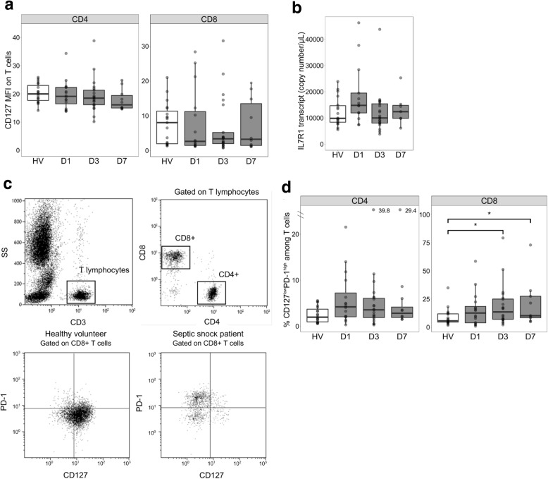
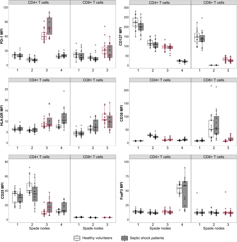
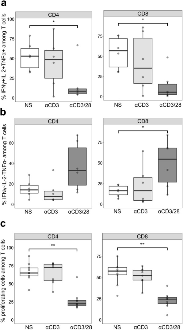
Results: In patients, neither CD127 expression nor its corresponding mRNA transcript level was modified compared with normal values. However, the percentage of CD127lowPD-1high T cells was increased while T cells also presented functional alterations. CD127lowPD-1high T cells co-expressed HLA-DR, an activation marker, suggesting a role for T-cell activation in the development of this phenotype. Indeed, T-cell receptor (TCR) activation of normal T lymphocytes ex vivo reproduced the increase of CD127lowPD-1high T cells and functional alterations following a second stimulation, as observed in patients. Finally, in this model, as observed in patients, IL-7 could improve T-cell proliferation.

Conclusions: The proportion of CD127lowPD-1high T cells in patients was increased compared with healthy volunteers, although no global CD127 regulation was observed. Our results suggest that TCR activation participates in the occurrence of this T-cell population and in the development of T-cell alterations in septic shock. Furthermore, we provide an ex vivo model for the investigation of the pathophysiology of sepsis-induced T-cell immunosuppression and the testing of innovative immunostimulant treatments.

Keywords: CD127; Exhaustion; IL-7; Immunosuppression; PD-1; Sepsis; T-cell activation.


Conflict of interest statement

Ethics approval and consent to participate

This project was approved by our Institutional Review Board for Ethics (“Comité de Protection des Personnes Sud-Est II”), which waived the need for informed consent, as the study was observational and performed on residual blood, after the completion of routine follow-up (#IRB 11236). This study is registered at the French Ministry of Research and Teaching (#DC-2008-509), at the Commission Nationale de l’Informatique et des Libertés, and on ClinicalTrials.gov (ClinicalTrials.gov Identifier: NCT02803346). Non-opposition to inclusion in the study was registered for each patient.

Consent for publication

Not applicable.

Competing interests

FV, GM, EP, and JT are co-inventors in three patent families covering IL-7 receptor biomarkers. This does not alter the authors’ adherence to all the Critical Care policies on sharing data and materials. JM, JT, GM, EP, and FV work in a joint research unit, co-funded by the Hospices Civils de Lyon, bioMérieux, and Lyon 1 University.

Publisher’s Note

Springer Nature remains neutral with regard to jurisdictional claims in published maps and institutional affiliations.

Figures

Fig. 1
Fig. 1
Cytokine production in T cells of patients with septic shock compared with those of healthy volunteers (HVs). Intracellular interleukin-2 (IL-2), tumor necrosis factor alpha (TNFα), and interferon gamma (IFNγ) staining was performed after 3 hours of T-cell activation using phorbol 12-myristate 13-acetate (PMA)/ionomycin in whole blood samples from patients with septic shock at day 1 (D1, n = 14), day 3 (D3, n = 18), and day 7 (D7, n = 10) after the onset of shock in comparison with HVs (n = 18). The percentages of cells producing each cytokine (IFNγ: left panel, IL-2: middle panel, and TNFα: right panel) are represented among CD4+ (a) and CD8+ (b) T cells. The percentages of polyfunctional cells simultaneously producing the three cytokines (c) or none of the three cytokines (d) are represented among CD4+ (left panel) and CD8+ (right panel) T cells. Data are presented as Tukey boxplots. Mann–Whitney tests were used to compare values between patients with septic shock and HVs, *P <0.05, **P <0.01
Fig. 2
Fig. 2
CD127 expression and CD127lowPD-1high T cells in patients with septic shock and healthy volunteers (HVs). a Cell surface expression of CD127 was measured by flow cytometry on septic shock patients’ CD4+ (left panel) and CD8+ (right panel) T cells at day 1 (D1, n = 15), day 3 (D3, n = 21), and day 7 (D7, n = 10) after the onset of shock in comparison with HVs (n = 20). Results are expressed as median of fluorescence intensity (MFI) on selected CD4+ and CD8+ T cells. b Expression of IL7R1 mRNA transcript, coding for cell surface CD127, was measured by reverse transcription quantitative polymerase chain reaction using RNA extracted from purified T cells at D1 (n = 13), D3 (n = 20), and D7 (n = 7) for patients with septic shock in comparison with HVs (n = 18). c Gating strategy for the determination of the percentage of CD127lowPD-1high T cells. Sequential gating was used to select first the CD3+ T-cell population among all leukocytes and then CD3+ CD4+ and CD3+ CD8+ T-cell subsets among CD3+ cells. PD-1 and CD127 expression thresholds were defined using isotype controls. One example of a CD127 and PD-1 staining among CD8+ T cells in one HV and one septic shock patient are represented. d The percentage of CD127lowPD-1high among CD4+ (left panel) and CD8+ (right panel) T cells at day 1 (D1, n = 15), day 3 (D3, n = 21), and day 7 (D7, n = 10) after the onset of shock in comparison with HVs (n = 20) is represented. Data are presented as Tukey boxplots. Mann–Whitney tests were used to compare values between patients with septic shock and HV, *P <0.05.
Fig. 3
Fig. 3
SPADE (Spanning-tree Progression Analysis of Density-normalized Events) analysis. T-cell phenotype was evaluated by using a SPADE algorithm based on the expression of different markers measured by flow cytometry on whole blood samples from patients with septic shock at day 3 after the onset of shock (D3, n = 17) and in healthy volunteers (HVs) (n = 14). CD4+ and CD8+ T cells from patients and donors were clustered in nodes on the basis of their similarities of expressions of CD127, CD38, HLA-DR, PD-1, FoxP3, and CD25. One tree containing four nodes was built for CD4+ T lymphocytes and one tree containing three nodes for CD8+ T cells. The medians of fluorescence intensity (MFIs) of the different markers for each node are represented on CD4+ and CD8+ T cells in patients and in HVs. Data are presented as Tukey boxplots. Node 4 of the CD4+ T cells, with a CD127lowCD25highFoxP3high phenotype, corresponds to regulatory T cells. Node 3 corresponding to CD127lowPD-1high cells has been highlighted in both CD4+ and CD8+ T cells
Fig. 4
Fig. 4
Occurrence of CD127lowPD-1high T cells in an ex vivo model of T-cell receptor activation of purified T cells from healthy volunteers (n = 9). Purified T cells were activated with anti-CD3/28 antibody-coated beads (αCD3/28, 1:1 bead-to-cell ratio) or anti-CD3 antibody-coated beads (αCD3, 1:1 bead-to-cell ratio) or not stimulated (NS) during 5 days. The percentage of CD127lowPD-1high (a) as well as the percentage of HLA-DR (b) positive cells were measured by flow cytometry among CD4+ (left panel) and CD8+ (right panel) T cells. Data are presented as Tukey boxplots. Mann–Whitney paired tests were used to compare values between non-stimulated and activated conditions, *P <0.05, **P <0.01, ***P <0.001
Fig. 5
Fig. 5
Altered T-cell functionality in an ex vivo model of T-cell receptor activation of purified T cells from healthy volunteers (n = 9). Purified T cells were activated with anti-CD3/28 antibody-coated beads (αCD3/28, 1:1 bead-to-cell ratio) or anti-CD3 antibody-coated beads (αCD3, 1:1 bead-to-cell ratio) or not stimulated (NS) for 5 days. T cells were then activated a second time with anti-CD2/3/28 antibody-coated beads (αCD2/3/28, 1:1 bead-to-cell ratio) for 3 days. Intracellular interleukin 2 (IL-2), tumor necrosis factor alpha (TNFα), and interferon gamma (IFNγ) staining was performed. The percentages of cells producing the three cytokines simultaneously (a) or none of the three cytokines (b) are represented for CD4+ (left panel) and CD8+ (right panel) T cells. c The percentages of proliferating cells are represented for CD4+ (left panel) and CD8+ (right panel) T cells. Data are presented as Tukey boxplots. Mann–Whitney paired tests were used to compare values between non-stimulated and stimulated conditions, *P <0.05, **P <0.01
Fig. 6
Fig. 6
Effect of interleukin-7 (IL-7) treatment on T-cell proliferation in an ex vivo model of T-cell receptor activation of purified T cells from healthy volunteers (n = 9). T-cell proliferation was evaluated on αCD3/28 pre-activated T cells after 3 days of activation using αCD2/3/28 antibody-coated beads in the presence and absence of IL-7. Results are presented for CD4+ (left panel) and CD8+ (right panel) T cells. Mann–Whitney paired tests were used to compare values with IL-7 with those without, *P <0.05

References

    1. Singer M, Deutschman CS, Seymour CW, Shankar-Hari M, Annane D, Bauer M, et al. The Third International Consensus Definitions for Sepsis and Septic Shock (Sepsis-3) JAMA. 2016;315:801. doi: 10.1001/jama.2016.0287. - DOI - PMC - PubMed
    1. Angus DC, van der Poll T. Severe Sepsis and Septic Shock. N Engl J Med. 2013;369:840–851. doi: 10.1056/NEJMra1208623. - DOI - PubMed
    1. Shankar-Hari M, Phillips GS, Levy ML, Seymour CW, Liu VX, Deutschman CS, et al. Developing a new definition and assessing new clinical criteria for septic shock: For the third international consensus definitions for sepsis and septic shock (sepsis-3) JAMA. 2016;315:775–787. doi: 10.1001/jama.2016.0289. - DOI - PMC - PubMed
    1. Lagu T, Lindenauer PK, Rothberg MB, Nathanson BH, Pekow PS, Steingrub JS, et al. Development and validation of a model that uses enhanced administrative data to predict mortality in patients with sepsis. Crit Care Med. 2011;39:2425–2430. doi: 10.1097/CCM.0b013e31822572e3. - DOI - PubMed
    1. Hotchkiss RS, Coopersmith CM, McDunn JE, Ferguson TA. Tilting toward immunosuppression. Nat Med. 2009;15:496–497. doi: 10.1038/nm0509-496. - DOI - PMC - PubMed