. 2019 Apr 17;11(488):eaav8375.
doi: 10.1126/scitranslmed.aav8375.

In utero gene editing for monogenic lung disease

Affiliations

In utero gene editing for monogenic lung disease

Deepthi Alapati et al. Sci Transl Med. .

Abstract

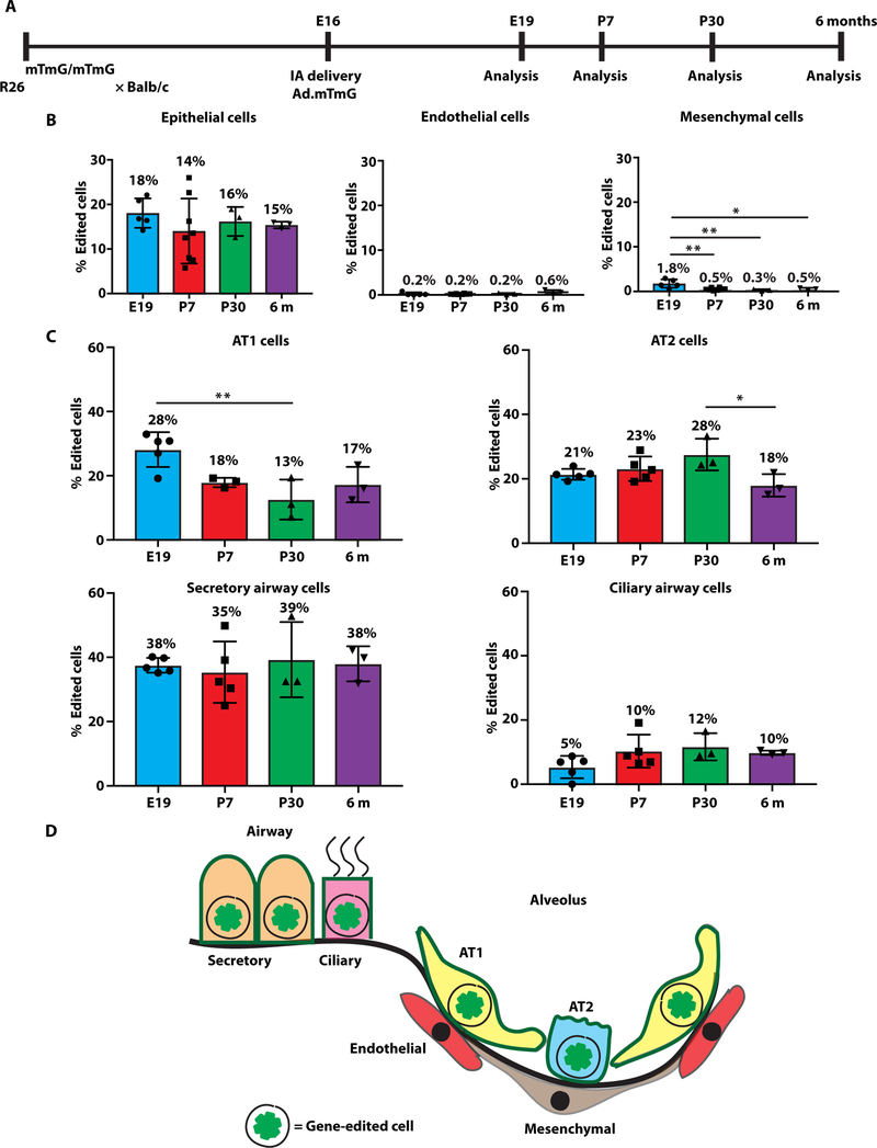
Monogenic lung diseases that are caused by mutations in surfactant genes of the pulmonary epithelium are marked by perinatal lethal respiratory failure or chronic diffuse parenchymal lung disease with few therapeutic options. Using a CRISPR fluorescent reporter system, we demonstrate that precisely timed in utero intra-amniotic delivery of CRISPR-Cas9 gene editing reagents during fetal development results in targeted and specific gene editing in fetal lungs. Pulmonary epithelial cells are predominantly targeted in this approach, with alveolar type 1, alveolar type 2, and airway secretory cells exhibiting high and persistent gene editing. We then used this in utero technique to evaluate a therapeutic approach to reduce the severity of the lethal interstitial lung disease observed in a mouse model of the human SFTPCI73T mutation. Embryonic expression of SftpcI73T alleles is characterized by severe diffuse parenchymal lung damage and rapid demise of mutant mice at birth. After in utero CRISPR-Cas9-mediated inactivation of the mutant SftpcI73T gene, fetuses and postnatal mice showed improved lung morphology and increased survival. These proof-of-concept studies demonstrate that in utero gene editing is a promising approach for treatment and rescue of monogenic lung diseases that are lethal at birth.


Figures

Fig. 1.
Fig. 1.. Intra-amniotic delivery of CRISPR-Cas9 results in pulmonary gene editing.
(A) Schematic representation of intra-amniotic route of fetal lung gene editing. (B) Experimental design of gene editing in R26mTmG/+ mice. (C) Fluorescent stereomicroscopy, using a filter to detect tdTomato and EGFP, of lungs from R26mTmG/+ mice injected with Ad.Cre, Ad.mTmG, or Ad.Null. (D) IHC for EGFP and tdTomato expression in the proximal airway and distal air saccules of lungs from R26mTmG/+ mice injected with Ad.Cre, Ad.mTmG, or Ad.Null. White arrowheads indicate EGFP staining. (E) PCR assay using primers to detect the on-target editing in DNA isolated from E19 lungs of R26mTmG/+ mice injected with Ad.Cre, Ad.mTmG, or Ad.Null. Edited band, 545 base pairs (bp); unedited band, 2951 bp. n = 2 to 6 per group. One fetus that was injected with Ad.mTmG and lacked notable EGFP fluorescence (GFP) was also negative for gene editing by PCR, indicating a likely technical failure at the time of injection. (F) Sanger sequencing of the 545-bp edited mTmG PCR product from an R26mTmG/+ mouse injected with Ad.mTmG. (G) Sanger sequencing of the 545-bp Cre-recombined mTmG PCR product from an R26mTmG/+ mouse injected with Ad.Cre. Scale bars, 1000 μm (C) and 50 μsm (D). IA, intra-amniotic; E, gestational day.
Fig. 2.
Fig. 2.. Intra-amniotic delivery of CRISPR-Cas9 targets pulmonary epithelial cells for gene editing.
(A) FACS plots of lungs harvested at E19 after intra-amniotic injection of Ad.mTmG, Ad.Cre, or Ad.Null at E16. Each row shows representative FACS plots from a single lung. (B) Quantitation of cell type–specific gene editing using FACS analysis for EGFP+ cells within each major pulmonary cell type after intra-amniotic injection of Ad.mTmG and Ad.Cre. n = 5 per group. (C) EGFP+ gene-edited and Cre-recombined cells depicted by white arrowheads within subsets of pulmonary epithelial cells marked by AQP5, SFTPC, SCGB1A1, and FOXJ1. (D) Quantification of gene-edited airway and alveolar epithelial cells after Ad.mTmG intra-amniotic delivery. (E) Quantification of Cre-recombined airway and alveolar epithelial cells after Ad.Cre intra-amniotic delivery. n = 2 to 5 per group. Epi, epithelial; Endo, endothelial; Mes, mesenchymal. Scale bars, 50 μm.
Fig. 3.
Fig. 3.. Pulmonary epithelial cell gene editing is stable over time.
(A) Experimental design for longer-term analysis of pulmonary epithelial cell gene editing after intra-amniotic Ad.mTmG delivery at E16. (B) Quantification of edited pulmonary epithelial, endothelial, and mesenchymal cell types at E19, P7, P30, and 6 months by FACS analysis. (C) Quantification of gene editing in individual pulmonary cell types at E19, P7, P30, and 6 months by IHC. (D) Schematic summary of fetal pulmonary cells that underwent gene editing after intra-amniotic delivery of CRISPR-Cas9 targeting the mT gene. n = 3 to 5 per group; **P < 0.01 and *P < 0.05 by one-way analysis of variance (ANOVA) followed by Tukey’s multiple comparison test.
Fig. 4.
Fig. 4.. Prenatal gene editing in SftpcI73T mice decreases mutant SP-CI73T proprotein and improves lung alveolarization.
(A) Schematic representation of SftpcI73T mutation causing intracellular accumulation of SP-CI73T proprotein resulting in AT2 cell injury and potential cell rescue with CRISPR-Cas9–mediated excision of SftpcI73T. (B) Fluorescent stereomicroscopy, using a filter to detect EGFP, of an E19 fetus (outlined by white dashed line) after intra-amniotic injection of Ad.Sftpc.GFP at E16 shows green fluorescence in the chest region. (C) Fluorescent stereomicroscopy, using a filter to detect EGFP, of lungs at E19 after E16 intra-amniotic injection of Ad.Sftpc.GFP. (D) IHC for EGFP of lung parenchyma at E19 after E16 intra-amniotic injection of Ad.Sftpc.GFP. (E) FACS analysis to assess EGFP expression in all pulmonary cells and pulmonary epithelial cells (EPCAM+ cells) from E19 fetuses after E16 intra-amniotic injection of Ad.Sftpc.GFP. n = 10 to 11 per group. (F) PCR analysis of DNA from E19 lung epithelial cells (EPCAM+-sorted cells) of E16 Ad.Sftpc.GFP intra-amniotic injected fetuses. Edited Sftpc band, 605 bp; −C and +C, negative and positive controls consisting of nontransfected mouse neuro-2a cells and mouse N2a cells cotransfected with plasmids containing spyCas9 or sgRNA1-A and sgRNA5-B, respectively. (G) Schematic of SftpcI73T experimental design. (H) Excision of the mutant Sftpc allele in AT2 cells was assessed by IHC. Lungs of E19 SftpcI73T/WT mice were assessed for expression of surfactant protein B (SFTPB) and hemagglutinin (HA) after E16 intra-amniotic injection of Ad.Null.GFP or Ad.Sftpc.GFP. SFTPB+HA (yellow arrowheads indicate representative cells), excision; SFTPB+HA+ (white arrowheads indicate representative cells), no excision; control, uninjected WT E19 lungs. (I) The percentage of SFTPB+HA cells on IHC was quantified. (J) Lung IHC for homeodomain only protein X (HOPX) at E19 to assess AT1 cell morphology and spreading in SftpcI73T/WT mice injected with Ad.Null.GFP or Ad.Sftpc.GFP at E16. (K) The internuclear distance was measured to quantify AT1 spreading. (L) Hematoxylin and eosin (H&E) staining of lungs from E19 SftpcI73T/WT mice injected at E16 with Ad.Null.GFP or Ad.Sftpc.GFP to assess alveolarization and sacculation. (M) The MLI was calculated to assess alveolarization. n = 3 to 4 per group; ##P < 0.0001, **P < 0.01, and *P < 0.05 by one-way ANOVA followed by Tukey’s multiple comparison test. WT, wild-type. Scale bars, 50 μm.
Fig. 5.
Fig. 5.. Prenatal gene editing in SftpcI73Tmutant mice improves survival.
(A) Schematic of experimental design for survival analysis of SftpcI73T mutant mice. (B) Survival of C57BL/6 mice injected at E16 with Ad.Sftpc.GFP (blue), gene-edited SftpcI73T/WT mice injected with Ad.Sftpc.GFP at E16 (red), SftpcI73T/WT mice injected with Ad.Null.GFP at E16 (green), and uninjected SftpcI73T/WT mice (purple). (C) The survival frequency of Ad.Sftpc.GFP-treated SftpcI73T/WT mice was normalized to the survival rate of control C57BL/6-treated mice at 1 week of age. n = 20 to 87 per group; **P < 0.01 by log-rank (Mantel-Cox) test for comparison of survival curves. (D) H&E staining of lungs from 1-week-old SftpcI73T/WT mice and C57BL/6 mice injected with Ad.Sftpc.GFP at E16 and uninjected WT C57BL/6 mice was performed to assess alveolarization. (E) The MLI was calculated to assess alveolarization. (F) IHC for SFTPB and HA was performed on lungs from 1-week-old SftpcI73T/WT mice and C57BL/6 mice injected with Ad.Sftpc.GFP at E16 and uninjected WT C57BL/6 mice to assess AT2 cell morphology and excision of the mutant Sftpc allele in AT2 cells. SFTPB+HA (yellow arrowheads indicate representative cells), excision; SFTPB+HA+ (white arrowheads indicate representative cells), no excision. (G) The percentage of SFTPB+HA cells, indicative of gene-edited cells in Ad.Sftpc.GFP injected SftpcI73T/WT mice, was quantified on IHC. (H) IHC for HOPX was performed to assess AT1 cell morphology in 1-week-old SftpcI73T/WT mice and C57BL/6 mice injected with Ad.Sftpc.GFP at E16 and uninjected WT C57BL/6 mice. (I) The internuclear distance was calculated to assess AT1 cell spreading. n = 3 to 4 per group; ##P < 0.0001 by one-way ANOVA followed by Tukey’s multiple comparison test. Scale bars, 50 μm.

Comment in

References

    1. Hamvas A, Cole FS, Nogee LM, Genetic disorders of surfactant proteins. Neonatology 91, 311–317 (2007). - PubMed
    1. Whitsett JA, Wert SE, Weaver TE, Alveolar surfactant homeostasis and the pathogenesis of pulmonary disease. Annu. Rev. Med 61, 105–119 (2010). - PMC - PubMed
    1. Rowe SM, Miller S, Sorscher EJ, Cystic fibrosis. N. Engl. J. Med 352, 1992–2001 (2005). - PubMed
    1. Crystal RG, Alpha 1-antitrypsin deficiency, emphysema, and liver disease. Genetic basis and strategies for therapy. Genetic basis and strategies for therapy. J. Clin. Invest 85, 1343–1352 (1990). - PMC - PubMed
    1. Spoonhower KA, Davis PB, Epidemiology of cystic fibrosis. Clin. Chest Med 37, 1–8 (2016). - PubMed

Publication types

Substances