. 2019 Apr 17;11(488):eaau9748.
doi: 10.1126/scitranslmed.aau9748.

Filamentous bacteriophages are associated with chronic Pseudomonas lung infections and antibiotic resistance in cystic fibrosis

Affiliations

Filamentous bacteriophages are associated with chronic Pseudomonas lung infections and antibiotic resistance in cystic fibrosis

Elizabeth B Burgener et al. Sci Transl Med. .

Abstract

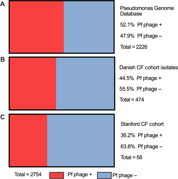
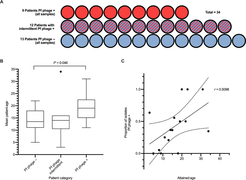
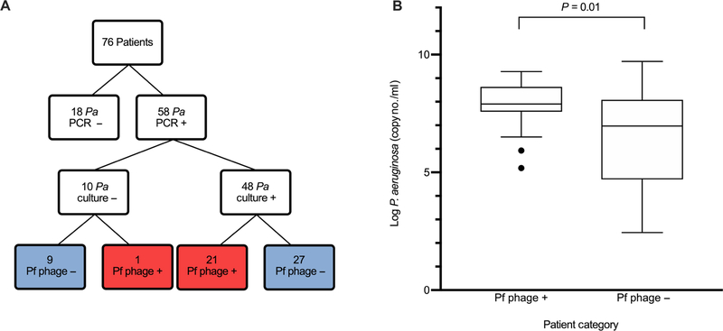
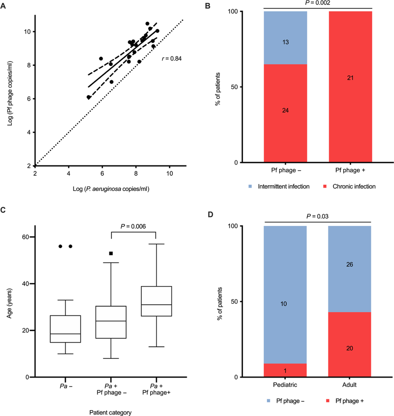
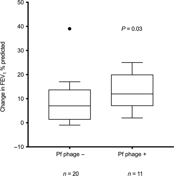
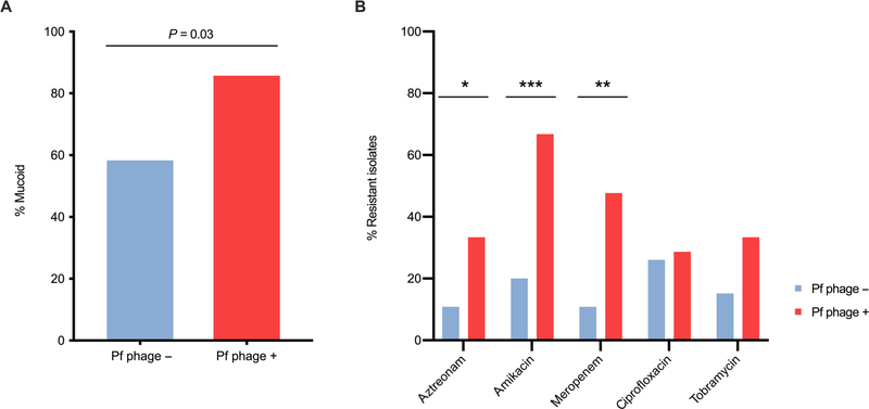
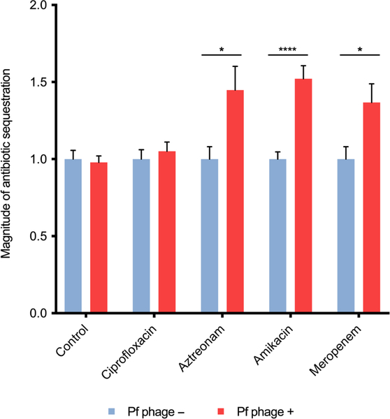
Filamentous bacteriophage (Pf phage) contribute to the virulence of Pseudomonas aeruginosa infections in animal models, but their relevance to human disease is unclear. We sought to interrogate the prevalence and clinical relevance of Pf phage in patients with cystic fibrosis (CF) using sputum samples from two well-characterized patient cohorts. Bacterial genomic analysis in a Danish longitudinal cohort of 34 patients with CF revealed that 26.5% (n = 9) were consistently Pf phage positive. In the second cohort, a prospective cross-sectional cohort of 58 patients with CF at Stanford, sputum qPCR analysis showed that 36.2% (n = 21) of patients were Pf phage positive. In both cohorts, patients positive for Pf phage were older, and in the Stanford CF cohort, patients positive for Pf phage were more likely to have chronic P. aeruginosa infection and had greater declines in pulmonary function during exacerbations than patients negative for Pf phage presence in the sputum. Last, P. aeruginosa strains carrying Pf phage exhibited increased resistance to antipseudomonal antibiotics. Mechanistically, in vitro analysis showed that Pf phage sequesters these same antibiotics, suggesting that this mechanism may thereby contribute to the selection of antibiotic resistance over time. These data provide evidence that Pf phage may contribute to clinical outcomes in P. aeruginosa infection in CF.


Conflict of interest statement

Competing interests: The remainder of the authors have declared that no conflict of interest exists.

Figures

Fig. 1.
Fig. 1.. Pf phage is prevalent in the Pseudomonas Genome Database, a Danish CF cohort and the Stanford CF cohort.
(A) The prevalence of the detection of the Pf prophage in P. aeruginosa logged in the Pseudomonas Genome Database, an internet- and community-based genome project containing the genome sequences and annotations of both clinical and environmental isolates of P. aeruginosa. (B) The prevalence of the detection of Pf prophage in P. aeruginosa isolates from the Danish CF cohort previously described by Marvig et al. (40). Four hundred seventy-four P. aeruginosa whole-genome sequences were collected from a cohort of 34 Danish patients with CF who were followed longitudinally. There are multiple isolates from each patient represented in this figure. (C) The prevalence of Pf phage detected by quantitative polymerase chain reaction (qPCR) in the sputum of patients also with P. aeruginosa in their sputum from the Stanford CF cohort. Each patient contributed only one sputum sample.
Fig. 2.
Fig. 2.. In a Danish CF cohort, the prevalence of Pf phage rises with increasing patient age.
(A) Patients were categorized as having all isolates with the Pf prophage detected (Pf phage +), intermittently having the Pf prophage detected (Pf phage intermittent), or all isolates with no Pf prophage detected (Pf phage −). (B) Mean age of patients with CF in each category. Age was unavailable for three subjects (n = 12, n = 9, n = 9). Boxes represent interquartile range (IQR) with whiskers showing 1.5 × IQR and dots as outliers. Comparison was made by Mann-Whitney test. The outliers on graph were included in analysis. Data are available in table S1. (C) Correlation between subject attained age at the date of the last isolate versus the proportion of total isolates in which Pf prophage was detected(r = 0.5098, P = 0.048). Data for n = 31 patients are included with each dot representing a specific age category and assessed by Pearson correlation.
Fig. 3.
Fig. 3.. P. aeruginosa concentration is higher in patients with Pf phage detected in the sputum within the Stanford CF cohort.
(A) Patients enrolled at the Stanford CF Center with detection of P. aeruginosa by both qPCR and respiratory culture and detection of Pf phage by qPCR. Of the 10 patients with P. aeruginosa detected by PCR but not grown on respiratory culture, 9 had history of P. aeruginosa on previous respiratory culture and 1 patient had no previous records available. Pa, P. aeruginosa. (B) P. aeruginosa concentrations in patients with Pf phage–positive sputum and Pf phage–negative sputum (P = 0.01). Boxes represent IQR with whiskers showing 1.5 × IQR and dots as outliers. Comparison was done by Wilcoxon ranked sum. Outliers on graph were included in analysis.
Fig. 4.
Fig. 4.. Pf phage concentration correlates with P. aeruginosa burden, chronic infection and patient age in the Stanford CF cohort.
(A) Correlation between the concentrations of Pf phage and P. aeruginosa in patients positive for Pf phage (r = 0.84, P < 0.001). Dotted line, line for 1:1 correlation for reference. All samples were run in duplicate. The area between dashed lines represents the 95% CI. Correlation calculated by Kendall correlation. (B) Percentage of patients meeting the Leeds criteria for chronic P. aeruginosa infection in Pf phage–positive and Pf phage–negative samples (P = 0.002). Comparison by Pearson’s χ2 was used. (C) Age of patients with P. aeruginosa–negative sputum, P. aeruginosa–positive and Pf phage–negative sputum, and P. aeruginosa–positive and Pf phage–positive sputum. Boxes represent IQR with whiskers showing 1.5 × IQR and dots as outliers. Comparison was done by Wilcoxon ranked sum. (D) Prevalence of Pf phage in adult versus pediatric patients with CF infected with P. aeruginosa (P = 0.03). Comparison by Pearson’s χ2. Outliers on graph were included in analysis.
Fig. 5.
Fig. 5.. Patients positive for Pf phage have reduced lung function during exacerbations in the Stanford CF cohort.
The difference between FEV1% predicted at time of sampling and at presentation of subsequent exacerbation in patients with Pf phage–positive sputum and in those with Pf phage–negative sputum (P = 0.03). Only patients who were enrolled while at baseline health and who had exacerbation after enrollment were included in this analysis (n = 11 patients positive for Pf phage, n = 20 patients negative for Pf phage). These data include only patients with P. aeruginosa detected in their sputum. Boxes represent IQR with whiskers showing 1.5 × IQR and dots as outliers. Comparisons were made by Mann-Whitney test. Outlier on graph was included in analysis. Data are available in table S2.
Fig. 6.
Fig. 6.. Clinical isolates of P. aeruginosa from patients with Pf phage–positive sputum exhibit increased antibiotic resistance and are more likely to be mucoid.
(A) Proportion of isolates that display mucoid phenotype in isolates from Pf phage–negative sputum and Pf phage–positive sputum (P = 0.03). Comparison by Pearson χ2 test. (B) The proportion of isolates that were resistant to antibiotics listed. n = 28 Pf phage–negative samples, n = 20 Pf phage–positive samples. Comparison was made by Pearson’s χ2 test. *P = 0.0499, **P = 0.002, ***P = 0.0009.
Fig. 7.
Fig. 7.. Antibiotics aztreonam, amikacin, and meropenem can be sequestered by Pf phage.
The magnitude of sequestered antibiotic (bacterial proliferation normalized to control conditions) is displayed for each antibiotic when run through dialysis cassette containing either Pf phage and DNA or DNA alone (control). Samples were run in triplicate (N = 2 experiments). Error bars indicate mean ± SEM. Comparisons were made by Student’s t test with an α level of 0.05. ****P < 0.0001, *P < 0.05.

References

    1. Riordan JR, Rommens JM, Kerem B, Alon N, Rozmahel R, Grzelczak Z, Zielenski J, Lok S, Plavsic N, Chou JL, Drumm ML, Iannuzzi MC, Collins FS, Tsui L-C, Identification of the cystic fibrosis gene: Cloning and characterization of complementary DNA. Science 245, 1066–1073 (1989). - PubMed
    1. Welsh MJ, Ramsey BW, Accurso F, Cutting GR, in The Online Metabolic and Molecular Bases of Inherited Disease, Beaudet AL, Vogelstein B, Kinzler KW, Antonarakis SE, Ballabio A, Gibson KM, Mitchell G, Eds. (The McGraw-Hill Companies Inc, 2014).
    1. Poulsen JH, Fischer H, Illek B, Machen TE, Bicarbonate conductance and pH regulatory capability of cystic fibrosis transmembrane conductance regulator. Proc. Natl. Acad. Sci. U.S.A 91, 5340–5344 (1994). - PMC - PubMed
    1. Khan TZ, Wagener JS, Bost T, Martinez J, Accurso FJ, Riches DW, Early pulmonary inflammation in infants with cystic fibrosis. Am. J. Respir. Crit. Care Med 151, 1075–1082 (1995). - PubMed
    1. Davis PB, Drumm M, Konstan MW, Cystic fibrosis. Am. J. Respir. Crit. Care Med 154, 1229–1256 (1996). - PubMed

Publication types

Substances