. 2019 Jun 14;93(13):e02244-18.
doi: 10.1128/JVI.02244-18. Print 2019 Jul 1.

Regulation of Human Cytomegalovirus Secondary Envelopment by a C-Terminal Tetralysine Motif in pUL71

Affiliations

Regulation of Human Cytomegalovirus Secondary Envelopment by a C-Terminal Tetralysine Motif in pUL71

Clarissa Read et al. J Virol. .

Abstract

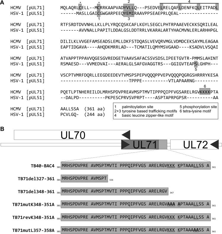
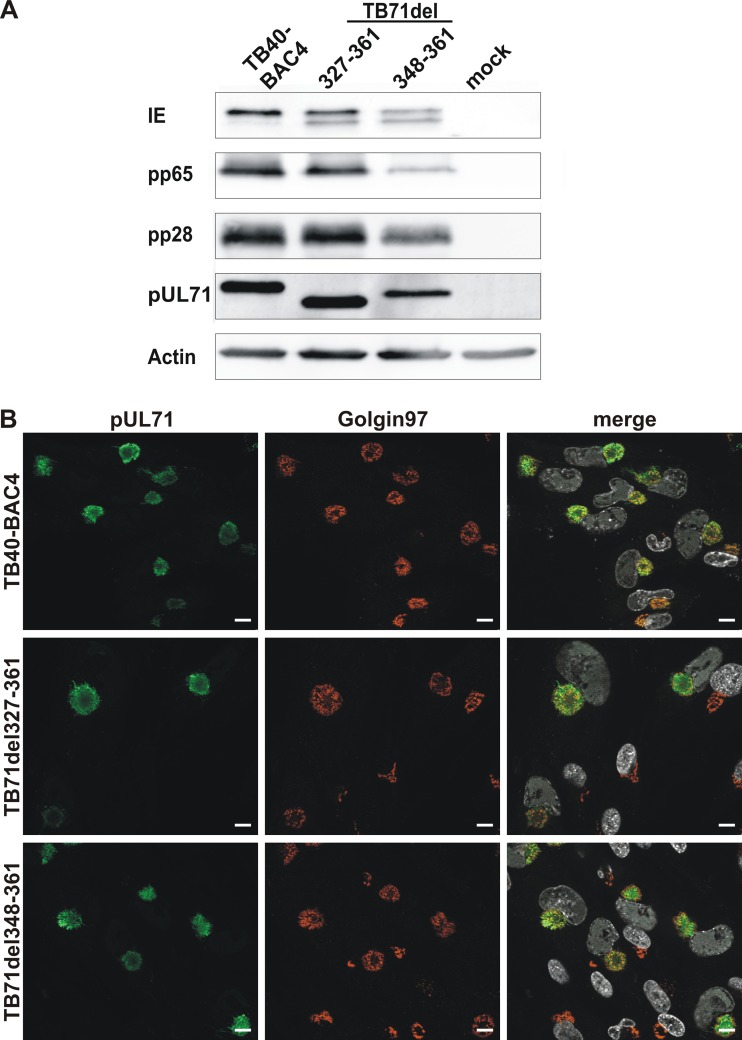
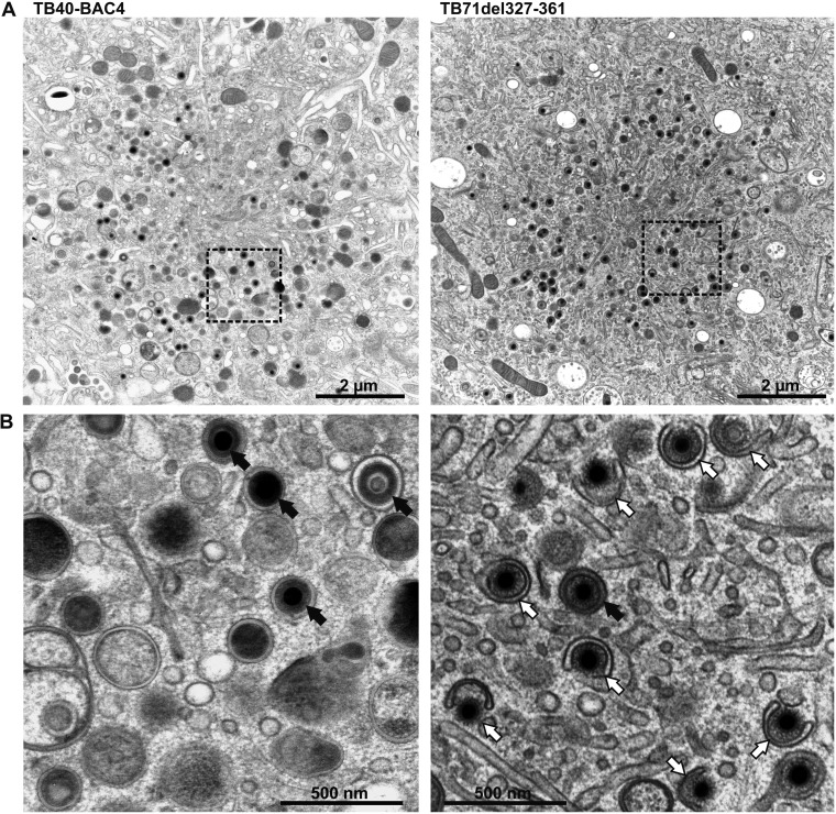
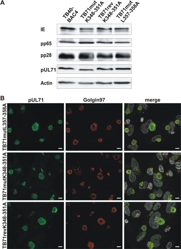
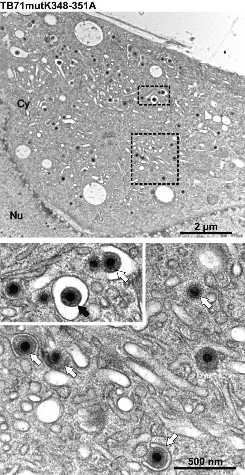
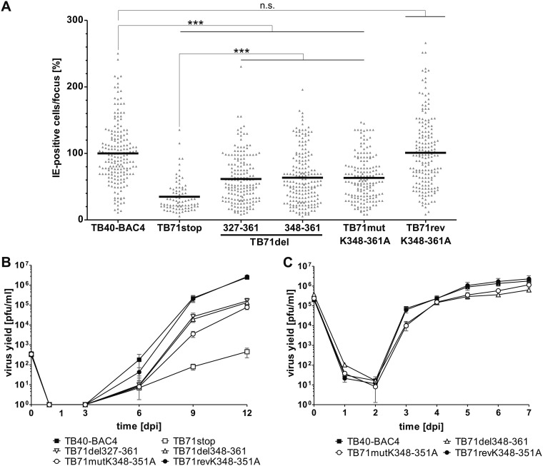
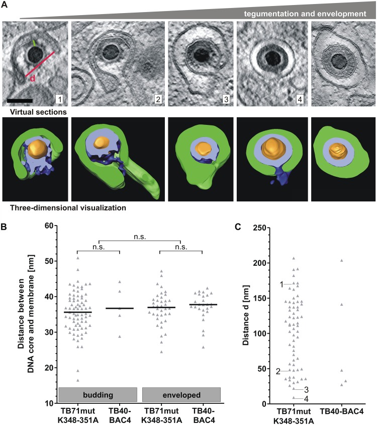
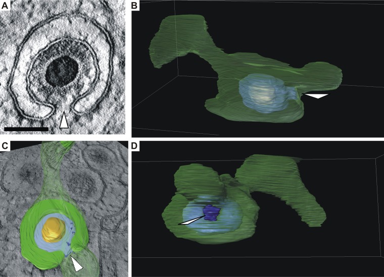
Human cytomegalovirus (HCMV) secondary envelopment requires the viral tegument protein pUL71. The lack of pUL71 results in a complex ultrastructural phenotype with increased numbers of viral capsids undergoing envelopment at the cytoplasmic virus assembly complex. Here, we report a role of the pUL71 C terminus in secondary envelopment. Mutant viruses expressing C-terminally truncated pUL71 (TB71del327-361 and TB71del348-351) exhibited an impaired secondary envelopment in transmission electron microscopy (TEM) studies. Further mutational analyses of the C terminus revealed a tetralysine motif whose mutation (TB71mutK348-351A) resulted in an envelopment defect that was undistinguishable from the defect caused by truncation of the pUL71 C terminus. Interestingly, not all morphological alterations that define the ultrastructural phenotype of a TB71stop virus were found in cells infected with the C-terminally mutated viruses. This suggests that pUL71 provides additional functions that modulate HCMV morphogenesis and are harbored elsewhere in pUL71. This is also reflected by an intermediate growth defect of the C-terminally mutated viruses compared to the growth of the TB71stop virus. Electron tomography and three-dimensional visualization of different stages of secondary envelopment in TB71mutK348-351A-infected cells showed unambiguously the formation of a bud neck. Furthermore, we provide evidence for progressive tegument formation linked to advancing grades of capsid envelopment, suggesting that tegumentation and envelopment are intertwined processes. Altogether, we identified the importance of the pUL71 C terminus and, specifically, of a positively charged tetralysine motif for HCMV secondary envelopment.IMPORTANCE Human cytomegalovirus (HCMV) is an important human pathogen that causes severe symptoms, especially in immunocompromised hosts. Furthermore, congenital HCMV infection is the leading viral cause of severe birth defects. Development of antiviral drugs to prevent the production of infectious virus progeny is challenging due to a complex and multistep virion morphogenesis. The mechanism of secondary envelopment is still not fully understood; nevertheless, it represents a potential target for antiviral drugs. Our identification of the role of a positively charged motif in the pUL71 C terminus for efficient HCMV secondary envelopment underlines the importance of pUL71 and, especially, its C terminus for this process. It furthermore shows how cell-associated spread and virion release depend on secondary envelopment. Ultrastructural analyses of different stages of envelopment contribute to a better understanding of the mechanisms underlying the process of secondary envelopment. This may bring us closer to the development of novel concepts to treat HCMV infections.

Keywords: HCMV; Human betaherpesvirus 5; TEM analysis; UL71; secondary envelopment; tetralysine motif.


Figures

FIG 1
FIG 1
(A) Sequence alignment of HCMV pUL71 (APG57260.1) and HSV pUL51 (ALM22621.1) by the online tool MUSCLE (68). Asterisks mark two predicted palmitoylation sites of pUL71 (CSS-Palm 4.0). Motifs that have been shown to be functional during virus morphogenesis are the following: 1, palmitoylation site of pUL51 (33); 2 and 3, tyrosine-based trafficking motifs in pUL71 and pUL51 (25, 34); 4, basic leucine zipper-like motif in pUL71 (26); 5, phosphorylation site of pUL51 (69); 6, tetralysine motif of pUL71 (this study). (B) Generation of C-terminal pUL71 mutant viruses. Shown is the genomic organization up- and downstream of the UL71 gene in parental virus TB40-BAC4 and the amino acid sequence of pUL71 in the indicated mutant viruses. TB71del327-361 and TB71del348-361 express C-terminally truncated versions of pUL71. Mutation of the underlined amino acids to the amino acids depicted in bold resulted in the point mutants TB71mutK348-351A and TB71mutL357-358A. The revertant virus TB71revK348-351A was generated by restoring the amino acid sequence of TB71mutK348-351A to the sequence of the parental virus TB40-BAC4.
FIG 2
FIG 2
Characterization of pUL71 C-terminal deletion mutants. (A) Cell lysates of TB40-BAC4-, TB71del327-361-, TB71del348-361-, and mock-infected HFFs (120 h postinfection) were processed for Western blot analysis. The blot was probed with antibodies against viral IE, pp65, pp28, pUL71, and cellular actin. (B) Representative confocal images of the subcellular localization of pUL71 (green) and trans-Golgi network protein golgin-97 (red) in HFFs infected with the indicated viruses (MOI of 1) at 120 h postinfection. Nuclei were stained with DAPI (white). Scale bars, 10 μm.
FIG 3
FIG 3
(A) Electron micrographs of the vAC of HCMV TB40-BAC4- (left) and TB71del327-361-infected (right) HFFs at 120 h postinfection. (B) Higher magnification of selected areas from panel A shows fully enveloped virus capsids (black arrows) in TB40-BAC4-infected cells (left), whereas TB71del327-361-virus-infected cells (right) contain a high number of capsids in the process of secondary envelopment (white arrows).
FIG 4
FIG 4
Characterization of pUL71 C-terminal point mutants. (A) Western blot analysis of cell lysates of TB40-BAC4, TB71mutL357-358A, TB71mutK348-351A, and TB71revK348-351A (120 h postinfection). The blot was probed with antibodies against IE, pp65, pp28, pUL71, and cellular actin. (B) Representative confocal images of the subcellular localization of pUL71 (green) and trans-Golgi network protein golgin-97 (red) in HFFs infected with the indicated viruses (MOI of 1) at 120 h postinfection. Nuclei were stained with DAPI (white). Scale bars, 10 μm.
FIG 5
FIG 5
Electron micrographs of the vAC of HCMV TB71mutK348-351A-infected HFFs at 120 h postinfection. Higher magnifications below show many nonenveloped capsids (white arrows) and one enveloped virus particle (black arrow).
FIG 6
FIG 6
Growth of C-terminal pUL71 mutants. (A) Focus expansion assay of the indicated viruses in HFFs under methylcellulose overlay. HCMV-infected cells were detected by indirect immunofluorescence staining for IE1/2 antigen at 9 days postinfection (dpi). Each data point represents the relative number of IE1/2-positive nuclei per focus. Shown is the mean focus size for each virus (black line) normalized to the mean focus size of TB40-BAC4. At least 80 foci from three independent experiments were determined for each virus. Significance testing was performed by a Kruskal-Wallis test followed by a Dunn’s multiple-comparison test (P < 0.05). Significance is given compared to TB40-BAC4 and TB71stop. ***, P < 0.0001; n.s., not significant. (B) Multistep growth kinetics experiments of the indicated viruses were performed by infecting HFFs with an infection rate of 0.3%. Virus yields in the supernatants of infected cells were determined at the indicated times by titration on HFFs. Growth curves show the mean virus yields and standard deviations from three independent virus supernatants. Virus yields at time zero represent the starting infection rates determined at 24 h postinfection. (C) Single-step growth kinetics experiments of the indicated viruses were performed on HFFs with an MOI of 3. Virus yields in the supernatants of infected cells were determined at the indicated times by titration on HFFs. Growth curves show the mean virus yields and standard deviations from three independent virus supernatants. Virus yields at time zero represent the virus yields of the inocula.
FIG 7
FIG 7
Stages of HCMV secondary envelopment. (A) TB71mutK348-351A virus capsids at different stages of secondary envelopment based on three-dimensional STEM tomography. Images from left to right show representative capsids with increasing amounts of tegument in correlation with different stages of secondary envelopment. In three-dimensional visualizations, membranes are segmented in green, tegument in blue, and capsids in orange. The cross sections through the tegument are shown in bright blue. Scale bar, 100 nm. (B) The distance between the outline of the DNA core and the enwrapping membrane (green line in image A1) of budding and enveloped capsids was measured in tomograms of TB71mutK348-351A- and TB40-BAC4-infected cells. Every triangle represents the distance measurement for a single virus particle. The black lines indicate the median distance. Significance was calculated by using the Kruskal-Wallis test followed by a Dunn’s multiple-comparison test (P < 0.05). (C) Distance d (red line in image A1) was determined for 62 TB71mutK348-351A capsids and 5 TB40-BAC4 capsids. Each data point represents d of a single budding capsid. The numbers 1 to 4 in the diagram indicate the distance of the respective capsids shown in A.
FIG 8
FIG 8
Formation of a bud neck (white arrowheads). (A) Virtual section of a representative budding HCMV capsid based on three-dimensional STEM tomography showing a bud neck. Scale bar, 100 nm. (B and C) Three-dimensional visualizations of the capsid in panel A which show that the vesicle membrane (green, semitransparent) has a large tubular shape. Orange, capsid; blue, tegument. (C) Cross section through the vesicle as shown in image A. The cross section through the tegument is shown in light blue. (B and D) Views of the vesicle from different angles (see Movie S1 in the supplemental material).

Similar articles

Cited by

References

    1. Griffiths P, Baraniak I, Reeves M. 2015. The pathogenesis of human cytomegalovirus. J Pathol 235:288–297. doi:10.1002/path.4437. - DOI - PubMed
    1. Mocarski E, Shenk T, Pass R. 2006. Cytomegaloviruses, p. 2701–2771. In Knipe DM, Howley PM, Griffin DE, Lamb RA, Martin MA (ed), Fields virology 5th ed Lippincott Williams & Wilkins, Philadelphia, PA.
    1. Schauflinger M, Villinger C, Mertens T, Walther P, von Einem J. 2013. Analysis of human cytomegalovirus secondary envelopment by advanced electron microscopy: HCMV morphogenesis. Cell Microbiol 15:305–314. doi:10.1111/cmi.12077. - DOI - PubMed
    1. Varnum SM, Streblow DN, Monroe ME, Smith P, Auberry KJ, Pasa-Tolic L, Wang D, Camp DG, Rodland K, Wiley S, Britt W, Shenk T, Smith RD, Nelson JA. 2004. Identification of proteins in human cytomegalovirus (HCMV) particles: the HCMV proteome. J Virol 78:10960–10966. doi:10.1128/JVI.78.20.10960-10966.2004. - DOI - PMC - PubMed
    1. Sanchez V, Greis KD, Sztul E, Britt WJ. 2000. Accumulation of virion tegument and envelope proteins in a stable cytoplasmic compartment during human cytomegalovirus replication: characterization of a potential site of virus assembly. J Virol 74:975–986. doi:10.1128/JVI.74.2.975-986.2000. - DOI - PMC - PubMed

Publication types