. 2019 Jun 14;93(13):e00237-19.
doi: 10.1128/JVI.00237-19. Print 2019 Jul 1.

Oral Vaccination with Replication-Competent Adenovirus in Mice Reveals Dissemination of the Viral Vaccine beyond the Gastrointestinal Tract

Affiliations

Oral Vaccination with Replication-Competent Adenovirus in Mice Reveals Dissemination of the Viral Vaccine beyond the Gastrointestinal Tract

Emeline Goffin et al. J Virol. .

Abstract

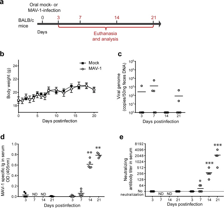
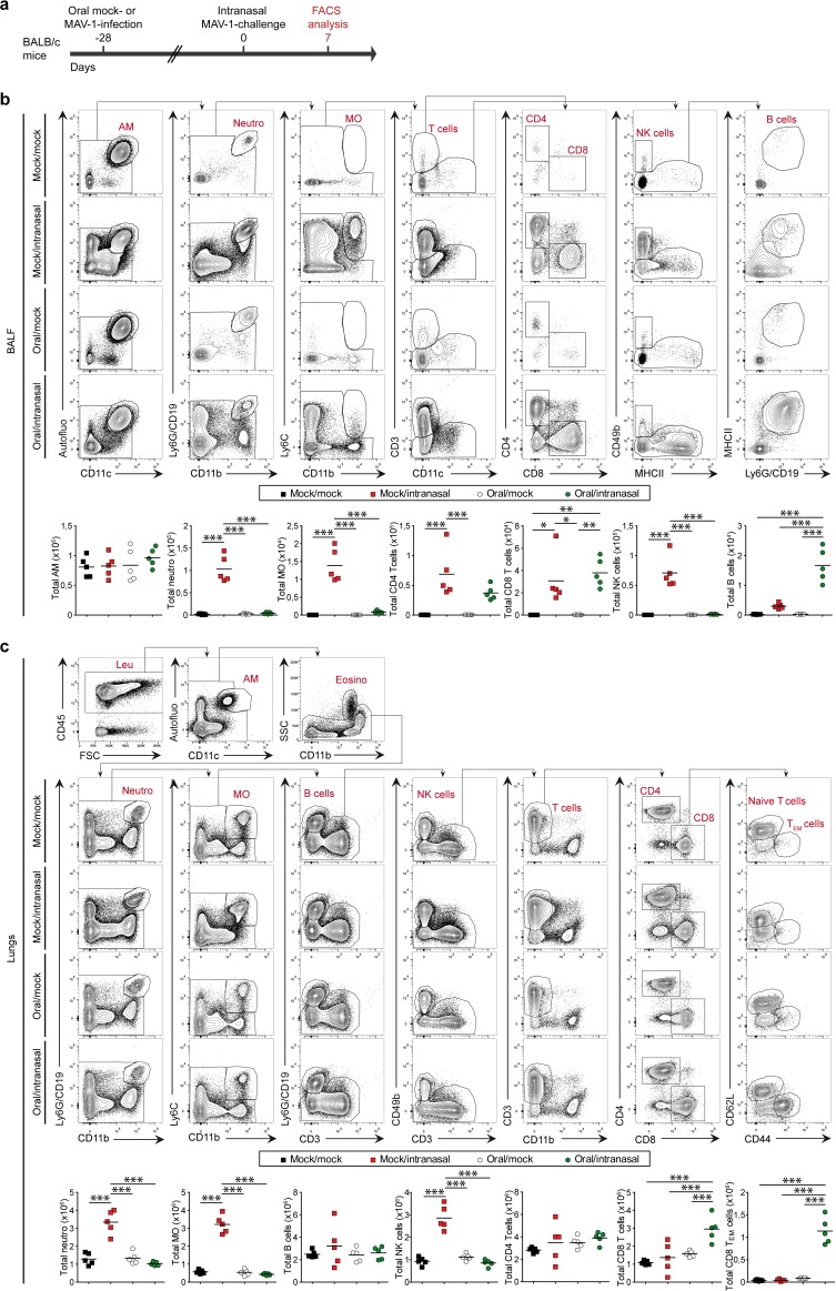
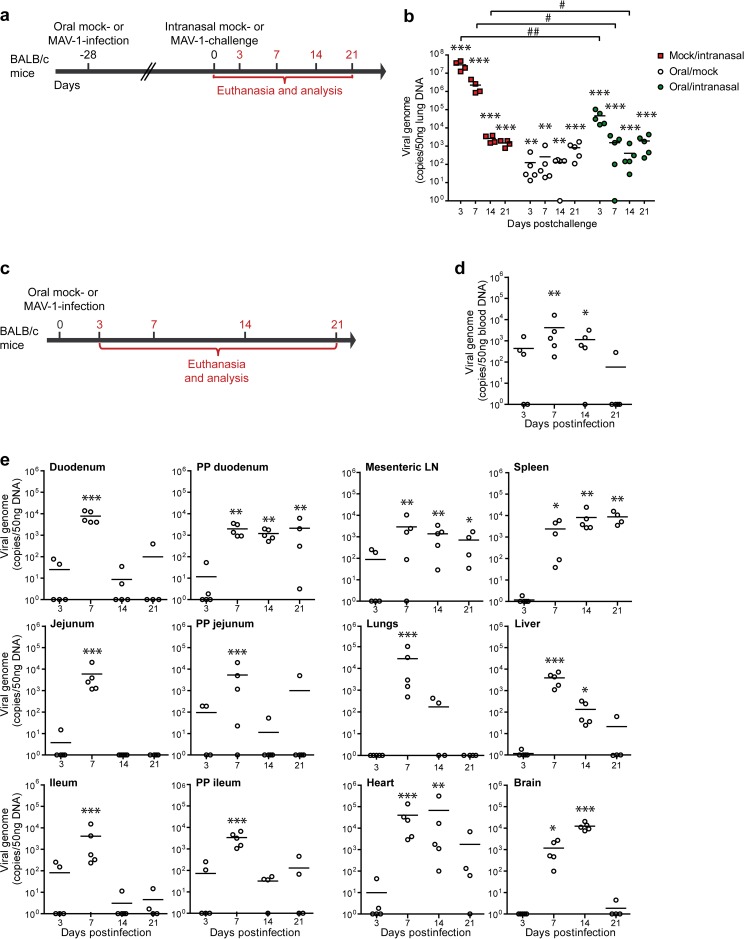
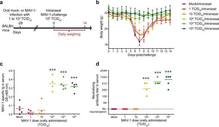
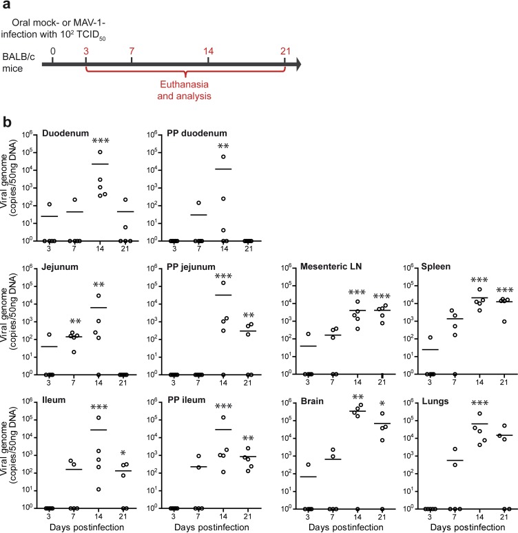
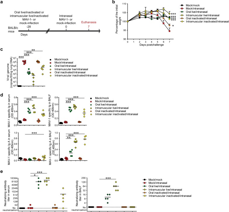
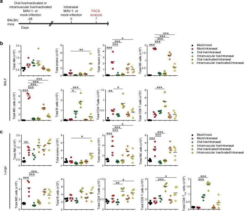
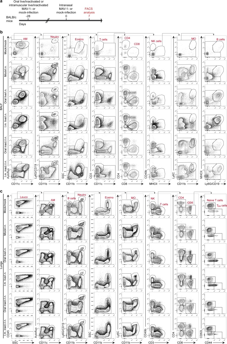
Since the 1970s, replication-competent human adenoviruses 4 and 7 have been used as oral vaccines to protect U.S. soldiers against the severe respiratory diseases caused by these viruses. These vaccines are thought to establish a digestive tract infection conferring protection against respiratory challenge through antibodies. The success of these vaccines makes replication-competent adenoviruses attractive candidates for use as oral vaccine vectors. However, the inability of human adenoviruses to replicate efficiently in laboratory animals has hampered the study of such vectors. Here, we used mouse adenovirus type 1 (MAV-1) in mice to study oral replication-competent adenovirus-based vaccines. We show that MAV-1 oral administration provides protection that recapitulates the protection against homologous respiratory challenge observed with adenovirus 4 and 7 vaccines. Moreover, live oral MAV-1 vaccine better protected against a respiratory challenge than inactivated vaccines. This protection was linked not only with the presence of MAV-1-specific antibodies but also with a better recruitment of effector CD8 T cells. However, unexpectedly, we found that such oral replication-competent vaccine systemically spread all over the body. Our results therefore support the use of MAV-1 to study replication-competent oral adenovirus-based vaccines but also highlight the fact that those vaccines can disseminate widely in the body.IMPORTANCE Replication-competent adenoviruses appear to be promising vectors for the development of oral vaccines in humans. However, the study and development of these vaccines suffer from the lack of any reliable animal model. In this study, mouse adenovirus type 1 was used to develop a small-animal model for oral replication-competent adenovirus vaccines. While this model reproduced in mice what is observed with human adenovirus oral vaccines, it also highlighted that oral immunization with such a replication-competent vaccine is associated with the systemic spread of the virus. This study is therefore of major importance for the future development of such vaccine platforms and their use in large human populations.

Keywords: adenovirus; mouse model; oral vaccination.


Figures

FIG 1
FIG 1
Characterization of MAV-1 oral infection in BALB/c mice. (a) Eight-week-old female BALB/c mice were orally infected with 104 TCID50 of MAV-1 or PBS (mock) and euthanized at day 3, 7, 14, or 21 p.i. for analysis (n = 5 mice in each group). The numbers above the arrow indicate the day postinfection. (b) Weight of mice. (c) MAV-1 genome copy numbers in feces. (d) MAV-1-specific antibodies. OD, optical density. (e) MAV-1-neutralizing antibody titers. The data presented are either the means for 5 mice ± SEM (b) or individual data and means (c to e). Pooled data from naive mice at days 3 and 21 provided the negative control (n = 10). P values are for comparison between mock-infected mice and infected mice at the indicated time points. *, P < 0.05; **, P < 0.01; ***, P < 0.001; ND, not determined.
FIG 2
FIG 2
Evaluation of the protection induced by MAV-1 oral administration against a respiratory homologous challenge. (a) Eight-week-old female BALB/c mice were orally infected with 104 TCID50 of MAV-1 or PBS as a control (mock). At 28 days p.i., the mice were challenged or not by intranasal administration of 105 TCID50 of MAV-1 while they were under mild isoflurane anesthesia. Mice were euthanized at days 3, 7, 14, and 21 after intranasal challenge. Mice that did not receive MAV-1 either orally or intranasally (mock/mock) were euthanized at days 3 and 21 as negative controls (n = 5 in each group). (b) Weight of mice through day 21 postchallenge. For each time point, means were compared to the mean for the mock/mock group. (c) Weight of lungs at euthanasia. (d) Hematoxylin-eosin-stained sections of lungs at 7 days postchallenge. The images are representative of those from 3 animals. Bar, 100 μm. Mock/mock (top left in panel d), mice that were not infected either orally or intranasally; mock/intranasal (top right in panel d), mice that were mock infected orally and then infected intranasally with MAV-1; oral/mock (bottom left in panel d), mice that were infected orally with MAV-1 and then mock infected intranasally; oral/intranasal (bottom right in panel d), mice that were infected orally with MAV-1 and then challenged intranasally with the same virus. The data presented are either the means for 5 mice ± SEM (b) or individual data and means (c). Pooled data from naive mice at days 3 and 21 provided the negative control (n = 10). P values are for comparisons between the indicated time points (#, P < 0.05; ##, P < 0.01; ###, P < 0.001) or between mock-infected and the other groups of mice at the indicated time points (*, P < 0.05; **, P < 0.01; ***, P < 0.001).
FIG 3
FIG 3
Characterization of lung immune cell populations after MAV-1 oral immunization and challenge. (a) Eight-week-old female BALB/c mice (n = 5 per group) were orally infected with 104 TCID50 of MAV-1 or PBS as a control (mock). At 28 days p.i., mice were challenged by intranasal administration of 105 TCID50 of MAV-1 while they were under mild isoflurane anesthesia. Mice were euthanized at day 7 after intranasal challenge. (b) Gating strategy (top) and total numbers (bottom) of immune cell populations in BALF. (c) Gating strategy (top) and total numbers (bottom) of immune cell populations in lungs. P values are for comparisons between all pairs of groups. *, P < 0.05; **, P < 0.01; ***, P < 0.001. SSC, side scatter; FSC, forward scatter; AM, alveolar macrophages; Neutro, neutrophils; MO, monocytes; Leu, leukocytes; Eosino, eosinophils; TEM cells, effector memory T cells.
FIG 4
FIG 4
MAV-1 distribution after oral immunization and challenge. (a) Eight-week-old female BALB/c mice (n = 5 per group) were orally infected with 104 TCID50 of MAV-1 or PBS as a control (mock). At 28 days p.i., mice were challenged by intranasal administration of 105 TCID50 of MAV-1 while they were under mild isoflurane anesthesia. Mice were euthanized at day 7 after intranasal challenge. (b) MAV-1 genome copy numbers in lungs. (c) Eight-week-old female BALB/c mice were orally infected with 104 TCID50 of MAV-1 or PBS (mock) and euthanized at days 3, 7, 14, or 21 p.i. for analysis (n = 5 in each group). The numbers above the arrow indicate the day p.i. (d) MAV-1 genome copy numbers in blood. (e) MAV-1 genome copy numbers in organs. PP, Peyer’s patches; LN, lymph nodes. The data presented are individual data and means. Pooled data from naive mice at days 3 and 21 provided the negative control (n = 10). P values are for comparisons between the indicated time points (#, P < 0.05; ##, P < 0.01) or between the mock-infected group and the other groups at the indicated time points (*, P < 0.05; **, P < 0.01; ***, P < 0.001).
FIG 5
FIG 5
Comparison of the protection induced by different doses of MAV-1 against a respiratory homologous MAV-1 challenge. (a) Eight-week-old female BALB/c mice (n = 5 per group) were orally infected with 1, 10, 102, 103, or 104 TCID50 of MAV-1 or PBS as a control (mock). At 28 days p.i., mice were challenged by intranasal administration of 105 TCID50 of MAV-1 while they were under mild isoflurane anesthesia. Mice were euthanized at day 14 after intranasal challenge. (b) Weight of mice from day 0 to day 14 postchallenge. The data presented are the average for 5 mice ± SEM. For each time point, means were compared to the mean for mice in the mock/intranasal group. Statistically significant differences were observed between mice challenged with 102, 103, and 104 TCID50 and the mock/intranasal group at days 6, 7, 8, and 9 p.i.; for clarity, this is not represented on the graph. (c and d) MAV-1-specific antibodies (c) and MAV-1-neutralizing antibody titers (d) after oral immunization but before challenge (day 0). OD, optical density. Individual data are presented, and means are shown by a line. P values are for comparison between mock-infected mice and mice receiving the indicated MAV-1 doses. ***, P < 0.001.
FIG 6
FIG 6
Dissemination of MAV-1 in BALB/c mice after a low-dose oral infection. (a) Eight-week-old female BALB/c mice were orally infected with 102 TCID50 of MAV-1 or PBS (mock) and euthanized at day 3, 7, 14, or 21 p.i. for analysis (n = 5 mice in each group). The numbers above the arrow indicate the day postinfection. (b) MAV-1 genome copy numbers in organs. Individual data and means are presented. Pooled data from naive mice at days 3 and 21 provided the negative control (n = 10). P values are for comparison between the mock-infected group and the other groups of mice at the indicated time points. *, P < 0.05; **, P < 0.01; ***, P < 0.001. PP, Peyer’s patches; LN, lymph nodes.
FIG 7
FIG 7
Comparison of the protection induced by live or inactivated MAV-1 oral or intramuscular immunization against a respiratory homologous challenge. (a) Eight-week-old female BALB/c mice (n = 5 per group) were orally or intramuscularly immunized with 104 TCID50 of live MAV-1 or the same dose of formalin-inactivated MAV-1. At 28 days p.i., mice were challenged by intranasal administration of 105 TCID50 of live MAV-1 while they were under mild isoflurane anesthesia. Mice were euthanized at day 7 postchallenge. (b) Weight of mice after challenge. (c) MAV-1 genome copy numbers in lungs at day 7 postchallenge. (d) MAV-1-specific total immunoglobulin (Ig) and IgA in serum and BALF at day 7 postchallenge. (e) MAV-1-neutralizing antibodies in serum and BALF at day 7 postchallenge. Mock/mock, mice that were not infected either orally or intranasally; mock/intranasal, mice that were mock infected orally and then infected intranasally with live MAV-1; oral live/intranasal, mice that were infected orally with live MAV-1 and then challenged intranasally; intramuscular live/intranasal, mice that were infected intramuscularly with live MAV-1 and then challenged intranasally; oral inactivated/intranasal, mice that were infected orally with inactivated MAV-1 and then challenged intranasally; intramuscular inactivated/intranasal, mice that were infected intramuscularly with inactivated MAV-1 and then challenged intranasally. The data presented are either the means for 5 mice ± SEM (b) or individual data and means (c, d, e). P values are for comparison between all pairs of groups. For clarity, only differences between the mock/intranasal and the other groups are shown and only differences for day 7 postchallenge are shown in the weight curve. *, P < 0.05; **, P < 0.01; ***, P < 0.001.
FIG 8
FIG 8
Characterization of lung immune cell populations after live or inactivated MAV-1 oral or intramuscular immunization and challenge. (a) Eight-week-old female BALB/c mice (n = 5 per group) were orally or intramuscularly infected with 104 TCID50 of live MAV-1 or the same dose of formalin-inactivated MAV-1. At 28 days p.i., mice were challenged by intranasal administration of 105 TCID50 of live MAV-1 while they were under mild isoflurane anesthesia. Mice were euthanized at day 7 postchallenge. (b) Total numbers of immune cell populations in BALF. (c) Total numbers of immune cell populations in lung parenchyma. Mock/mock, mice that were not infected either orally or intranasally; mock/intranasal, mice that were mock infected orally and then infected intranasally with live MAV-1; oral live/intranasal, mice that were infected orally with live MAV-1 and then challenged intranasally; intramuscular live/intranasal, mice that were infected intramuscularly with live MAV-1 and then challenged intranasally; oral inactivated/intranasal, mice that were infected orally with inactivated MAV-1 and then challenged intranasally; intramuscular inactivated/intranasal, mice that were infected intramuscularly with inactivated MAV-1 and then challenged intranasally. The data presented are individual data and means. P values are for comparison between all pairs of groups. For clarity, only differences between the mock/intranasal and the other groups are shown. *, P < 0.05; **, P < 0.01; ***, P < 0.001. AM, alveolar macrophages; neutro, neutrophils; MO, monocytes; eosino, eosinophils; TEM cells, effector memory T cells.
FIG 9
FIG 9
Gating strategy used for the characterization of lung immune cell populations of MAV-1-challenged mice after different immunization protocols (a) Eight-week-old female BALB/c mice (n = 5 per group) were orally or intramuscularly infected with 104 TCID50 of live MAV-1 or the same dose of formalin-inactivated MAV-1. At 28 days p.i., mice were challenged by intranasal administration of 105 TCID50 of live MAV-1 while they were under mild isoflurane anesthesia. Mice were euthanized at day 7 postchallenge. (b) Representative dot plots in BALF. (c) Representative dot plots in lung parenchyma. Mock/i.n., mice that were mock infected orally and then infected intranasally with live MAV-1; oral live/i.n., mice that were infected orally with live MAV-1 and then challenged intranasally; i.m. live/i.n., mice that were infected intramuscularly with live MAV-1 and then challenged intranasally; oral inact./i.n., mice that were infected orally with inactivated MAV-1 and then challenged intranasally; i.m. inactivat./i.n., mice that were infected intramuscularly with inactivated MAV-1 and then challenged intranasally; AM, alveolar macrophages; Neutro, neutrophils; MO, monocytes; Leuco, leucocytes; Eosino, eosinophils; TEM cells, effector memory T cells.

Similar articles

Cited by

References

    1. Holmgren J, Czerkinsky C. 2005. Mucosal immunity and vaccines. Nat Med 11:S45–S53. doi:10.1038/nm1213. - DOI - PubMed
    1. Neutra MR, Kozlowski PA. 2006. Mucosal vaccines: the promise and the challenge. Nat Rev Immunol 6:148–158. doi:10.1038/nri1777. - DOI - PubMed
    1. Levine MM. 2010. Immunogenicity and efficacy of oral vaccines in developing countries: lessons from a live cholera vaccine. BMC Biol 8:129. doi:10.1186/1741-7007-8-129. - DOI - PMC - PubMed
    1. Lycke N. 2012. Recent progress in mucosal vaccine development: potential and limitations. Nat Rev Immunol 12:592–605. doi:10.1038/nri3251. - DOI - PubMed
    1. Dubois B, Goubier A, Joubert G, Kaiserlian D. 2005. Oral tolerance and regulation of mucosal immunity. Cell Mol Life Sci 62:1322–1332. doi:10.1007/s00018-005-5036-0. - DOI - PMC - PubMed

Publication types