. 2019 Apr 18;4(8):e127291.
doi: 10.1172/jci.insight.127291.

The female-biased factor VGLL3 drives cutaneous and systemic autoimmunity

Affiliations

The female-biased factor VGLL3 drives cutaneous and systemic autoimmunity

Allison C Billi et al. JCI Insight. .

Abstract

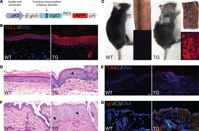
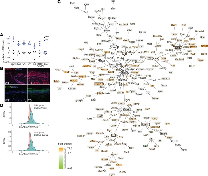
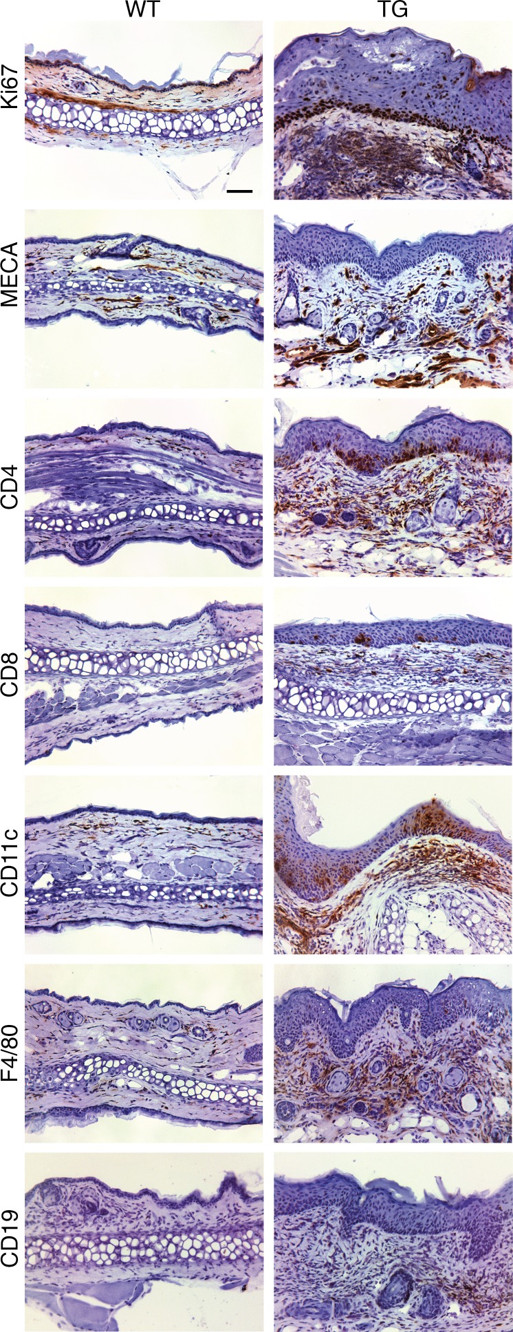
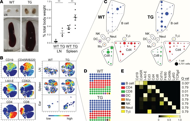
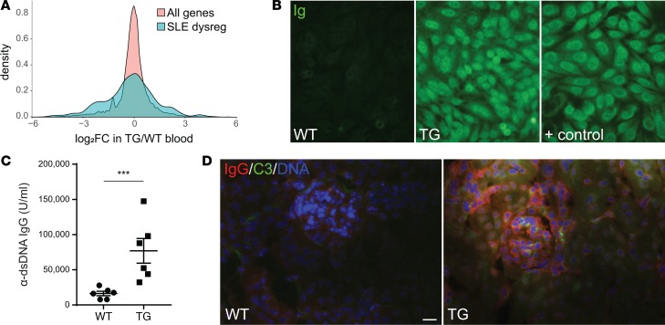
Autoimmune disease is 4 times more common in women than men. This bias is largely unexplained. Female skin is "autoimmunity prone," showing upregulation of many proinflammatory genes, even in healthy women. We previously identified VGLL3 as a putative transcription cofactor enriched in female skin. Here, we demonstrate that skin-directed overexpression of murine VGLL3 causes a severe lupus-like rash and systemic autoimmune disease that involves B cell expansion, autoantibody production, immune complex deposition, and end-organ damage. Excess epidermal VGLL3 drives a proinflammatory gene expression program that overlaps with both female skin and cutaneous lupus. This includes increased B cell-activating factor (BAFF), the only current biologic target in systemic lupus erythematosus (SLE); IFN-κ, a key inflammatory mediator in cutaneous lupus; and CXCL13, a biomarker of early-onset SLE and renal involvement. Our results demonstrate that skin-targeted overexpression of the female-biased factor VGLL3 is sufficient to drive cutaneous and systemic autoimmune disease that is strikingly similar to SLE. This work strongly implicates VGLL3 as a pivotal orchestrator of sex-biased autoimmunity.

Keywords: Autoimmune diseases; Autoimmunity; Dermatology; Lupus; Mouse models.


Conflict of interest statement

Conflict of interest: The authors have declared that no conflict of interest exists.

Figures

Figure 1
Figure 1. Overexpression of VGLL3 in the epidermis produces a skin phenotype with gross and histologic features of cutaneous lupus.
(A) Transgenic (TG) cassette. The bovine keratin 5 (K5) promoter drives polycistronic expression of the full-length mouse VGLL3 and mCherry red fluorescent protein linked by an internal ribosome entry site (IRES). β-glob, rabbit β-globin intronic sequence; pA, polyadenylation signal. (B) Detection of VGLL3 protein (red) by immunofluorescence (IF) in skin of female WT and TG mice. Scale bar: 20 μm. Images are representative of sections from 3 WT and 3 TG animals examined. (C) Left: WT mouse compared with age-matched TG mouse with lupus-like skin rash. Right: Bright-field and fluorescence images of WT and lesional TG tail skin. Scale bar: 2 mm. (D) H&E staining of WT and TG volar skin sections, demonstrating epidermal hyperplasia, basal cell vacuolization, apoptotic keratinocytes (arrowhead, magnified on inset), and dermal inflammatory infiltrate. Scale bar: 20 μm. (E) TUNEL (red) staining of WT and TG tail skin sections. Scale bar: 50 μm. (F) Periodic acid–Schiff staining of WT and TG dorsal skin sections. Arrowheads indicate subtle basement membrane thickening. Scale bar: 20 μm. (G) Detection of IgG and complement factor C3 by IF in WT and TG nonlesional neck skin. Scale bar: 50 μm. In EG, images are representative of sections from 3 WT and 3 TG animals examined.
Figure 2
Figure 2. Overexpression of VGLL3 in the epidermis recapitulates the autoimmunity-prone genetic signature of human female skin.
(A) Detection of Vgll3 and enriched immune transcripts versus example nonenriched transcript Ifnb (IFN-β) by quantitative reverse transcription PCR (qRT-PCR) in skin of WT (n = 3) and TG mice (n = 2) with high Vgll3 expression (more than tenfold WT average). Horizontal bars represent the mean. *P < 0.05 by 2-tailed Student’s t test. (B) Detection of VGLL3 targets CXCL13 (top, red) and IFN-κ (bottom, green) by IF in WT and TG skin. Blue, DNA. Scale bar: 20 μm. Images are representative of sections from 3 WT and 3 TG animals examined. (C) Literature-based network analysis of genes differentially expressed in nonlesional, normal-appearing TG skin relative to WT skin by RNA-seq. (D) Expression in nonlesional TG versus WT skin of genes dysregulated (dysreg) in discoid lupus erythematosus (DLE; P = 4.0 × 10⁻¹⁰) or subacute cutaneous lupus erythematosus (SCLE; P = 2.3 × 10⁻⁸) versus all genes. x axis, log2 fold change (FC) in TG versus WT. See Methods for additional statistical details.
Figure 3
Figure 3. Overexpression of VGLL3 in the epidermis causes skin inflammation with features of cutaneous lupus.
Detection of the indicated cell markers by immunohistochemistry in WT and TG ear sections. Ki67, cell proliferation marker; MECA, mouse endothelial cell antigen. Scale bar: 50 μm. Images are representative of sections from 4 WT and 4 TG animals examined.
Figure 4
Figure 4. Skin-directed VGLL3 overexpression drives a systemic inflammatory response with B cell expansion.
(A) Left: Representative images of WT and TG skin-draining lymph nodes (LN) and spleens. Right: LN and spleen weights represented as a percentage of total body weight. Error bars represent mean ± SEM. **P < 0.01 by 2‑tailed Student’s t test (LN, n = 6 WT and 3 TG; spleen, n = 8 WT and 8 TG). (B) CyTOF data derived from a single experiment consisting of 3 age- and sex-matched WT and TG mice visualized by viSNE. Left: viSNE maps depicting expression of select markers. Each dot is a cell. Color reflects the level of expression of marker from low (blue) to high (red). Right: Contour plot of viSNE maps colored by density of cells isolated from the specified tissues in WT and TG mice. Here, color reflects cell density from low (blue) to high (red). Data shown correspond to 1 WT and 1 TG mouse that are representative of the experiment. LN and spleen data represent approximately 20,000 CD45+ live singlets per sample. Ear skin samples represent all recorded CD45+ live singlets (WT, 262 and TG, 264) for each sample. The complete viSNE analysis for all markers and samples for the experiment can be found in Supplemental Figure 4. Data are representative of 2 independent CyTOF experiments. (C) SPADE tree of LN samples depicted in B. Node size represents the number of cells in the population derived from the experiment represented in B and Supplemental Figure 4B. Mϕ, macrophage; Neut, neutrophil; Tγδ, γ δ T cell. (D) Percentage dot plot showing the proportion of each cell type in aggregated WT and TG lymph node samples for the CyTOF experiment. Colors correspond to the populations indicated in C and E. (E) Heatmap showing marker expression of the SPADE populations specified in C across aggregated LN, spleen, and skin samples included in the CyTOF experiment. Q val, q values for differential detection in WT versus TG LN. q = 6.2 × 10⁻⁴, significantly upregulated in TG LN.
Figure 5
Figure 5. Mice with skin-directed VGLL3 overexpression develop manifestations of systemic autoimmune disease.
(A) Differential expression in WT (n = 4) and TG (n = 4) mouse blood of genes dysregulated in SLE peripheral blood mononuclear cells (aqua) versus all genes (peach). (B) Detection at 1:50 dilution of circulating antinuclear antibodies (Ig) by indirect IF in HEp-2 cells in WT, TG, and positive control (+ control, aged NZM2328) serum. In total, 7 TG and 3 WT animals were evaluated; all TG mouse sera showed positive autoantibody staining at a dilution of 1:25 or higher, whereas all WT mouse sera were negative. (C) Detection of circulating anti–double-stranded DNA antibodies (α-dsDNA) by ELISA. Error bars represent mean ± SEM. ***P = 0.0005 by 2-tailed Student’s t test. n = 6 WT and 6 TG. (D) Detection of IgG (red) and complement factor C3 (green) by IF in WT and TG glomeruli. Blue, DNA. Scale bar: 20 μm. Images are representative of glomeruli visualized on sections from 6 WT and 6 TG animals examined. 0 of 6 WT and 6 of 6 transgenic mice scored positive for renal immune complex deposition.

References

    1. Fish EN. The X-files in immunity: sex-based differences predispose immune responses. Nat Rev Immunol. 2008;8(9):737–744. doi: 10.1038/nri2394. - DOI - PMC - PubMed
    1. Tedeschi SK, Bermas B, Costenbader KH. Sexual disparities in the incidence and course of SLE and RA. Clin Immunol. 2013;149(2):211–218. doi: 10.1016/j.clim.2013.03.003. - DOI - PubMed
    1. Liang Y, et al. A gene network regulated by the transcription factor VGLL3 as a promoter of sex-biased autoimmune diseases. Nat Immunol. 2017;18(2):152–160. - PMC - PubMed
    1. Ramírez A, Bravo A, Jorcano JL, Vidal M. Sequences 5’ of the bovine keratin 5 gene direct tissue- and cell-type-specific expression of a lacZ gene in the adult and during development. Differentiation. 1994;58(1):53–64. - PubMed
    1. Sarkar MK, et al. Photosensitivity and type I IFN responses in cutaneous lupus are driven by epidermal-derived interferon kappa. Ann Rheum Dis. 2018;77(11):1653–1664. doi: 10.1136/annrheumdis-2018-213197. - DOI - PMC - PubMed

Publication types

MeSH terms

Substances