Review
. 2019 Aug 1;58(15):2173-2177.
doi: 10.2169/internalmedicine.1992-18. Epub 2019 Apr 17.

Endoscopic Ultrasound-guided Drainage of a Mediastinal Abscess Caused by an Ingested Fish Bone

Affiliations
Review

Endoscopic Ultrasound-guided Drainage of a Mediastinal Abscess Caused by an Ingested Fish Bone

Hitoshi Shibuya et al. Intern Med. .

Abstract

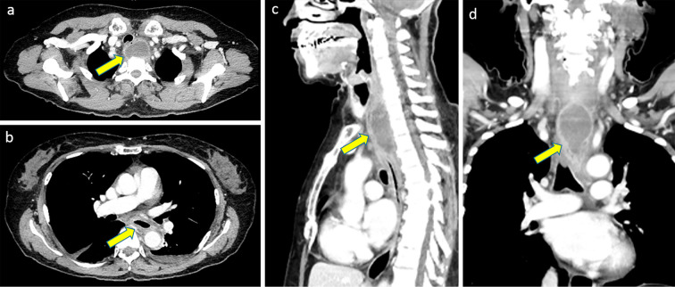
Cases of foreign body ingestion are encountered relatively often in clinical settings; however, serious complications are rare. In such cases, mediastinal abscess due to esophageal perforation can become a life-threatening complication. Although highly invasive, surgery is often used as the first-line treatment. We herein report the case of a 65-year-old woman who presented with complaints of progressive odynophagia and dysphagia for 2 weeks following a fish meal. Enhanced cervicothoracic computed tomography demonstrated an enhanced round mass with peripheral contrasted margins. The mass was diagnosed as a mediastinal abscess resulting from esophageal perforation caused by a fish bone. Endoscopic ultrasound-guided abscess drainage (EUS-AD) was performed using a nasobiliary drainage tube (NDT). Two weeks later, the abscess had completely disappeared. EUS-AD was safe and effective in this case; furthermore, external drainage using NDT was suitable for this abscess located very close to the upper esophageal sphincter.

Keywords: endoscopic ultrasound-guided abscess drainage; fish bone; mediastinal abscess.


Conflict of interest statement

The authors state that they have no Conflict of Interest (COI).

Figures

Figure 1.
Figure 1.
Imaging findings. a: Axial image of contrast-enhanced computed tomography (CT) showing a round low-density lesion with rim enhancement on the back side of the esophagus. No foreign body was detected. The lesion was located in front of the thoracic vertebrae. b: The wall of the inferior esophagus was thick. c, d: Coronal and sagittal images of enhanced CT. The lesion was located in front of the thoracic vertebrae (C7-Th4). There were no significant findings in the thoracic vertebrae and no direct connection to the pleural cavity.
Figure 2.
Figure 2.
The endoscopic examination and treatment. a: Upper endoscopy revealing that the esophageal lumen was severely compressed by the abscess. b: Convex endoscopic ultrasound images revealing an abscess with a clear marginal mixed echoic pattern with acoustic enhancement, implying floating abscess fluid. c: Mobile C-arm fluoroscopic X-ray system showing the nasogastric drainage tube advanced over the guidewire. d: High-viscosity pus aspirated from the abscess.
Figure 3.
Figure 3.
Patient’s clinical course after admission. Treatment details and transitive graphs of the WBC count and CRP level after admission. ABPC/SBT: ampicillin/sulbactam, ABPC: ampicillin, PPN: peripheral parenteral nutrition, OED: oral elemental diet, TPN: total parenteral nutrition, EUS-AD: endoscopic ultrasound-guided abscess drainage, NT: nasogastric tube, OI: oral intake, WBC: white blood cell, CRP: C-reactive protein
Figure 4.
Figure 4.
Imaging findings after the procedure. a, b: Contrast-enhanced computed tomography images (a, coronal; b, sagittal) seven days after admission. The size of the abscess decreased, but the pus persisted. A nasogastric drainage tube (NDT) was advanced through the esophageal wall at the upper esophageal sphincter (yellow arrow). c, d: Fluoroscopic guidance in the front and oblique directions. The NDT was coiled in the abscess (c, frontal presentation; d, oblique presentation). e, f: Contrast revealing shrinkage of the abscess. After shrinkage was confirmed, the NDT loop was decreased.

References

    1. Eisen GM, Baron TH, Dominitz JA, et al. . Guideline for the management of ingested foreign bodies. Gastrointest Endosc 55: 802-806, 2002. - PubMed
    1. Sung SH, Jeon SW, Son HS, et al. . Factors predictive of risk for complications in patients with esophageal foreign bodies. Dig Liver Dis 43: 632-635, 2011. - PubMed
    1. Kim JP, Kwon OJ, Shim HS, et al. . Analysis of clinical feature and management of fish bone ingestion of upper gastrointestinal tract. Clin Exp Otorhinolaryngol 8: 261-267, 2015. - PMC - PubMed
    1. Grodinsky M, Holyoke EA. The fasciae and fascial spaces of the head, neck and adjacent regions. Am J Anat 63: 367-408, 1938.
    1. Ahmad R, Ishlah W, Shaharudin MH, et al. . Posterior mediastinal abscess secondary to esophageal perforation following fish bone ingestion. Med J Malaysia 63: 162-163, 2008. - PubMed