. 2019 Apr 17;9(1):6196.
doi: 10.1038/s41598-019-42549-4.

Visualizing flow in an intact CSF network using optical coherence tomography: implications for human congenital hydrocephalus

Affiliations

Visualizing flow in an intact CSF network using optical coherence tomography: implications for human congenital hydrocephalus

Priya Date et al. Sci Rep. .

Erratum in

Abstract

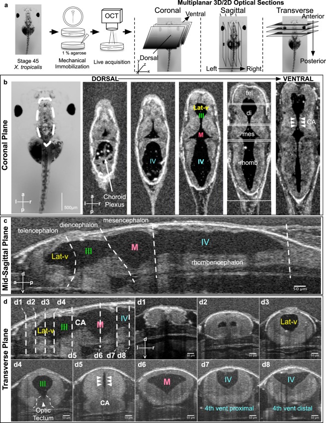
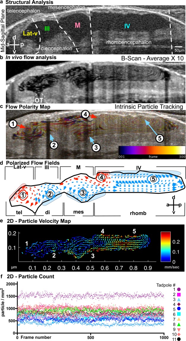
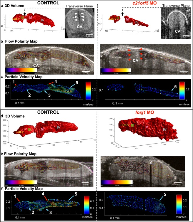
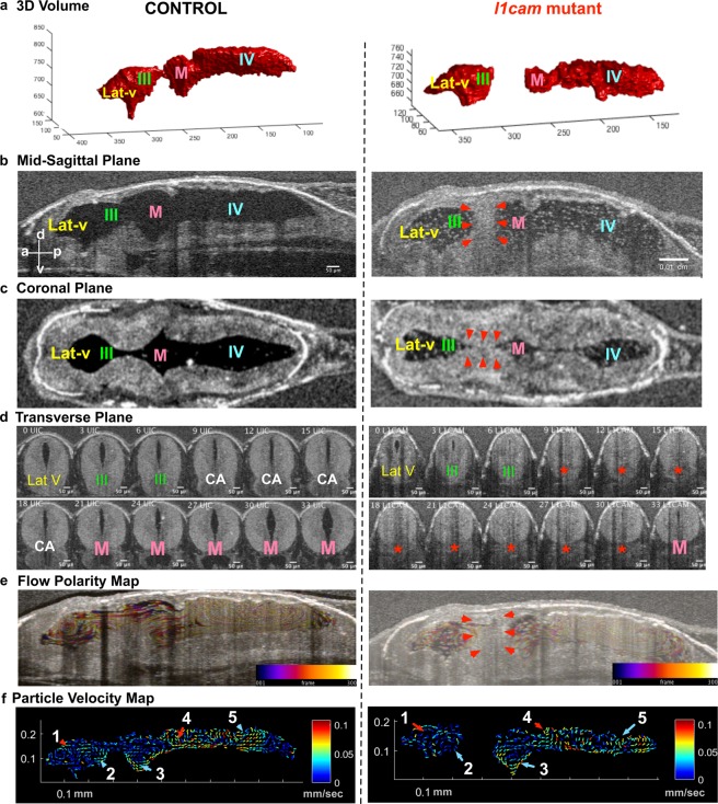
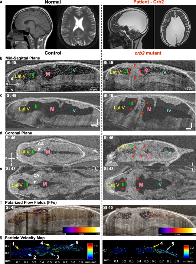
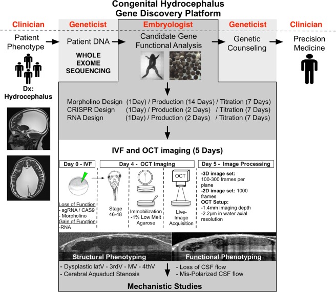
Cerebrospinal fluid (CSF) flow in the brain ventricles is critical for brain development. Altered CSF flow dynamics have been implicated in congenital hydrocephalus (CH) characterized by the potentially lethal expansion of cerebral ventricles if not treated. CH is the most common neurosurgical indication in children effecting 1 per 1000 infants. Current treatment modalities are limited to antiquated brain surgery techniques, mostly because of our poor understanding of the CH pathophysiology. We lack model systems where the interplay between ependymal cilia, embryonic CSF flow dynamics and brain development can be analyzed in depth. This is in part due to the poor accessibility of the vertebrate ventricular system to in vivo investigation. Here, we show that the genetically tractable frog Xenopus tropicalis, paired with optical coherence tomography imaging, provides new insights into CSF flow dynamics and role of ciliary dysfunction in hydrocephalus pathogenesis. We can visualize CSF flow within the multi-chambered ventricular system and detect multiple distinct polarized CSF flow fields. Using CRISPR/Cas9 gene editing, we modeled human L1CAM and CRB2 mediated aqueductal stenosis. We propose that our high-throughput platform can prove invaluable for testing candidate human CH genes to understand CH pathophysiology.


Conflict of interest statement

The authors declare no competing interests.

Figures

Figure 1
Figure 1
In Vivo OCT imaging of the Xenopus tadpole ventricular system. (a) Stage 45 tadpoles are embedded in 1% low-melt agarose with the dorsal side of the animal facing the OCT beam. 2D and 3D images are taken. (b) A stage 45 tadpole as seen under light microscopy. Far left panel - white dotted line outlines the brain. A series of OCT images taken at progressively more ventral planes show the ventricular system with different parts of the brain. (c) A mid-sagittal view of the ventricular system at the midline. White dotted lines show the boundaries between different brain regions. (d) Transverse sections through the ventricular system along the anterior to posterior axis of the animal (d1–d8). The position of each section corresponds to the white dotted lines shown on the mid-sagittal view in the top left panel. (Lat-V: lateral ventricle, III: 3rd ventricle, M: Midbrain ventricle, IV: 4th ventricle, tel: Telencephalon, di: Diencephalon, mes: Mesencephalon, rhomb: Rhombencephalon, CA: Cerebral aqueduct).
Figure 2
Figure 2
Imaging ventricular CSF flow with OCT. (a) Mid-sagittal view of the brain allowing simultaneous visualization of the four ventricles. (b) B-scan image acquisition using OCT: Ventricles of a stage 45 tadpole harbor native free floating particles. In this image, the spatial position of the particles are recorded over time to demonstrate discrete flow patterns within each ventricle (OT: optical tectum). (c) Intrinsic Particle Tracking: Using ImageJ, we tracked particles using 300 frames of 2D images. Temporal color coding depicts their trajectory over time. Color bar represents color versus corresponding frame number in the color-coded image. Based on the trajectory map, 5 distinct flow fields were observed: FF1-FF5. We numbered flow fields 1 to 5 along the anterior-posterior axis. FF 1-Telencephalic flow and FF 2-Diencephalic flow are in the lateral ventricle. FF 3-Mesencephalic flow is in the 3rd ventricle. Finally, the 4th ventricle has 2 flow fields: FF 4-Anterior Rhombencephalic flow and FF 5-Posterior Rhombencephalic flow. (d) Outline of the ventricular system and vector maps of the CSF flow fields. Based on the trajectory map and real time observation (Movie 3); FF1 and FF4 are clockwise and FF2, 3 and 5 are counter-clockwise. (e) 2D Particle Velocity Map: Particle trajectories averaged across all frames (1000) to form flow velocities (see methods for details). Based on the particle velocity map and real time observations FF1 and FF2 are relatively slower compared to FF 3, 4 and 5. The fastest flow is recorded within the 4th ventricle. (f) 2D Particle Count (n = 11): Over 1000 frames, total particle number are counted for 11 different wild type tadpoles and plotted for each frame.
Figure 3
Figure 3
Characterization of c21orf59 and foxj1 morphants. (a) 3D rendering of the tadpole ventricular system shows aqueductal stenosis and a smaller ventricular system in c21orf59 morphants compared to controls and (b,c) flow polarity and particle velocity maps confirm loss of FFs 1-4 as well as diminished flow in FF5. (d) 3D rendering of the tadpole ventricular system shows ventriculomegaly in foxj1 morphants and (e,f) flow polarity and particle velocity map confirm loss of FFs1-4 and slow FF5. (Lat-V: lateral ventricle, III: 3rd ventricle, M: Midbrain ventricle, IV: 4th ventricle).
Figure 4
Figure 4
F0 CRISPR mutation in L1CAM causes cerebral aqueduct stenosis. The left column shows control and right column shows l1cam F0 CRISPR mutant images for all panels. (a) 3D rendering of the tadpole ventricular system shows aqueductal stenosis and a smaller ventricular system in l1cam F0 CRISPR mutant. (b) Mid sagittal view and (c) Coronal view of control and l1cam F0 CRISPR mutant, the later showing stenosis of the cerebral aqueduct (red arrowheads). (d) Transverse view of the control and l1cam F0 CRISPR mutant, starting at the end of the lateral ventricle through the cerebral aqueduct and ending in the midbrain ventricle. Control embryo shows normal opening of the duct whereas the mutant shows complete blockage (red star). (e) Relatively normal ciliary flow fields in the control and mutant animals. (f) 2D Particle Velocity Map shows intact FFs 1-5. (Lat-V: lateral ventricle, III: 3rd ventricle, M: Midbrain ventricle, IV: 4th ventricle).
Figure 5
Figure 5
Hydrocephaly phenotype with CRB2 mutations. (a) CT scan of a normal brain and patient brain, with enlargement of the brain ventricles. (b) Mid-sagittal view (d) Coronal view using OCT shows reduction in ventricular size along with CA stenosis in crb2 F0 CRISPR mutant tadpole brain at stage 45 as compared to controls. (c) Mid-sagittal view (e) Coronal view of control and crb2 F0 CRISPR mutant tadpole raised to stage 48 shows enlargement of the ventricles as compared to controls. This result was seen in 3 animals that survived to this later stage. (White arrow head – CA, red arrowheads – stenosis of CA). (f) Stage 45 tadpole particle tracking shows the impairment of the flow fields in the lateral 3rd and midbrain ventricles, but normal flow in the fourth ventricle for the crb2 F0 CRISPR mutant as compared to the control. (g) 2D Particle Velocity Map shows loss of FFs 1-3 and intact FFs 4-5. (Lat-V: lateral ventricle, III: 3rd ventricle, M: Midbrain ventricle, IV: 4th ventricle).
Figure 6
Figure 6
Schematic representation of the gene discovery platform for screening novel developmental hydrocephaly genes using Xenopus and OCT as a model system.

Similar articles

Cited by

  • Camel regulates development of the brain ventricular system.
    Yang S, Emelyanov A, You MS, Sin M, Korzh V. Yang S, et al. Cell Tissue Res. 2021 Feb;383(2):835-852. doi: 10.1007/s00441-020-03270-1. Epub 2020 Sep 9. Cell Tissue Res. 2021. PMID: 32902807 Free PMC article.
  • DLG5 variants are associated with multiple congenital anomalies including ciliopathy phenotypes.
    Marquez J, Mann N, Arana K, Deniz E, Ji W, Konstantino M, Mis EK, Deshpande C, Jeffries L, McGlynn J, Hugo H, Widmeier E, Konrad M, Tasic V, Morotti R, Baptista J, Ellard S, Lakhani SA, Hildebrandt F, Khokha MK. Marquez J, et al. J Med Genet. 2021 Jul;58(7):453-464. doi: 10.1136/jmedgenet-2019-106805. Epub 2020 Jul 6. J Med Genet. 2021. PMID: 32631816 Free PMC article.
  • A novel SMARCC1 BAFopathy implicates neural progenitor epigenetic dysregulation in human hydrocephalus.
    Singh AK, Allington G, Viviano S, McGee S, Kiziltug E, Ma S, Zhao S, Mekbib KY, Shohfi JP, Duy PQ, DeSpenza T Jr, Furey CG, Reeves BC, Smith H, Sousa AMM, Cherskov A, Allocco A, Nelson-Williams C, Haider S, Rizvi SRA, Alper SL, Sestan N, Shimelis H, Walsh LK, Lifton RP, Moreno-De-Luca A, Jin SC, Kruszka P, Deniz E, Kahle KT. Singh AK, et al. Brain. 2024 Apr 4;147(4):1553-1570. doi: 10.1093/brain/awad405. Brain. 2024. PMID: 38128548 Free PMC article.
  • CC2D1A causes ciliopathy, intellectual disability, heterotaxy, renal dysplasia, and abnormal CSF flow.
    Kim AH, Sakin I, Viviano S, Tuncel G, Aguilera SM, Goles G, Jeffries L, Ji W, Lakhani SA, Kose CC, Silan F, Oner SS, Kaplan OI; MarmaRare Group; Ergoren MC, Mishra-Gorur K, Gunel M, Sag SO, Temel SG, Deniz E. Kim AH, et al. Life Sci Alliance. 2024 Aug 21;7(10):e202402708. doi: 10.26508/lsa.202402708. Print 2024 Oct. Life Sci Alliance. 2024. PMID: 39168639 Free PMC article.
  • Cilia-driven flows in the brain third ventricle.
    Eichele G, Bodenschatz E, Ditte Z, Günther AK, Kapoor S, Wang Y, Westendorf C. Eichele G, et al. Philos Trans R Soc Lond B Biol Sci. 2020 Feb 17;375(1792):20190154. doi: 10.1098/rstb.2019.0154. Epub 2019 Dec 30. Philos Trans R Soc Lond B Biol Sci. 2020. PMID: 31884922 Free PMC article. Review.

References

    1. Johanson CE, et al. Multiplicity of cerebrospinal fluid functions: New challenges in health and disease. Cerebrospinal Fluid Res. 2008;5:10. doi: 10.1186/1743-8454-5-10. - DOI - PMC - PubMed
    1. Hladky SB, Barrand MA. Mechanisms of fluid movement into, through and out of the brain: evaluation of the evidence. Fluids Barriers CNS. 2014;11:26. doi: 10.1186/2045-8118-11-26. - DOI - PMC - PubMed
    1. Ibanez-Tallon I, et al. Dysfunction of axonemal dynein heavy chain Mdnah5 inhibits ependymal flow and reveals a novel mechanism for hydrocephalus formation. Hum Mol Genet. 2004;13:2133–2141. doi: 10.1093/hmg/ddh219. - DOI - PubMed
    1. Banizs B, et al. Dysfunctional cilia lead to altered ependyma and choroid plexus function, and result in the formation of hydrocephalus. Development (Cambridge, England) 2005;132:5329–5339. doi: 10.1242/dev.02153. - DOI - PubMed
    1. Abdelhamed Zakia, Vuong Shawn M., Hill Lauren, Shula Crystal, Timms Andrew, Beier David, Campbell Kenneth, Mangano Francesco T., Stottmann Rolf W., Goto June. A mutation in Ccdc39 causes neonatal hydrocephalus with abnormal motile cilia development in mice. Development. 2018;145(1):dev154500. doi: 10.1242/dev.154500. - DOI - PMC - PubMed

Publication types