Review
. 2019 Jun;18(2):164-179.
doi: 10.1007/s12663-018-01182-w. Epub 2019 Jan 28.

Fungal Rhinosinusitis: Unravelling the Disease Spectrum

Affiliations
Review

Fungal Rhinosinusitis: Unravelling the Disease Spectrum

Virendra Singh. J Maxillofac Oral Surg. 2019 Jun.

Abstract

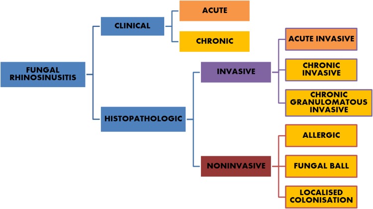
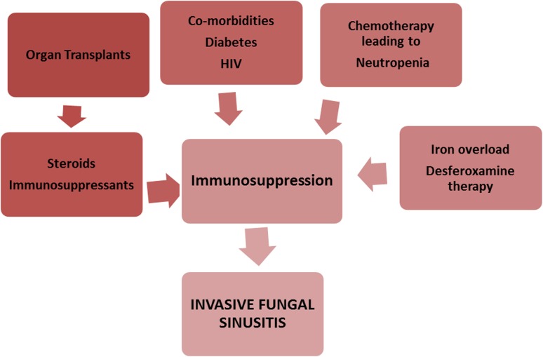
Fungal rhinosinusitis (FRS), once considered a rare disease, has seen a steep rise in incidence in recent times. This global rise in the burden of fungal disease is a consequence of an increment in the population with weakened immune systems. Increased life expectancy with rise in conditions like diabetes mellitus, medical advancements with invasive interventions, use of immunosuppressive drugs and chemo-radiotherapy all lead to unique risk situations. The situation becomes more alarming with the fact that there has been a significant rise in cases in immune-competent hosts with no predisposing factors. FRS represents a wide spectrum of disease ranging from the mild form of superficial colonization, allergic manifestations to life threatening extensive invasive disease. The categorization of disease into acute and chronic and invasive or noninvasive is important factor with implications in disease management and prognosis and this has been emphasized greatly in recent years. Diagnosis of FRS has been a challenge as the presenting clinical signs and symptoms and radiographic manifestations are often nonspecific. Definitive diagnosis requires direct fungi identification and hence culture and microscopic examination remain the gold standard. Availability of advanced and rapid diagnostic techniques is rare in majority of developing nations. Therapeutic dilemmas are another aspect of the management of FRS as in spite of the availability of new antifungal drugs, treatment is often empirical due to non-availability of early diagnosis, rapid disease progression and high costs of antifungal drugs. A description of the different types of FRS, their diagnosis and management has been presented in this review.

Keywords: Fungal rhinosinusitis; Invasive fungal sinusitis; Mucormycosis.


Figures

Fig. 1
Fig. 1
Classification suggested by consensus workshop
Fig. 2
Fig. 2
Risk factors for invasive fungal rhinosinusitis
Fig. 3
Fig. 3
Clinical staging of Acute invasive fungal rhinosinusitis (AIFRS) based on disease extension
Fig. 4
Fig. 4
Clinico-radiologic presentations of FRS. (i) A Extensive cheek necrosis, B palatal necrosis, C exposed necrotic bone on intraoral examination, D CT scan of a patient diagnosed with Allergic FRS. Coronal section showing hyperdensity of mucin within right ethmoid and maxillary sinuses, E coronal CT image showing mixed lytic sclerotic changes with bony destruction involving left zygoma and walls of left maxillary sinus. Also there is soft tissue attenuation with air loculi. (ii) A A patient at clinical presentation with very mild diffuse bilateral swelling, B, C intraoral pictures showing exposed necrotic bone and generalized mobility of maxillary teeth stabilized with Essig’s wiring, D, E intraoperative pictures for sequestrectomy and curettage, F CT scan image shows mucosal thickening of the maxillary sinuses with erosion of bone. (iii) A A clinical presentation of case of CIFRS with extensive orbital involvement mimicking malignancy, B, C, D radiographic picture of bone erosion localized to the area of extra-sinus component of the disease more extensive than intra-sinus component
Fig. 5
Fig. 5
Photographic series of management of a patient with FRS. A preoperative clinical presentation of immunocompetent patient showing diffuse swelling on left side of face and orbital involvement. B Histopathology showing PAS-positive wall of a negatively stained section showing fungal hyphae(× 400), C Axial CT scan shows diffusely enhancing mass (arrow) involving the left maxillary sinus and the subcutaneous plane of the left cheek and upper lip, D Axial CT scan showing left eye proptosis caused by a mass involving the temporal fossa and orbit extending to involve the ethmoid air cells with bone erosion(arrows), E Weber- Fergusson incision, F Zygomatic swing osteotomy to get an access to orbital floor, G Zygoma swung laterally, H Surgical debridement extending to ethmoid sinus, I Realignment, J Closure, K 2 months post operative picture after a course of Amphotericin B, L 1 year postoperative patient had vision loss and was given Voriconazole after which disease no longer progressed M 5 years post treatment patient had another relapse, N Patient responded well to a course of Voriconazole again

References

    1. Hawksworth DL. The magnitude of fungal diversity: the 1.5 million species estimate revisited. Mycol Res. 2001;105:1422–1432. doi: 10.1017/S0953756201004725. - DOI
    1. Blackwell M. The fungi: 1, 2, 3… 5.1 million species? Am J Bot. 2011;98:426–438. doi: 10.3732/ajb.1000298. - DOI - PubMed
    1. Taylor LH, Latham SM, Woolhouse ME. Risk factors for human disease emergence. Philos Trans R Soc Lond B Biol Sci. 2001;356:983–998. doi: 10.1098/rstb.2001.0888. - DOI - PMC - PubMed
    1. Chakrabarti A, Sethuraman N. Introduction to medical mycology. In: Mora-Montes H, Lopes-Bezerra L, editors. Current progress in medical mycology. Cham: Springer; 2017.
    1. Chakrabarti A, Chatterjee SS, Shivaprakash MR. Overview of opportunistic fungal infections in India. J Med Mycol. 2008;49:165–172. doi: 10.3314/jjmm.49.165. - DOI - PubMed

LinkOut - more resources