

Case Reports
doi: 10.1002/ccr3.2007.
eCollection 2019 Apr.
Cutaneous lymphangioma circumscriptum: The relevance of clinical, dermoscopic, radiological, and histological assessments
Affiliations
- PMID: 30997047
- PMCID: PMC6452527
- DOI: 10.1002/ccr3.2007
Item in Clipboard
Case Reports
Cutaneous lymphangioma circumscriptum: The relevance of clinical, dermoscopic, radiological, and histological assessments
Clin Case Rep.
.
Abstract
Cutaneous lymphangioma circumscriptum (CLC) is a rare congenital malformation of the superficial cutaneous lymphatic ducts. Case report: A 7-year-old boy presented plaque with grouped vesicles resembling a "frog spawn" on the upper left thigh, since 4 months old. The clinical, dermatoscopic and histopathological correlation is of great importance.
Keywords: cystic tumor; dermoscopy; lymphangioma; vascular malformations.
Conflict of interest statement
None declared.
Figures
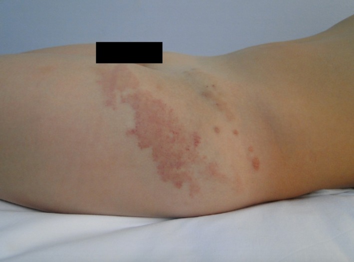
Cutaneous lymphangioma circumscriptum. Well‐defined, irregular, brownish plaque of 15 × 4 cm, surmounted by small grouped translucent vesicles with a serohematic content and satellite lesions with the same characteristics on the upper left thigh

Cutaneous lymphangioma circumscriptum. Dermoscopy examination: Brown‐orange lacunes in the upper portion and red‐violet in the lower portion (hypopio‐like), wrapped by pale septa

Cutaneous lymphangioma circumscriptum. MRI of the left thigh: Serpiginous‐like structures in the subcutaneous tissue on the anterolateral face of the thigh root on the left, which are isointense in relation to musculature in T1
References
-
- Fatima S, Uddin N, Idrees R, et al. Lymphangioma circumscriptum: clinicopathological spectrum of 29 cases. J Coll Physicians Surg Pak. 2015;25:658‐661. - PubMed
-
- Patel GA, Schwartz RA. Cutaneous lymphangioma circumscriptum: frog spawn on the skin. Int J Dermatol. 2009;48:1290‐1295. - PubMed
-
- Zaballos P, del Pozo LJ, Argenziano G, et al. Dermoscopy of lymphangioma circumscriptum: A morphological study of 45 cases. Australas J Dermatol. 2018;59:e189‐93. - PubMed
-
- Wegener G. Ueber lymphangiome. Arch Klin Chir. 1877;20:641–707.
-
- Fox T, Fox TC. On a case of lymphangiectodes with an account of the histology of the growth. Trans Pathol Soc London. 1878;30:470‐476.
Publication types
LinkOut - more resources
Full Text Sources
