

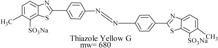
Demethylation and desulfonation of textile industry dye, Thiazole Yellow G by Aspergillus niger LAG
- PMID: 30997348
- PMCID: PMC6451163
- DOI: 10.1016/j.btre.2019.e00327
Demethylation and desulfonation of textile industry dye, Thiazole Yellow G by Aspergillus niger LAG
Erratum in
-
Erratum regarding missing Declaration of Competing Interest statements in previously published articles.Biotechnol Rep (Amst). 2021 Mar 19;29:e00582. doi: 10.1016/j.btre.2020.e00582. eCollection 2021 Mar. Biotechnol Rep (Amst). 2021. PMID: 33786326 Free PMC article.
Abstract
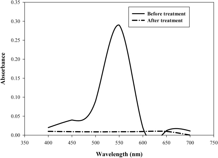
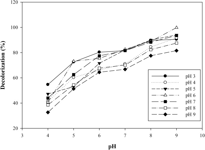
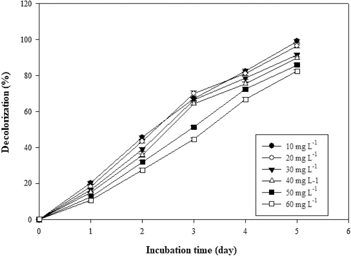
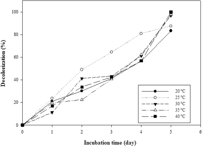
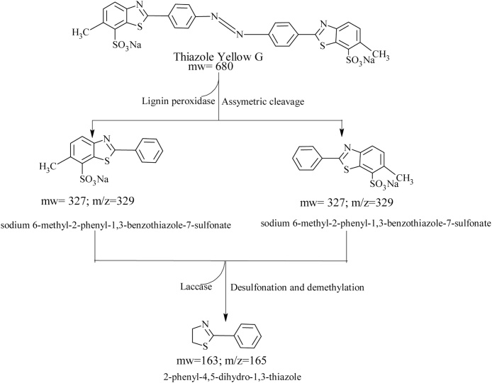
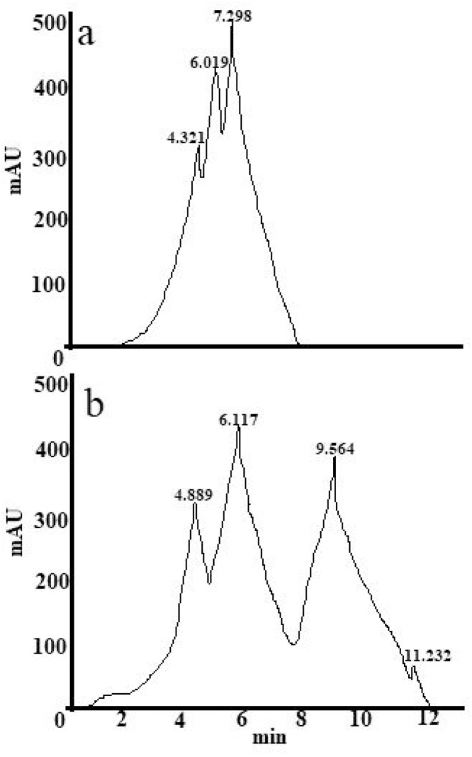
Filamentous fungi perform tremendously in adsorption of dyes from polluted environment. In this study, Aspergillus niger LAG decolorized thiazole yellow G dye within 5 days. Scale up studies done revealed that maximum decolorization (98%) was achieved at a concentration (10 mg L-1), temperature (35 °C) and pH 6. The fungus exhibited significant inductions in laccase (71%) and lignin peroxidase (48%) respectively. Spectrometric analysis (UV-vis, HPLC and gas chromatography-mass spectrometry) was used in analyzing the degraded products of the dye. The GCMS analysis revealed the production of two metabolites; sodium 6-methyl-2-phenyl-1,3-benzothiazole-7-sulfonate and 2-phenyl-4,5-dihydro-1,3-thiazole after degradation of thiazole yellow G dye. A metabolic pathway of thiazole yellow G dye degradation by Aspergillus niger was proposed. Significant growth in plumule and radicle couple with an attendant increase in germination further confirmed the detoxified status of the dye after degradation.
Keywords: Aspergillus niger; Biodegradation; Detoxification; Thiazole yellow G dye.
Figures
References
-
- Crini G. Non-conventional low-cost adsorbents for dye removal: a review. Bioresour. Technol. Rep. 2006;97:1061–1085. - PubMed
-
- Hadibarata T., Kristanti R. Effect of environmental factors in the decolourisation of Remazol Brillant Blue R by Polyporus sp. S133. J. Chilean Chem. Soc. 2012;57:1095–1998.
-
- Mishra G., Tripathy M. A critical review of the treatments for decolourization of textile effluent. Colourage. 1993;40:35–38.
-
- Aksu Z. Application of biosorption for the removal of organic pollutants: a review. Process Biochem. 2005;40:997–1026.
-
- Zhao G., Li M., Hu Z., Hu H. Dissociation and removal of complex chromium ions contained in dye wastewaters. Separat. Purif. Technol. 2005;43:227–232.
LinkOut - more resources
Full Text Sources