

Pancreaticobiliary metastasis presenting as primary mucinous ovarian neoplasm: A systematic literature review
- PMID: 30997376
- PMCID: PMC6453658
- DOI: 10.1016/j.gore.2019.03.012
Pancreaticobiliary metastasis presenting as primary mucinous ovarian neoplasm: A systematic literature review
Abstract
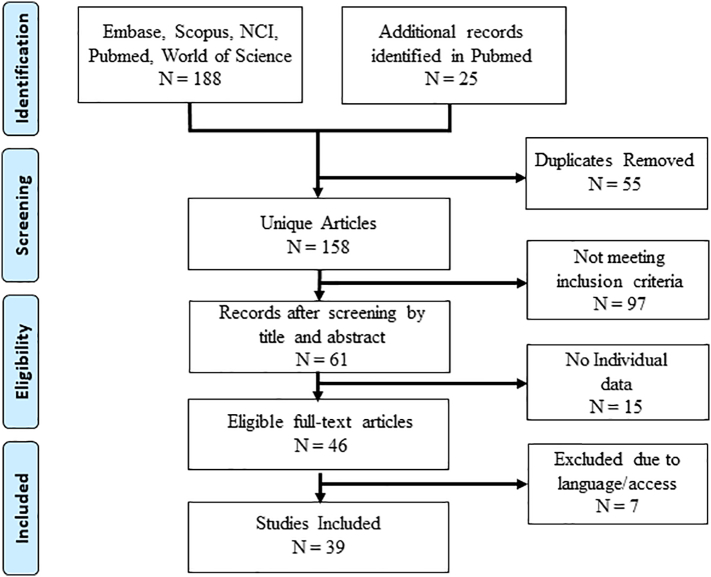
True primary mucinous ovarian carcinomas are rarer than originally thought and their clinical behavior and treatment response are different than more common epithelial ovarian carcinomas. Secondary ovarian neoplasms often mimic the clinical and histological features of mucinous ovarian cancer making their diagnosis, and therefore treatment, more difficult. Misdiagnosis can have a significant impact on both treatment and prognosis. The majority of these secondary ovarian neoplasms arise from the gastrointestinal tract, with mucinous histology often of pancreaticobiliary origin. Our study objective was to review current evidence distinguishing pancreaticobiliary ovarian metastasis from primary mucinous ovarian carcinoma. We utilized a PubMed search using MeSH terms and selected articles were reviewed, synthesized and summarized. Thirty-nine articles were included in the review. The clinical, gross, histological and immunohistochemical features distinguishing primary mucinous ovarian carcinomas from pancreaticobiliary ovarian metastasis were identified. Compared to primary mucinous ovarian carcinoma, metastatic pancreaticobiliary tumors are more often bilateral, <10 cm, have irregular external surface and surface implants, display an infiltrative pattern of invasion and stain for MUC1 and CK17. Primary ovarian mucinous tumors rarely (<3%) have signet ring cells or involvement of the hilum. Metastatic mucinous tumors mimic their primary mucinous ovarian counterparts and their clinical and histopathological features overlap in many ways. However, these metastatic tumors have features that can help differentiate them from primary mucinous carcinoma. With a high index of suspicion and knowledge of the reviewed features, distinguishing these tumors will continue to become easier.
Keywords: Immunohistochemistry; Metastatic pancreaticobiliary tumors; Ovarian mucinous tumors.
Figures
References
-
- Alvarado-Cabrero I., Rodríguez-Gómez A., Castelan-Pedraza J., Valencia-Cedillo R. Metastatic ovarian tumors-A clinicopathologic study of 150 cases. Anal. Quant. Cytol. Histol. 2013;35(5):241–248. - PubMed
-
- Corr B.R., Mantia-Smaldone G., Cantor J., Livolsi V.A., Furth E., Chu C.S. Metastatic cholangiocarcinoma to the ovary: a case series. Int. J. Gynecol. Pathol. 2013;32(6):562–565. - PubMed
-
- Garcia A., De la Torre J., Castellvi J., Gil A., Lopez M. Ovarian metastases caused by cholangiocarcinoma: a rare Krukenberg's tumour simulating a primary neoplasm of the ovary: a two-case study. Arch. Gynecol. Obstet. 2004;270(4):281–284. - PubMed
-
- Goldstein N.S., Bassi D., Uzieblo A. WT1 is an integral component of an antibody panel to distinguish pancreaticobiliary and some ovarian epithelial neoplasms. Am. J. Clin. Pathol. 2001;116(2):246–252. - PubMed
Publication types
LinkOut - more resources
Full Text Sources
Research Materials
Miscellaneous