Case Reports
. 2019 Jul;50(3):859-870.
doi: 10.1007/s42770-019-00082-6. Epub 2019 Apr 17.

Disseminated melanized fungal infection due to Cladosporium halotolerans in a dog coinfected with canine adenovirus-1 and canine parvovirus-2

Affiliations
Case Reports

Disseminated melanized fungal infection due to Cladosporium halotolerans in a dog coinfected with canine adenovirus-1 and canine parvovirus-2

Selwyn Arlington Headley et al. Braz J Microbiol. 2019 Jul.

Abstract

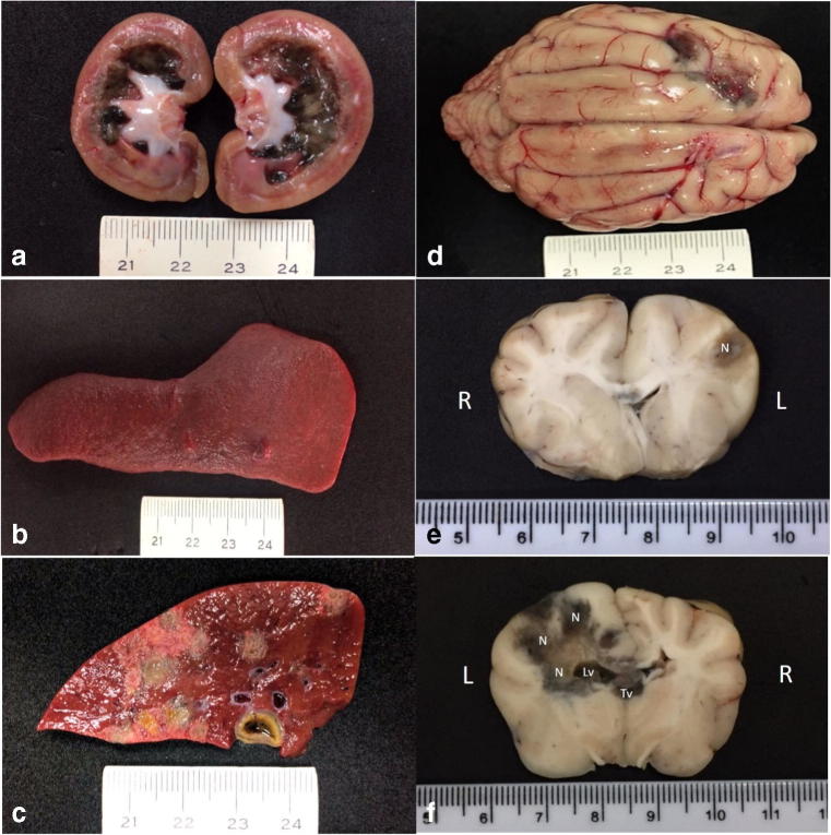
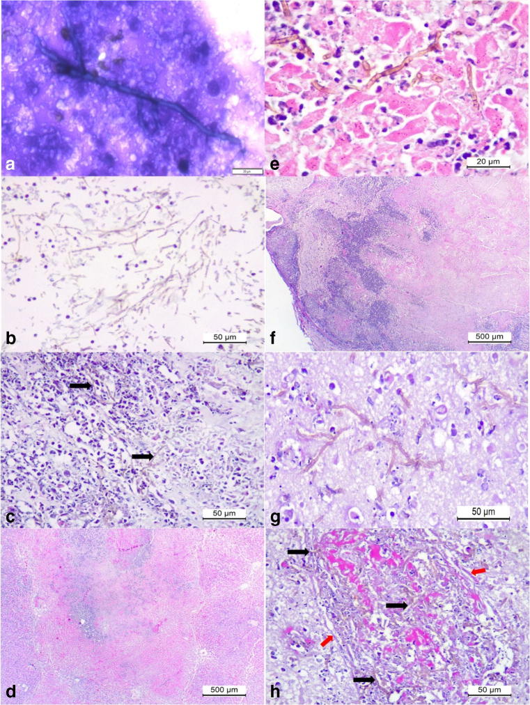
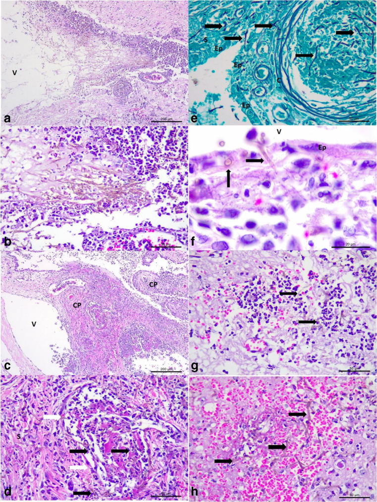
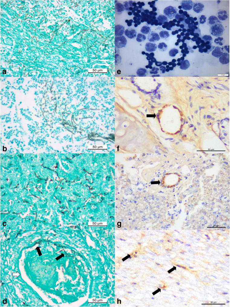
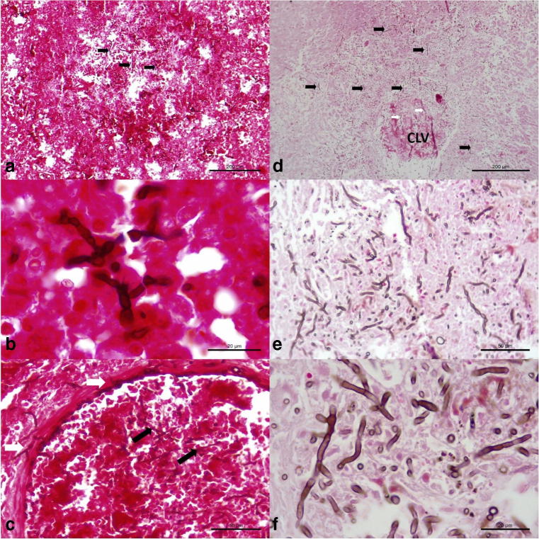
This report presents the pathologic findings associated with disseminated infection due to Cladosporium halotolerans in a dog that was simultaneously infected with canine adenovirus-1 (CAdV-1) and canine parvovirus-2 (CPV-2). A 12-year-old, mixed breed dog, with a clinical history of neurological manifestations was submitted for routine autopsy due to poor prognosis. The principal pathologic findings were mycotic necrotizing nephritis, hepatitis, and splenitis with embolic dissemination to the brain resulting in mycotic necrotizing meningoencephalitis, ventriculitis, choroid plexitis, and obstructive hydrocephalus associated with intralesional and intravascular septate pigmented fungi. PCR and sequencing of the ITS region of fungi revealed that the intralesional fungal organisms had 82% nucleotide identity with members of the Cladosporium sphaerospermum complex of organisms. However, a PCR assay and sequencing of the beta tubulin gene confirmed that the organism identified in this dog had 100% nucleotide sequence identity with C. halotolerans. Using immunohistochemistry, intralesional antigens of CAdV-1 were identified within the epithelial cells of the liver and lungs; there was positive immunolabeling for CPV-2 antigens in degenerated cardiomyocytes. These findings confirmed the active participation of C. halotolerans in the development of disseminated cladosporiosis in this dog and represent a rare occurrence of concomitant infection with CAdV-1 and CPV-2.

Keywords: Choroid plexitis; Cladosporiosis; Dematiaceous fungi; Diagnostic pathology; Dog; Pleocytosis; Thromboembolism; Ventriculitis.


Figures

Fig. 1
Fig. 1
Gross findings associated with disseminated phaeohyphomycosis in a mixed-breed dog. Observe the black-green accumulation within the medullary and the white substance at the pelvic regions of the left kidney (a) and the nodule at the spleen (b). There are multifocal to coalescing necrotic regions at the sectioned surface of the liver (c). Observe two green-black areas at the frontal lobe of the cerebrum (d). Transversal sections of the brain demonstrating the extension of the black-green necrotic (N) regions, which extended from the left frontal lobe (e) to the caudal part (f) of the brain (R, right; L, left). There is mild dilation of the left lateral ventricle (Lv) due to the accumulation of necrotic material; necrotic debris was also observed in the third ventricle (Tv). Scale in centimeters
Fig. 2
Fig. 2
Cytological and histopathologic findings associated with disseminated phaeohyphomycosis in a mixed-breed dog. Cytological smear from the splenic nodule revealed a septate fungal organism (a). Observe large accumulations of pigmented fungi (arrows) at the medullar region of the kidney admixed with cellular debris (b, c). There is massive hepatic necrosis (d) associated with intralesional-pigmented septate fungal hyphae (e). Observe severe cortical necrosis of the cerebrum (f) associated with intralesional pigmented septate fungal hyphae (g), and invasive vasculitis and thrombosis associated with intervascular accumulations of pigmented fungal hyphae (black arrows) within the severely destroyed wall (red arrows) of the vessel (h). Panoptic stain, a; hematoxylin and eosin stain, bh. Bar, a, 20 μm; b, c, e, f, and g, 50 μm; d and fh, 500 μm
Fig. 3
Fig. 3
Histopathologic and histochemical findings associated with mycotic ventriculitis and choroid plexitis in a mixed-breed dog. Mycotic ventriculitis: observe the dilated lateral ventricle (V) and severe accumulations of pigmented fungal septate hyphae (a); closer view showing the myriad of pigmented hyphae admixed with neutrophils and macrophages (b). Mycotic necrotizing choroid plexitis: there is severe necrosis of the stroma and ependymal cells of the choroid plexus (CP) within the lateral ventricle (V) of the brain (a). Closer view of the choroid plexus demonstrating a mycotic thrombosis at the stroma (S) of the choroid plexus; observe intravascular fungal hyphae (black arrows) within the stroma and a capillary (white arrows) of the choroid plexus (d). There is destruction of the ependymal membrane (Ep) and accumulations of fungal hyphae within a capillary (c) and the stroma of the choroid plexus (e). There are pigmented fungal organisms (black arrows) within the necrotic wall of the ependymal membrane (f), and within a microabscesses (g), and hemorrhage of the cerebrum (h). Hematoxylin and eosin stain, ad, fh; Gomori methenamine-silver histochemical stain, e. Bar, a, c, 200 μm; b, d, e, g, and h, 50 μm; f, 20 μm
Fig. 4
Fig. 4
Histochemical, cytological, and immunohistochemical findings of disseminated phaeohyphomycosis in a dog with simultaneous infections by canine adenovirus-1 (CAdV-1) and canine parvovirus-2 (CPV-2). Observe intralesional accumulations of septate fungal hyphae in the liver (a), kidney (b), and the cerebral cortex (c). There is vascular invasion of septate hyphae (black arrows) within the damaged vein (white arrow) of the cerebral cortex (d). Cytological evaluation of the cerebrospinal fluid revealing elevated number of fungal organisms (e). Immunohistochemical identification of antigens of CAdV-1 (arrows) within epithelial cells of mixed glands of the lungs (f) and epithelial cells of bile ducts within the liver (g). There is positive immunolabeling for antigens of CPV-2 (arrows) within degenerated cardiomyocytes of the myocardium (h). Gomori methenamine-silver histochemical stain, ad; panoptic stain, e; immunoperoxidase counterstained with hematoxylin, fh. Bar, ad, fh, 50 μm; e, 10 μm
Fig. 5
Fig. 5
Histochemical identification of intralesional septate fungal organisms in a dog with disseminated cladosporiosis. Observe intralesional pigmented fungi (black arrows) in the brain (a, b), and within an artery of the cerebrum (c). There are numerous accumulations of intralesional fungi in the parenchyma (black arrows) and within (white arrows) the affected central lobular vein (CLV) of the liver. Closer approximation (e and f) to demonstrate the intralesional fungal organisms within the liver. Fontana-Masson stain; bar, a, d, 200 μm; b, f, 20 μm; c, e, 50 μm

Similar articles

Cited by

References

    1. Brandt ME, Warnock DW. Epidemiology, clinical manifestations, and therapy of infections caused by dematiaceous fungi. J Chemother. 2003;15(sup2):36–47. doi: 10.1179/joc.2003.15.Supplement-2.36. - DOI - PubMed
    1. Wong EH, Revankar SG. Dematiaceous molds. Infect Dis Clin N Am. 2016;30(1):165–178. doi: 10.1016/j.idc.2015.10.007. - DOI - PubMed
    1. Revankar SG, Sutton DA. Melanized fungi in human disease. Clin Microbiol Rev. 2010;23(4):884–928. doi: 10.1128/CMR.00019-10. - DOI - PMC - PubMed
    1. Seyedmousavi S, Guillot J, de Hoog GS. Phaeohyphomycoses, emerging opportunistic diseases in animals. Clin Microbiol Rev. 2013;26(1):19–35. doi: 10.1128/CMR.00065-12. - DOI - PMC - PubMed
    1. Revankar SG, Sutton DA, Rinaldi MG. Primary central nervous system phaeohyphomycosis: a review of 101 cases. Clin Infect Dis. 2004;38(2):206–216. doi: 10.1086/380635. - DOI - PubMed

Publication types

MeSH terms