. 2019 Jul;38(7):880-887.
doi: 10.1097/ICO.0000000000001954.

Expressible Meibomian Glands Have Occult Fixed Obstructions: Findings From Meibomian Gland Probing to Restore Intraductal Integrity

Affiliations

Expressible Meibomian Glands Have Occult Fixed Obstructions: Findings From Meibomian Gland Probing to Restore Intraductal Integrity

Steven L Maskin et al. Cornea. 2019 Jul.

Abstract

Purpose: To describe and quantify findings of intraductal obstruction during probing expressible and nonexpressible meibomian glands (MGs) in patients with obstructive meibomian gland dysfunction using a 1-mm intraductal MG probe.

Methods: A retrospective study of probe findings from 108 consecutive patients. Nonparametric tests using SPSS software 25.0 to explore relationships between expressibility and probe findings.

Results: Of 11,776 probed glands of 404 lids, 84% showed mechanical resistance (MR) and 16% showed no resistance (NR). Fixed, firm, focal unyielding resistance (FFFUR) occurred in 79.5% of obstructed glands, and nonfixed, nonfocal easily yielding soft resistance (SFT) in 20.4%. FFFUR was characterized by an audible and tactile "firm pop" (FP) or "firm gritty" (FG) sensation. No significant difference in MR and FFFUR for lids between 0% and >90% gland expressibility was observed. FP correlated with increased expressibility (P = 0.011), lid tenderness (P = 0.045), and complete proximal obstruction (P = 0.037), whereas SFT correlated with reduced expressibility (P = 0.016). Upper lids showed greater incidence of MR (P < 0.001), FFFUR (P < 0.001), and FG (P < 0.001), whereas lower lids showed greater expressibility (P < 0.001) and NR (P < 0.001).

Conclusions: FFFUR was the most common probe finding in a large series of consecutively probed MGs, with an incidence of 67% of glands and 80% of obstructed glands. FFFUR was independent of gland expressibility, demonstrating expressible glands harbor FFFUR deep to at least one acinus. FP was associated with expressible gland occult obstruction and lid tenderness. SFT correlated with reduced expressibility, perhaps related to altered duct/duct contents. Upper lids correlated with increased MR, FFFUR, and FG and lower lids with increased expressibility and NR, possibly reflecting contrasting anatomy and blink-related microtrauma.


Figures

FIGURE 1.
FIGURE 1.
Representative examples of periductal fibroses and corresponding internal lumen stricture and altered diameters using confocal microscopy: (A and B) are images of the same gland orifice at different depths. A, shows an oval lumen at 67 μm depth with apparent fibroses (arrows), including a fibrotic sheet (star) which appears in (B) at 112 μm as a tight periductal fibrotic band (star and arrows) surrounding and pinching the external duct wall. B, also shows an oval lumen with the flattest border (bracket), corresponding to the tightest area of fibroses (open arrow). (C and D) are views of the same gland orifice at 64 and 112 μm depth (different gland orifice than A/B). C, shows scalloping and pinching of the external duct wall from apparent fibroses (arrows), with flattening of oval lumen at corresponding meridians (brackets). D, shows indentation of the external wall (arc), with corresponding lumen stricture (open arrow).
FIGURE 2.
FIGURE 2.
The top row of images represents a fixed obstruction or FFFUR (indicated by the orange ring around the gland duct) of the MG distal to all acini. This type of obstruction will initially present with LT because meibum is being produced without an escape, leading to the buildup of pressure behind the obstruction (this obstruction is termed CDO, as depicted in the first image of the top row). Eventually, the buildup of pressure will result in a loss of function of the gland (termed CDO-NF, as depicted in the middle image of the top row), ultimately resulting in MG dropout as the entire gland atrophies (top row, right). No meibum will be expressed from this gland. The first 2 images in the bottom row represent a fixed proximal obstruction or FFFUR (indicated by the orange ring around the more proximal gland duct) of the MG, in which 1 or more acini are in communication with the central duct and orifice. LT will be present because meibum is being produced without an escape, leading to the buildup of pressure behind the obstruction. However, this classification of MGs will have expressible meibum because the acini located distally to the obstruction still remains in communication with the central duct and orifice (this obstruction is termed CPO, as depicted in the first image of the bottom row). Eventually, the buildup of pressure will result in a loss of function of the gland proximal to the obstruction, ultimately resulting in a truncated MG (middle image, bottom row). The final classification of PDO (as depicted in the bottom row, right), is the result of a partial distally obstructed gland, such as orifice squamous metaplasia. This gland presents with no LT because there is not a complete obstruction and meibum is able to be expressed at the orifice. [Adapted from: Maskin S. Intraductal Meibomian Gland Probing: A Paradigm Shift for the Successful Treatment of Obstructive Meibomian Gland Dysfunction. In: Tsubota K, ed. Diagnosis and Treatment of Meibomian Gland Dysfunction. Tokyo, Japan: Kanehara & Co, Ltd; 2016:157 (Figure 6). Maskin SL, Testa WR. Growth of Meibomian gland tissue after intraductal Meibomian gland probing in patients with obstructive Meibomian gland dysfunction. Br J Ophthalmol. 2018;102:59–68.). Adaptations are themselves works protected by copyright. So in order to publish this adaptation, authorization must be obtained both from the owner of the copyright in the original work and from the owner of copyright in the translation or adaptation.
FIGURE 3.
FIGURE 3.
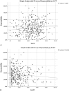
A, Scatter plot showing positive correlation between the percentage of FP and percentage expressibility observed in a lid by Spearman Rho at P = 0.011. B, Scatter plot showing negative correlation between the percentage of soft (SFT) obstruction and percentage expressibility observed in a lid by Spearman Rho at P = 0.016.

References

    1. Nichols KK, Foulks GN, Bron AJ, et al. The International Workshop on Meibomian gland dysfunction: executive summary. Invest Ophthalmol Vis Sci. 2011;52:1922–1929. - PMC - PubMed
    1. Lemp MA, Crews LA, Bron AJ, et al. Distribution of aqueous-deficient and evaporative dry eye in a clinic-based patient cohort: a retrospective study. Cornea. 2012;31:472–478. - PubMed
    1. Finis D, Ackermann P, Pischel N, et al. Evaluation of Meibomian gland dysfunction and local distribution of Meibomian gland atrophy by non-contact infrared meibography. Curr Eye Res. 2015;40:982–989. - PubMed
    1. Foulks GN, Bron AJ. Meibomian gland dysfunction: a clinical scheme for description, diagnosis, classification, and grading. Ocul Surf. 2003;1:107–126. - PubMed
    1. Geerling G, Tauber J, Baudouin C, et al. The international workshop on Meibomian gland dysfunction: report of the subcommittee on management and treatment of Meibomian gland dysfunction. Invest Ophthalmol Vis Sci. 2011;52:2050–2064. - PMC - PubMed