. 2019 May 30:667:14-21.
doi: 10.1016/j.abb.2019.04.003. Epub 2019 Apr 16.

Quantitative atomic force microscopy provides new insight into matrix vesicle mineralization

Affiliations

Quantitative atomic force microscopy provides new insight into matrix vesicle mineralization

Justin S Plaut et al. Arch Biochem Biophys. .

Abstract

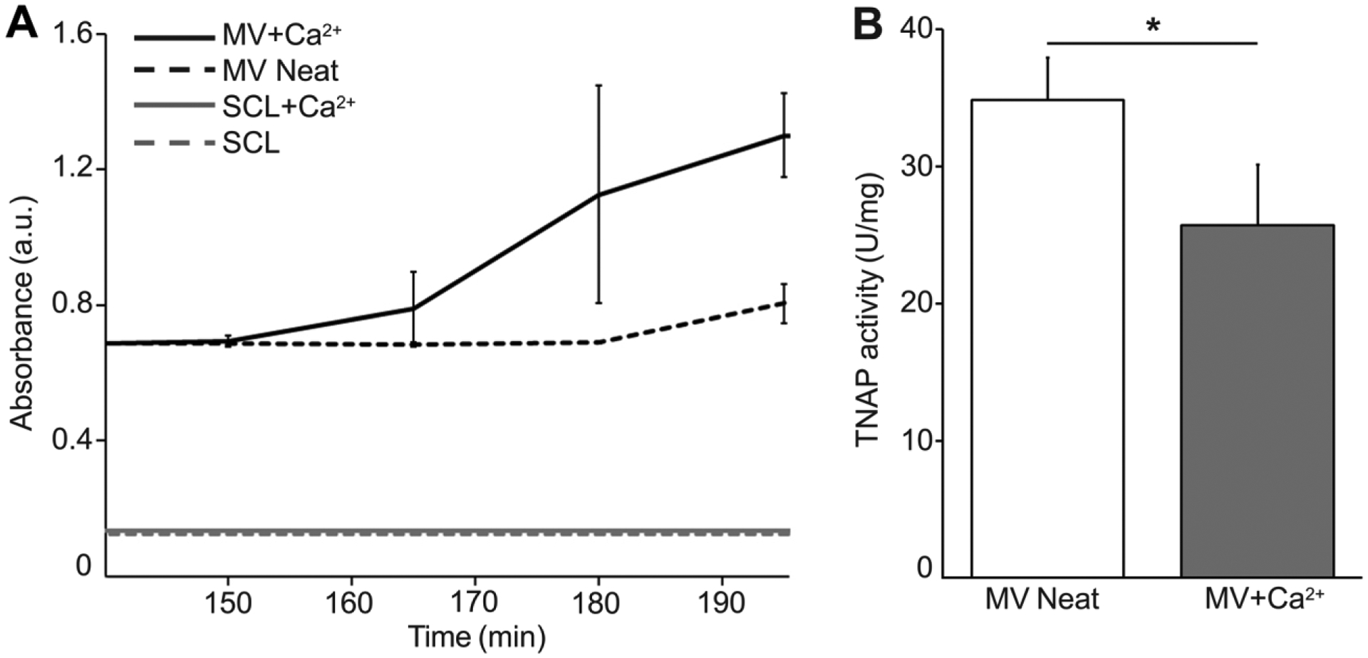
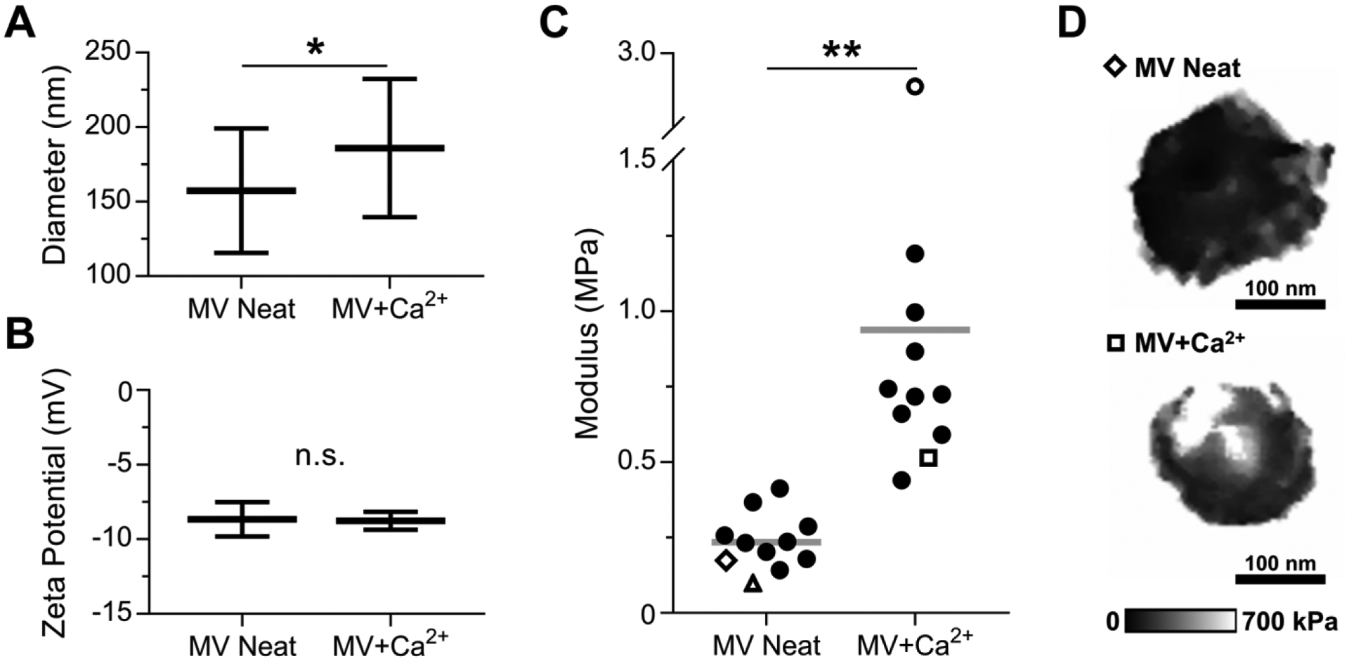
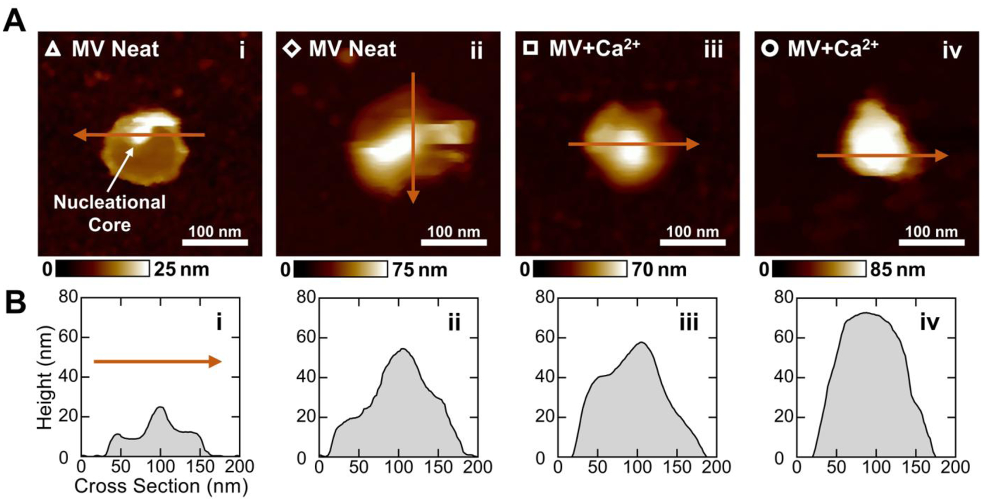
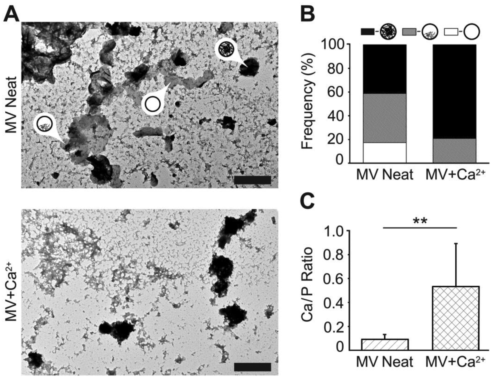
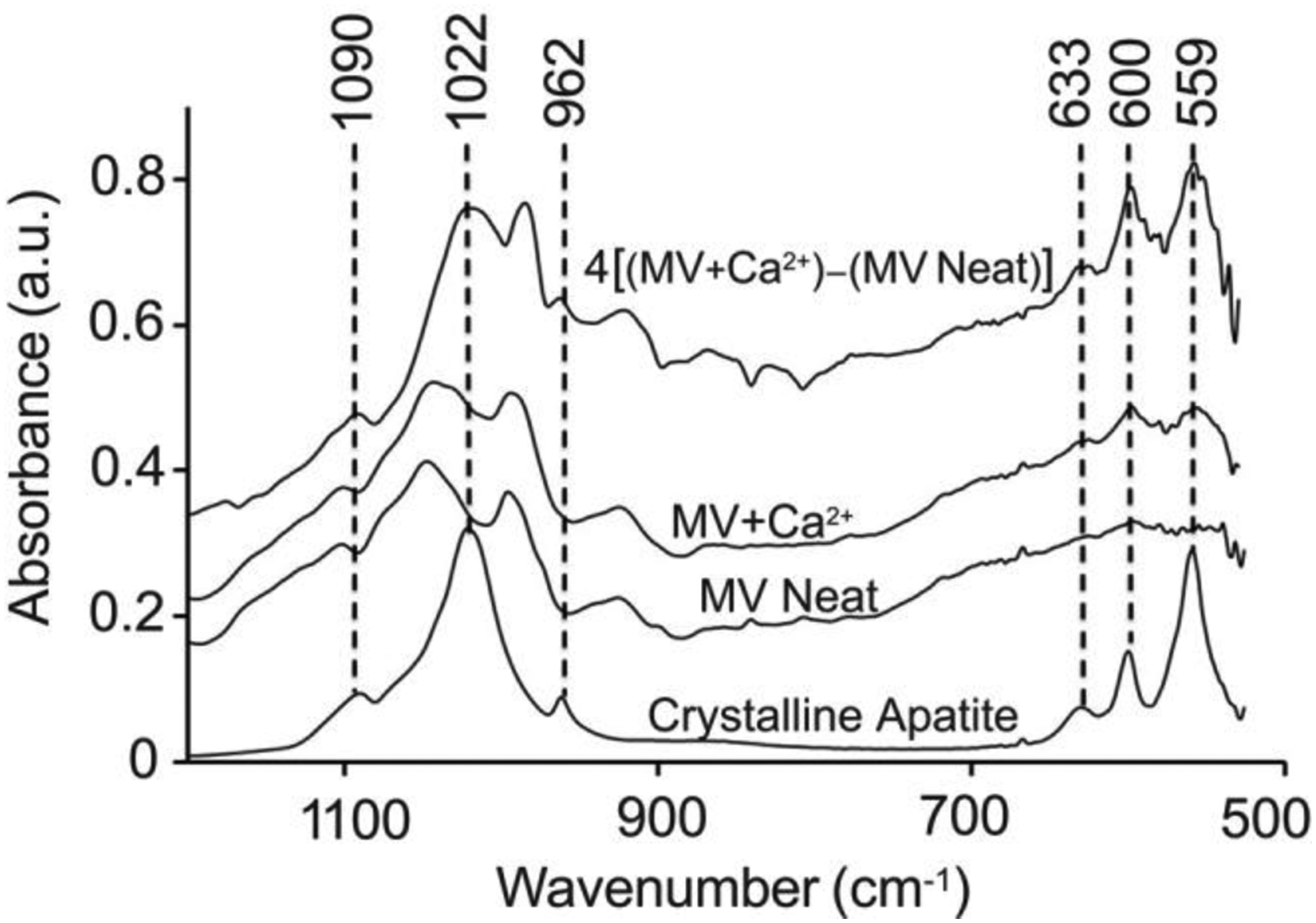
Matrix vesicles (MVs) are a class of extracellular vesicles that initiate mineralization in cartilage, bone, and other vertebrate tissues by accumulating calcium ions (Ca2+) and inorganic phosphate (Pi) within their lumen and forming a nucleation core (NC). After further sequestration of Ca2+ and Pi, the NC transforms into crystalline complexes. Direct evidence of the existence of the NC and its maturation have been provided solely by analyses of dried samples. We isolated MVs from chicken embryo cartilage and used atomic force microscopy peak force quantitative nanomechanical property mapping (AFM-PFQNM) to measure the nanomechanical and morphological properties of individual MVs under both mineralizing (+Ca2+) and non-mineralizing (-Ca2+) fluid conditions. The elastic modulus of MVs significantly increased by 4-fold after incubation in mineralization buffer. From AFM mapping data, we inferred the morphological changes of MVs as mineralization progresses: prior to mineralization, a punctate feature, the NC, is present within MVs and this feature grows and stiffens during mineralization until it occupies most of the MV lumen. Dynamic light scattering showed a significant increase in hydrodynamic diameter and no change in the zeta potential of hydrated MVs after incubation with Ca2+. This validates that crystalline complexes, which are strongly negative relative to MVs, were forming within the lumen of MVs. These data were substantiated by transmission electron microscopy energy dispersive X-ray and Fourier transform infrared spectroscopic analyses of dried MVs, which provide evidence that the complexes increased in size, crystallinity, and Ca/P ratio within MVs during the mineralization process.

Keywords: Atomic force microscopy; Elastic modulus; Matrix vesicles; Mineralization; Nucleation core.


Conflict of interest statement

The authors report no conflicts of interest.

Figures

Figure 1.
Figure 1.
Characterization of MV functionality by turbidity and TNAP activity analyses. (A) Turbidity time course data from MV solutions with (MV+Ca2+) and without (MV Neat) Ca2+. SCL buffer with (SCL+Ca2+) and without Ca2+ (SCL) were used as negative controls. (B) TNAP activity assay of MVs before Ca2+ addition (MV Neat) and 200 min after addition (MV+Ca2+).
Figure 2.
Figure 2.
Characterization of NC mineralization by DLS and AFM-PFQNM measurements of hydrated MVs. MVs were incubated in SCL devoid of Ca2+ (MV Neat) or supplemented with 2 mM Ca2+ (MV+Ca2+) for 24 h. (A) Hydrodynamic diameter (n = 25 measurements per group) and (B) zeta potential (n = 10 measurements per group) of MV samples. (C) Elastic modulus data calculated from AFM micrographs of individual vesicles acquired in PFQNM mode (n = 11 vesicles per group, data calculated from loading curves). Each marker corresponds to the average elastic modulus of a single MV and the horizontal bar is the mean of the group. Hollow markers correspond to individual MVs shown here and in Figure 3. (D) Modulus maps of representative individual vesicles.
Figure 3.
Figure 3.
Morphological characterization of different MVs before and after mineralization by AFM-PFQNM. (A) Height maps of representative MVs arranged in the order that mineralization is expected to proceed. The hollow markers correspond to the elastic modulus data points in Figure 2C. An intense peak believed to be the NC is indicated in panel i. (B) Two-dimensional cross sections of MVs taken across the maximum height of each vesicle. The arrows in (A) show the direction of each cross-sectional measurement.
Figure 4.
Figure 4.
Characterization of NC mineralization by TEM-EDX analyses of dried MVs. MVs were incubated in SCL devoid of Ca2+ (MV Neat) or supplemented with 2 mM Ca2+ (MV+Ca2+) for 24 h, then dried and analyzed by means of TEM-EDX. (A) TEM images (scale bars are 500 nm) of MVs devoid of, partially filled with, and fully filled with mineral deposits (arrows with labels). (B) Frequency of MVs devoid of (white area), partially filled with (grey area), and fully filled with (black area) mineral deposits. (C) The Ca/P ratio of mineral deposits found in MVs incubated in SCL devoid of Ca2+ (MV Neat, hatched area) or supplemented with 2 mM Ca2+ (MV+Ca2+, crosshatched area) as measured by TEM-EDX.
Figure 5.
Figure 5.
Characterization of NC mineralization by FTIR measurements of dried MVs. MVs were incubated in SCL devoid of Ca2+ (MV Neat) or supplemented with 2 mM Ca2+ (MV+Ca2+) for 24 h, then dried and analyzed by means of FTIR. The top trace is the spectrum for MV+Ca2+ after background subtraction of the MV Neat sample and subsequent amplification by a factor of four to better resolve the peaks. The spectrum of crystalline hydroxyapatite is reported as a control. All FTIR spectra were averaged from at least three independent samples.

References

    1. Wuthier RE, Lipscomb GF, Matrix vesicles: structure, composition, formation and function in calcification, Front. Biosci. (Landmark Ed.) 16(1) (2011) 2812–902. 10.2741/3887. - DOI - PubMed
    1. Anderson HC, Matrix vesicles and calcification, Curr. Rheumatol. Rep 5(3) (2003) 222–6. 10.1007/s11926-003-0071-z. - DOI - PubMed
    1. Golub EE, Biomineralization and matrix vesicles in biology and pathology, Semin. Immunopathol 33(5) (2011) 409–17. 10.1007/s00281-010-0230-z. - DOI - PMC - PubMed
    1. Bottini M, Mebarek S, Anderson KL, Strzelecka-Kiliszek A, Bozycki L, Simao AMS, Bolean M, Ciancaglini P, Pikula JB, Pikula S, Magne D, Volkmann N, Hanein D, Millan JL, Buchet R, Matrix vesicles from chondrocytes and osteoblasts: Their biogenesis, properties, functions and biomimetic models, Biochim. Biophys. Acta. Gen. Subj 1862(3) (2018) 532–546. 10.1016/j.bbagen.2017.11.005. - DOI - PMC - PubMed
    1. Anderson HC, Vesicles associated with calcification in the matrix of epiphyseal cartilage, J. Cell Biol 41(1) (1969) 59–72. 10.1083/jcb.41.1.59. - DOI - PMC - PubMed

Publication types

MeSH terms