. 2019 Apr 17;20(8):1890.
doi: 10.3390/ijms20081890.

Pitaya HpWRKY3 Is Associated with Fruit Sugar Accumulation by Transcriptionally Modulating Sucrose Metabolic Genes HpINV2 and HpSuSy1

Affiliations

Pitaya HpWRKY3 Is Associated with Fruit Sugar Accumulation by Transcriptionally Modulating Sucrose Metabolic Genes HpINV2 and HpSuSy1

Wei Wei et al. Int J Mol Sci. .

Abstract

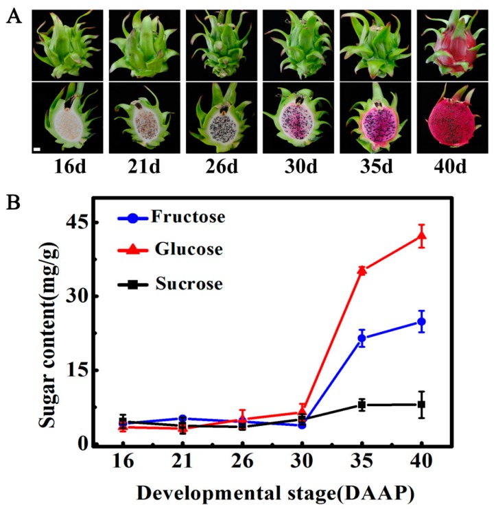
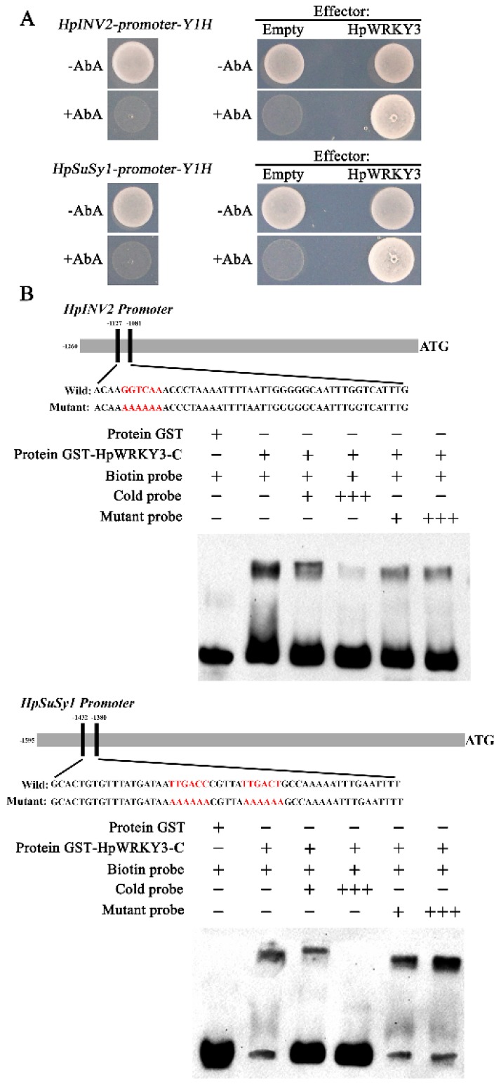
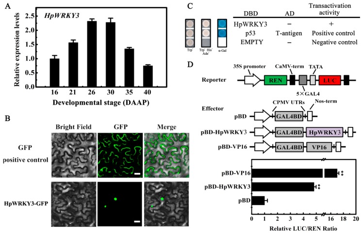
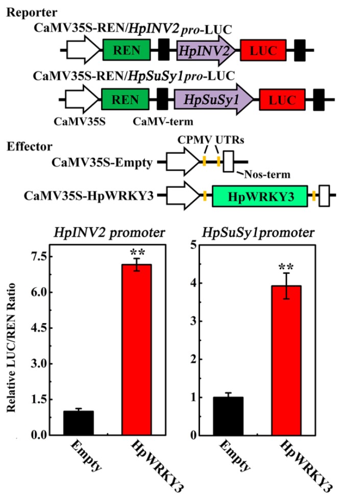
Sugar level is an important determinant of fruit taste and consumer preferences. However, upstream regulators that control sugar accumulation during fruit maturation are poorly understood. In the present work, we found that glucose is the main sugar in mature pitaya (Hylocereus) fruit, followed by fructose and sucrose. Expression levels of two sucrose-hydrolyzing enzyme genes HpINV2 and HpSuSy1 obviously increased during fruit maturation, which were correlated well with the elevated accumulation of glucose and fructose. A WRKY transcription factor HpWRKY3 was further identified as the putative binding protein of the HpINV2 and HpSuSy1 promoters by yeast one-hybrid and gel mobility shift assays. HpWRKY3 was localized exclusively in the nucleus and possessed trans-activation ability. HpWRKY3 exhibited the similar expression pattern with HpINV2 and HpSuSy1. Finally, transient expression assays in tobacco leaves showed that HpWRKY3 activated the expressions of HpINV2 and HpSuSy1. Taken together, we propose that HpWRKY3 is associated with pitaya fruit sugar accumulation by activating the transcriptions of sucrose metabolic genes. Our findings thus shed light on the transcriptional mechanism that regulates the sugar accumulation during pitaya fruit quality formation.

Keywords: fruit maturation; fruit quality; sucrose-hydrolyzing enzyme genes; transcriptional activation.


Conflict of interest statement

The authors declare no conflict of interest.

Figures

Figure 1
Figure 1
Glucose is the main sugar in mature pitaya fruit. (A) Photograph of pitaya fruit at different developmental stages. (B) Changes of soluble sugars (glucose, fructose, and sucrose) contents during fruit maturation. Fruit at 16, 21, 26, 30, 35, and 40 days after artificial pollination (DAAP) was sampled for analysis. Data represent mean values from three biological replicates (± S.E.).
Figure 2
Figure 2
The expression of HpINV2 and HpSuSy1 are correlated with glucose and fructose accumulation during pitaya fruit maturation. (A) The expression patterns of sucrose metabolic genes HpINVs and HpSuSy1 in pitaya fruit at different developmental stages. Fruit at 16, 21, 26, 30, 35, and 40 days after artificial pollination (DAAP) was sampled for analysis. Expression values are means ± S.E. of three biological replicates normalized using ACTIN as an internal control. (B) Correlation between HpINVs and HpSuSy1 expression levels and contents of glucose and fructose. * and ** represent significant correlation at 0.05 and 0.01 level respectively determined by SPSS Statistics 20.0.
Figure 3
Figure 3
HpWRKY3 directly bind to HpINV2 or HpSuSy1 promoter. (A) Y1H assay showing the physical interaction of the HpWRKY3 protein with HpINV2 or HpSuSy1 promoter. (Left) No basal expression of HpINV2- or HpSuSy1-pro was detected in yeast grown on synthetic dropout (SD) medium lacking Leu (SD/−Leu) in the presence of 500 ng/mL Aureobasidin A (AbA) for stringent selection. (Right) Yeast growth assays were performed after the Y1H reporter strains were transformed with plasmids carrying cassettes constitutively expressing HpWRKY3 effector or empty (pGADT7, negative control). Interaction was determined based on the ability of transformed yeast to grow on SD/−Leu in the presence of AbA. All suspensions of yeast cells in this assay were adjusted to OD600 = 0.1. (B) Electrophoretic mobility shift assay (EMSA) showing the binding of HpWRKY3 to the W-box of the HpINV2 or HpSuSy1 promoter. Purified glutathione-S-transferases (GST)-tagged HpWRKY3 protein were incubated with the biotin-labeled wild-type probe containing W-box, and the DNA–protein complexes were separated on native polyacrylamide gels. Sequences of both the wild-type and mutated probes are shown at the top of the image (wild-type and mutated W-box are marked with red letters). The probe with the mutated W-box was used to test binding specificity. Shifted bands, suggesting the formation of DNA–protein complexes, are indicated by arrows. ‘‘−’’ represents absence, ‘‘+’’ represents presence. ‘‘+++’’ indicates increasing amounts of unlabeled or mutated probes for competition experiments.
Figure 4
Figure 4
Multiple sequence alignment and phylogenetic analysis of HpWRKY3. (A) Multiple alignment of HpWRKY3 with other WRKY members. The following proteins were used for analysis: AtWRKY3 (NP_178433.1), BvWRKY3 (XP_010683088.1), AtWRKY26 (NP_196327.1), and AtWRKY33 (NP_181381.2). Identical and similar amino acids were shaded in black and grey, respectively. The WRKY domains and the zinc-finger structures are boxed and marked by asterisks, respectively. (B) Phylogenetic analysis of WRKYs. HpWRKY3 was highlighted with black circle. The phylogenetic tree was constructed with neighbor-joining test using MEGA program (version 5.0).
Figure 5
Figure 5
Molecular properties of HpWRKY3. (A) Temporal expression patterns of HpWRKY3 during pitaya fruit maturation. Fruit at 16, 21, 26, 30, 35, and 40 days after artificial pollination (DAAP) was sampled for analysis. Expression values are means ± S.E. of three biological replicates normalized using ACTIN as an internal control. (B) Subcellular localization of HpWRKY3 in epidermal cells of tobacco leaves. A plasmid harboring GFP or HpWRKY3-GFP was transformed into Nicotiana benthamiana leaves by Agrobacterium tumefaciens strain EHA105. GFP signals was observed with a fluorescence microscope after 2 d of infiltration. Bars, 30 μm. (C) Transcriptional activation of HpWRKY3 in yeast cells. The coding region of HpWRKY3 was inserted into the pGBKT7 (GAL4DBD) to create the pGBKT7-HpWRKY3 construct. The yeast cells of strain Y2HGold harboring the pGBKT7-HpWRKY3 plasmids were grown on SD plates without tryptophan (Trp) or without tryptophan, histidine, and adenine (TrpHisAde) for three days at 28 °C, followed by the α-galactosidase assay (α-Gal staining). pGBKT7 and pGBKT7-53 + pGADT7-T were used as negative and positive control, respectively. (D) Trans-activation of HpWRKY3 in Nicotiana benthamiana leaves. The trans-activation ability of HpWRKY3 was demonstrated by the ratio of luciferase (LUC) to renilla luciferase (REN). The LUC/REN ratio of the empty pBD vector (negative control) was used as a calibrator (set as 1). pBD-VP16 was used as a positive control. Data are means ± S.E. of six independent biological replicates. Asterisks represents significant differences at 0.01 level by student’s t-test, compared to pBD.
Figure 6
Figure 6
HpWRKY3 activates HpINV2 or HpSuSy1 transcriptions by dual-luciferase transient expression assay in Nicotiana benthamiana leaves. The reporter and effector vectors are illustrated in the top panel. Data are means ± S.E. of six independent biological replicates. Asterisks indicate significant differences by student’s t-test (** p < 0.01).

References

    1. Kim H., Choi H.K., Moon J.Y., Kim Y.S., Mosaddik A., Cho S.K. Comparative antioxidant and antiproliferative activities of red and white pitayas and their correlation with flavonoid and polyphenol content. J. Food Sci. 2011;76:C38–C45. doi: 10.1111/j.1750-3841.2010.01908.x. - DOI - PubMed
    1. Hua Q., Chen C., Tel Zur N., Wang H., Wu J., Chen J., Zhang Z., Zhao J., Hu G., Qin Y. Metabolomic characterization of pitaya fruit from three red-skinned cultivars with different pulp colors. Plant Physiol. Biochem. 2018;126:117–125. doi: 10.1016/j.plaphy.2018.02.027. - DOI - PubMed
    1. García-Cruz L., Dueñas M., Santos-Buelgas C., Valle-Guadarrama S., Salinas-Moreno Y. Betalains and phenolic compounds profiling and antioxidant capacity of pitaya (Stenocereus spp.) fruit from two species (S. Pruinosus and S. stellatus) Food Chem. 2017;234:111–118. doi: 10.1016/j.foodchem.2017.04.174. - DOI - PubMed
    1. Suh D.H., Lee S., Heo do Y., Kim Y.S., Cho S.K., Lee S., Lee C.H. Metabolite profiling of red and white pitayas (Hylocereus polyrhizus and Hylocereus undatus) for comparing betalain biosynthesis and antioxidant activity. J. Agric. Food Chem. 2014;62:8764–8771. doi: 10.1021/jf5020704. - DOI - PubMed
    1. Menezes Cordeiro M.H., Da Silva J.M., Mizobutsi G.P., Mizobutsi E.H., Da Mota W.F. Physical, chemical and nutritional characterization of pink pitaya of red pulp. Rev. Bras. Frutic. 2015;37:20–26.

LinkOut - more resources