. 2019 Apr 18;21(1):23.
doi: 10.1186/s12968-019-0533-8.

Sorted Golden-step phase encoding: an improved Golden-step imaging technique for cardiac and respiratory self-gated cine cardiovascular magnetic resonance imaging

Affiliations

Sorted Golden-step phase encoding: an improved Golden-step imaging technique for cardiac and respiratory self-gated cine cardiovascular magnetic resonance imaging

Liheng Guo et al. J Cardiovasc Magn Reson. .

Abstract

Background: Numerous self-gated cardiac imaging techniques have been reported in the literature. Most can track either cardiac or respiratory motion, and many incur some overhead to imaging data acquisition. We previously described a Cartesian cine imaging technique, pseudo-projection motion tracking with golden-step phase encoding, capable of tracking both cardiac and respiratory motion at no cost to imaging data acquisition. In this work, we describe improvements to the technique by dramatically reducing its vulnerability to eddy current and flow artifacts and demonstrating its effectiveness in expanded cardiovascular applications.

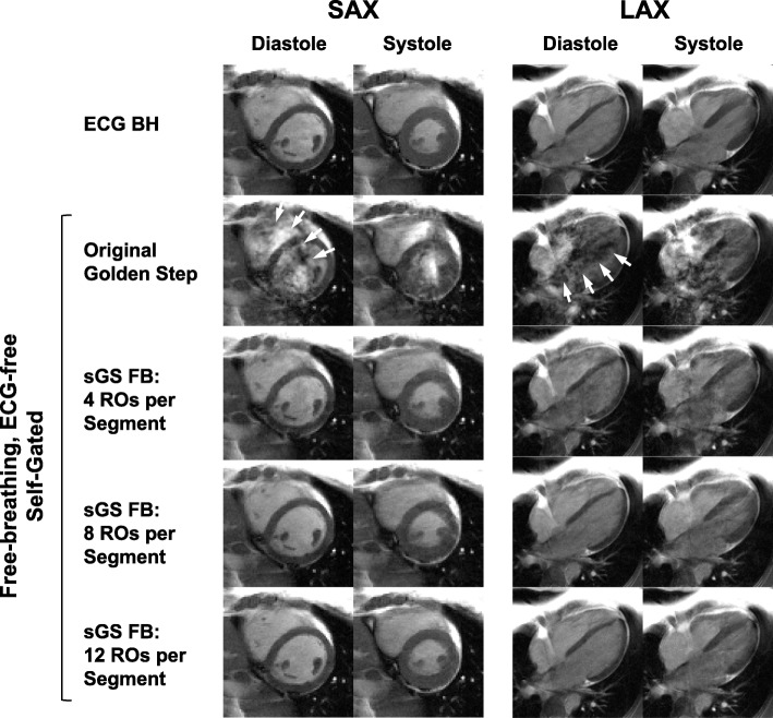
Methods: As with our previous golden-step technique, the Cartesian phase encodes over time were arranged based on the integer golden step, and readouts near ky = 0 (pseudo-projections) were used to derive motion. In this work, however, the readouts were divided into equal and consecutive temporal segments, within which the readouts were sorted according to ky. The sorting reduces the phase encode jump between consecutive readouts while maintaining the pseudo-randomness of ky to sample both cardiac and respiratory motion without comprising the ability to retrospectively set the temporal resolution of the original technique. On human volunteers, free-breathing, electrocardiographic (ECG)-free cine scans were acquired for all slices of the short axis stack and the 4-chamber view of the long axis. Retrospectively, cardiac motion and respiratory motion were automatically extracted from the pseudo-projections to guide cine reconstruction. The resultant image quality in terms of sharpness and cardiac functional metrics was compared against breath-hold ECG-gated reference cines.

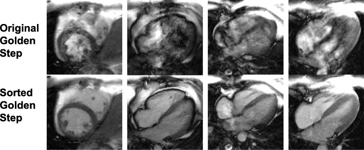
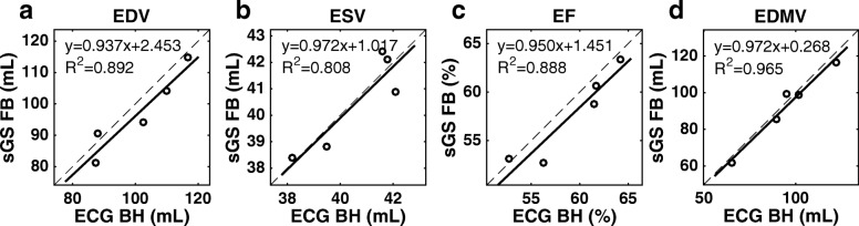
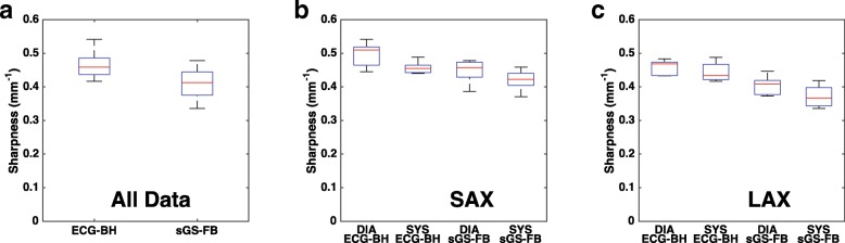
Results: With sorting, motion tracking of both cardiac and respiratory motion was effective for all slices orientations imaged, and artifact occurrence due to eddy current and flow was efficiently eliminated. The image sharpness derived from the self-gated cines was found to be comparable to the reference cines (mean difference less than 0.05 mm- 1 for short-axis images and 0.075 mm- 1 for long-axis images), and the functional metrics (mean difference < 4 ml) were found not to be statistically different from those from the reference.

Conclusions: This technique dramatically reduced the eddy current and flow artifacts while preserving the ability of cost-free motion tracking and the flexibility of choosing arbitrary navigator zone width, number of cardiac phases, and duration of scanning. With the restriction of the artifacts removed, the Cartesian golden-step cine imaging can now be applied to cardiac imaging slices of more diverse orientation and anatomy at greater reliability.

Keywords: Cine imaging; Dark flow artifacts; Golden step; Motion tracking; Pseudo-projections; Self-gating; Self-navigation.


Conflict of interest statement

Ethics approval and consent to participate

The studies were performed with written informed consent and under research protocol (NA00083408), approved by the institutional review board of the Johns Hopkins University School of Medicine.

Consent for publication

Publication consent was part of the IRB-approved research protocol.

Competing interests

The authors declare that they have no competing interests.

Publisher’s Note

Springer Nature remains neutral with regard to jurisdictional claims in published maps and institutional affiliations.

Figures

Fig. 1
Fig. 1
Comparison of Cartesian phase-encode (PE) schemes. In each row, a Cartesian ky grid with 144 evenly spaced PEs is covered exactly once by a particular PE scheme. In each column, a central-ky region of a specific width is defined as the “navigator zone” (red dashed). Low-PE readouts falling within it are considered “pseudo-projections” (red squares) and used for motion tracking. Row 1: the original integer golden-step scheme provides pseudorandom navigator-zone coverage that is approximately uniform in both time and ky. Row 2 and 3: the golden-step PEs are sorted into temporal segments of 8 and 12, respectively, significantly reducing PE jumps while preserving the pseudorandom coverage of the navigator zone in both time and ky. Row 4 and 5: for comparison, if the “interleaved” PE scheme, a commonly used segmented scheme, were to be used instead of the golden step, there would be large temporal gaps in the navigator zone coverage, i.e., there would be no PE falling inside the central-ky zone (red) to sample the motion for extended periods of time (e.g. row 4 columns 1 and 2, and row 5 column 1). Those that do fall inside would also have be temporally structured (slowly drifting from negative ky to positive in all scenarios in rows 4 and 5), causing slow time-varying bias in motion measurements
Fig. 2
Fig. 2
Streams of sorted pseudo-projections for motion tracking and effect of sorting-segment size. a The golden-step (GS) PEs are sorted by ky into segments (size of 8 shown) before execution. The sorting does not affect the original GS motion tracking technique, which follows: Cartesian readouts falling within a near-zero ky zone (known as the “navigator zone”) are considered “pseudo-projections.” To reveal motion, the pseudo-projections’ ky-dependent magnitude variation is corrected, and any remaining variation in time is smoothed. Cardiac motion and respiratory motion are extracted to guide the two-stage data selection, after which cine frames were reconstructed through simple inverse Fourier transform. b The effects of segment size and navigator zone width on pseudo-projections: sorted GS with various segment size are compared to the original GS at typical navigator zone widths. Both cardiac and respiratory cycles are visible at all practical segment sizes, but generally more clearly at lower navigator zone widths. Note that the streams shown here were processed to prioritize cardiac motion; respiratory motion could also be highlighted using the same data (see Figure 3). ROs: readouts
Fig. 3
Fig. 3
Cardiac and respiratory motion extraction from pseudo-projections. The central 5% phase encodes (pseudo-projections) were processed for optimal cardiac motion detection (a). From a group of automatically detected “cardiac pixels” (red “+” markers), the cardiac waveform was derived and was used to generate cardiac events (b) to replace ECG triggers (dashed, shown for reference). The same pseudo-projections were processed for optimal respiratory motion detection (c), from which a respiratory waveform was extracted using principal component analysis (PCA) to perform respiratory gating (d). In this experiment, the subject was instructed to breath-hold for several seconds before breathing freely. The plateau region at the beginning of (d) shows that the PCA can capture non-cyclical motion. AU: arbitrary unit
Fig. 4
Fig. 4
Comparison of images acquired at various number of readouts per segment. A balanced steady state free precession (bSSFP) off-resonance band near the imaging slice, which is not observed on the ECG-gated breath-hold reference cines (ECG BH, Row 1) due to its sequential PE, caused severe dark flow artifact (white arrows) with the original GS (Row 2) due to the compounding effects of large PE jumps and flow. The artifact is significantly reduced when readouts are sorted into segments of ascending or descending PEs (sGS FB, Row 3–5). At more than 4 readouts per segment, the artifact becomes essentially unnoticeable. bSSFP: balanced steady state free precession. PE: phase encode. RO: readout. SAX: short axis. LAX: long axis
Fig. 5
Fig. 5
Comparison of the golden step and sorted golden step acquisitions in artifact-prone slices. In some slices of some subjects, the main field inhomogeneity and blood flow are significant enough to compound with the eddy current induced by the original golden step’s large phase-encode jumps, forming the dark flow artifact of bSSFP (Row 1). However, the same slice can be imaged virtually free of the artifact using the sorted GS (Row 2, using 12 readouts/segment). The sorted golden step (sGS) has dramatically reduced, if not entirely eliminated, the artifact in all problematic slices encountered in this study. Four such slices from three subjects are shown here
Fig. 6
Fig. 6
Visual comparison of cine image quality. Eight frames from a 24-frames free-breathing ECG-free self-gated sorted golden-step cine (sGS FB) acquired with 12 readouts per segment are compared with the references cine (ECG BH) acquired with ECG gating, breath hold, and sequential PEs. Several representative slices of the SAX stack at different levels (apical, mid-ventricular, basal) and a 4-chamber LAX slice are shown. Mild ghosting and blurring may be visible on some sGS images but all are free from flow- and eddy current-induced dark flow artifacts. SAX: short axis. LAX: long axis
Fig. 7
Fig. 7
Comparison of LV functional metrics as measured from ECG BH and sGS FB images. As measured from the SAX stacks, the end-diastolic volume (EDV, a), end-systolic volume (ESV, b), ejection fraction (EF, c), and end-diastolic myocardial volume (EDMV, d) of the free-breathing self-gated sorted golden-step (sGS FB) images show good agreement with those of the ECG-gated breath-hold references (ECG BH), with average absolute errors of approximately 5% or less. LV: left ventricle
Fig. 8
Fig. 8
Comparison of LV blood-myocardium sharpness as measured from ECG BH and sGS FB images. Whether combined (a) or separated into SAX (b) and LAX (c) groups, the sGS FB images were in general slightly less sharp than the ECG BH scan, although the differences are not large. SAX: short axis. LAX: long axis. DIA: end-diastole. SYS: end-systole. LV: left ventricle

Similar articles

Cited by

References

    1. Polson MJ, Barker AT, Gardiner S. The effect of rapid rise-time magnetic fields on the ECG of the rat. Clin Phys Physiol Meas. 1982;3:231–234. doi: 10.1088/0143-0815/3/3/008. - DOI - PubMed
    1. Shetty AN. Suppression of radiofrequency interference in cardiac gated MRI: a simple design. Magn Reson Med. 1988;8:84–88. doi: 10.1002/mrm.1910080110. - DOI - PubMed
    1. Rokey R, Wendt RE, Johnston DL. Monitoring of acutely ill patients during nuclear magnetic resonance imaging: use of a time-varying filter electrocardiographic gating device to reduce gradient artifacts. Magn Reson Med. 1988;6:240–245. doi: 10.1002/mrm.1910060213. - DOI - PubMed
    1. Damji AA, Snyder RE, Ellinger DC, Witkowski FX, Allen PS. RF interference suppression in a cardiac synchronization system operating in a high magnetic field NMR imaging system. Magn Reson Imaging. 1988;6:637–640. doi: 10.1016/0730-725X(88)90086-0. - DOI - PubMed
    1. Fischer SE, Wickline SA, Lorenz CH. Novel real-time R-wave detection algorithm based on the vectorcardiogram for accurate gated magnetic resonance acquisitions. Magn Reson Med. 1999;42:361–370. doi: 10.1002/(SICI)1522-2594(199908)42:2<361::AID-MRM18>3.0.CO;2-9. - DOI - PubMed

Publication types