. 2019 Apr 18;10(1):123.
doi: 10.1186/s13287-019-1221-1.

Interleukin-33 regulates hematopoietic stem cell regeneration after radiation injury

Affiliations

Interleukin-33 regulates hematopoietic stem cell regeneration after radiation injury

Ping Huang et al. Stem Cell Res Ther. .

Abstract

Background: IL-33 is a pleiotropic cytokine of the IL-1 family, which has been reported to implicate in both innate and adaptive immune responses. Recent studies suggest IL-33 is crucial for regulation of myelopoiesis and myeloid cell activity. Here, we explore the potential effect of IL-33 against hematopoietic injury after total body irradiation (TBI).

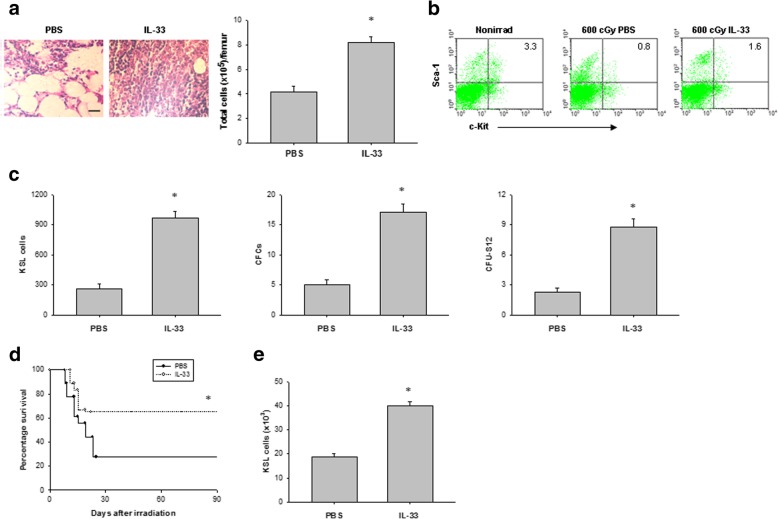
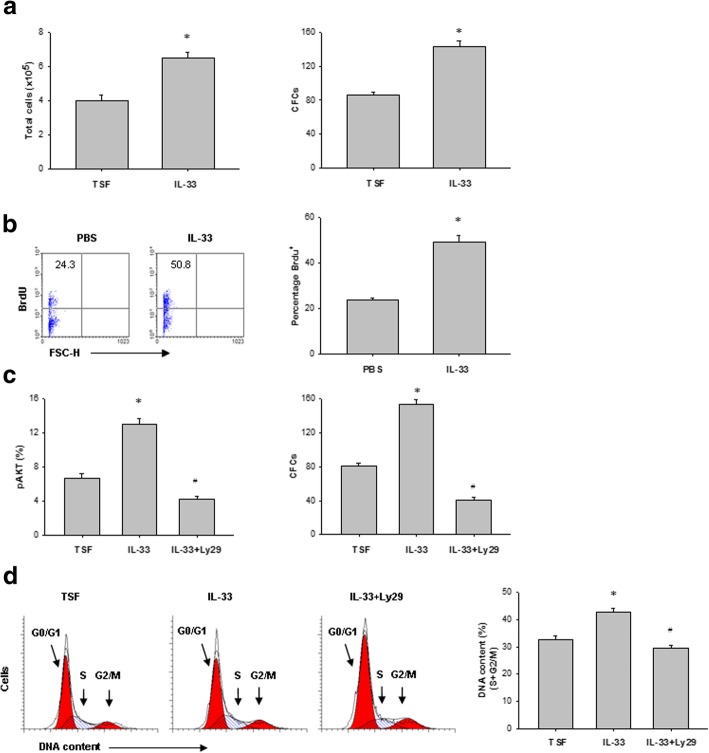
Methods: C57BL/6 mice were irradiated with a sublethal dose of radiation (600 cGy) and treated with IL-33 at a dose of 3 μg/dose i.p. once a day for seven consecutive days. H&E staining was used to determine the bone marrow cellularity. A flow cytometer was used to quantify the hematopoietic stem cell (HSC) population, cell proliferation, and apoptosis. The colony-forming assay was used to evaluate the clonogenic function of HSCs. RT-qPCR was used to determine the expression of apoptosis-associated genes.

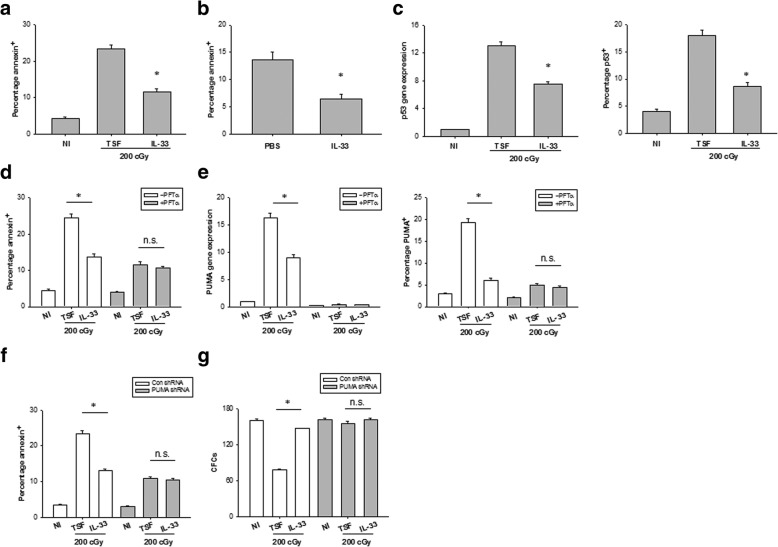
Results: Bone marrow HSCs from wild-type mice expressed functional IL-33 receptor (ST2), and treatment with IL-33 promoted the recovery of the HSC pool in vivo and improved the survival of mice after TBI. Conversely, mice with ST2 deficiency showed decreased HSC regeneration and mouse survival after TBI. Of note, IL-33 reduced radiation-induced apoptosis of HSCs and mediated this effect through repression of the p53-PUMA pathway.

Conclusions: IL-33 regulates HSC regeneration after myelosuppressive injury through protecting HSCs from apoptosis and enhancing proliferation of the surviving HSCs.

Keywords: Cell apoptosis; Hematopoietic stem cells; IL-33; Ionizing radiation; PUMA.


Conflict of interest statement

Ethics approval

All animal studies were approved by the Animal Care and Use Committee at the Guangdong Pharmaceutical University.

Consent for publication

Not applicable.

Competing interests

The authors declare that they have no competing interests.

Publisher’s Note

Springer Nature remains neutral with regard to jurisdictional claims in published maps and institutional affiliations.

Figures

Fig. 1
Fig. 1
IL-33 administration improves survival of mice after TBI. a Survival curves of C57BL/6 mice that were irradiated with 600-cGy TBI followed by daily IL-33 or PBS treatments for 7 days. Data are pooled from three experiments, n = 6 mice per group per experiment. *P < 0.05 compared to untreated controls. b Survival rate of untreated ST2−/− and WT mice was irradiated with 600-cGy. Data are pooled from three experiments, n = 6 mice per group per experiment. *P < 0.05 compared to ST2−/− controls. c sST2 and IL-33 concentration in the bone marrow serum of WT and ST2−/− mice before irradiation (Nonirrad) and at 7 days after 600-cGy irradiation. Data are mean ± SEM (n = 6 in each group). *P < 0.05 compared to nonirradiated controls. d Representative ST2 surface expression on bone marrow Lin cells and KSL cells from C57BL/6 mice before irradiation and at 6 h after 600-cGy irradiation. The numbers shown indicate the percentage of ST2 surface expression on the indicated cell population. e Survival rate of ST2−/− and WT mice was irradiated with 600-cGy and treated with IL-33 as in a
Fig. 2
Fig. 2
IL-33 signaling mediates HSC regeneration in vivo. a Left, representative H&E-stained femurs from irradiated mice treated with either PBS or IL-33 for 7 days. Scale bar, 100 μm. Right, bone marrow cell counts. Data are mean ± SEM (n = 6 in each group). *P < 0.05 compared to untreated controls. b Representative FACS analysis of bone marrow c-Kit+Sca-1+ cells within the Lin gate (KSL) from nonirradiated (Nonirrad) mice and at day 7 from irradiated mice treated with either PBS or IL-33. The numbers shown indicate the percentage of c-Kit+Sca-1+ cells within the Lin population. c Bone marrow KSL cells, CFCs, and CFU-S12 at day 7 in irradiated mice treated with either PBS or IL-33. Data are mean ± SEM (n = 6 in each group). *P < 0.05 compared to untreated controls. d Survival rates of lethally irradiated mice that were adoptively transferred with 5 × 105 BM cells from irradiated mice treated with or without IL-33. Data are pooled from three experiments, n = 6 mice per group per experiment. *P < 0.05 compared to untreated controls. e KSL cells in the BM of recipient mice at 10 weeks after transplantation of 5 × 105 bone marrow cells from irradiated and PBS-treated or irradiated and IL-33-treated donor mice. Data are mean ± SEM (n = 6 in each group). *P < 0.05 compared to untreated controls
Fig. 3
Fig. 3
IL-33 regulates HSC proliferation after irradiation. a Numbers of total cells and CFC at 72 h in irradiated bone marrow Linc-Kit+ cells cultured with either TSF or TSF plus IL-33. Data are mean ± SEM (n = 3 in each group). *P < 0.05 compared to untreated controls. b Representative BrdU incorporation in bone marrow KSL cells in vivo at day 7 after 600-cGy TBI and treatment with either PBS or IL-33 (left). Right, numbers indicate the percentage of BrdU+ cells within the total bone marrow KSL population. Data are mean ± SEM (n = 3 in each group). *P < 0.05 compared to untreated controls. c Left, percentage phosphorylated AKT (pAKT) in bone marrow Linc-Kit+ cells after 200-cGy irradiation and the culture conditions are shown. Right, CFCs from bone marrow Linc-Kit+ cells after 200-cGy irradiation and the culture conditions are shown. *P < 0.05 for TSF compared to IL-33, #P < 0.01 for IL-33 compared to IL-33 plus Ly294002 (Ly29) (mean ± SEM, n = 3 in each group). d Representative FACS analysis of the cell-cycle status of bone marrow Linc-Kit+ cells at 72 h after irradiation and the culture conditions shown. *P < 0.05 for TSF compared to IL-33, #P < 0.01 for IL-33 compared to IL-33 plus Ly294002 (Ly29) (mean ± SEM, n = 3 in each group)
Fig. 4
Fig. 4
IL-33 protects HSCs from apoptosis after irradiation. a The percentage of annexin+ bone marrow Linc-Kit+ cells at 72 h of culture with TSF or TSF plus IL-33 and after 200-cGy irradiation and the culture conditions are shown. Data are mean ± SEM (n = 3 in each group). *P < 0.05 compared to untreated controls. b The percentage of annexin+CD45+ cells in the bone marrow at day 7 after 600-cGy TBI and treatment with PBS or IL-33. Data are mean ± SEM (n = 6 in each group). *P < 0.05 compared to untreated controls. c Left, P53 mRNA expression in bone marrow Linc-Kit+ cells at 6 h of culture with TSF or TSF plus IL-33 and after 200-cGy irradiation and the culture conditions are shown. Right, mean percentages of P53 protein expression in Linc-Kit+ cells at 36 h of culture with TSF or TSF plus IL-33 and after 200-cGy irradiation and the culture conditions are shown. Data are mean ± SEM (n = 3 in each group). *P < 0.05 compared to untreated controls. d The percentage of annexin+ Linc-Kit+ cells with or without P53 inhibition at 72 h of culture with TSF or TSF plus IL-33 and after 200-cGy irradiation and the culture conditions are shown. Data are mean ± SEM (n = 3 in each group). *P < 0.05 compared to untreated controls. n.s. not significant. e Left, PUMA mRNA expression in bone marrow Linc-Kit+ cells at 6 h of culture with TSF or TSF plus IL-33 and after 200-cGy irradiation and the culture conditions are shown. Right, mean percentages of PUMA protein expression in Linc-Kit+ cells at 36 h of culture with TSF or TSF plus IL-33 and after 200-cGy irradiation and the culture conditions are shown. Data are mean ± SEM (n = 3 in each group). *P < 0.05 compared to untreated controls. n.s. not significant. f The percentage of annexin+ Linc-Kit+ cells with or without PUMA knockdown at 72 h of culture with TSF or TSF plus IL-33 and after 200-cGy irradiation and the culture conditions are shown. Data are mean ± SEM (n = 3 in each group). *P < 0.05 compared to untreated controls. n.s. not significant. g CFCs from bone marrow Linc-Kit+ cells with or without PUMA knockdown in irradiated TSF culture and irradiated TSF plus IL-33 culture groups. Data are mean ± SEM (n = 3 in each group). *P < 0.05 compared to untreated controls. n.s. not significant
Fig. 5
Fig. 5
Deficiency of ST2 impairs HSC regeneration after irradiation. a Representative FACS analysis of bone marrow c-Kit+Sca-1+ cells within the Lin gate (KSL) at day 7 in irradiated WT and ST2-deficient mice. The numbers shown indicate the percentage of c-Kit+Sca-1+ cells within the Linpopulation. b Bone marrow KSL cells and CFCs at day 7 in irradiated WT and ST2-deficient mice. Data are mean ± SEM (n = 6 in each group). *P < 0.05 compared to ST2−/− mice. c The percentage of annexin+CD45+ cells in the bone marrow at day 7 in irradiated WT and ST2-deficient mice. Data are mean ± SEM (n = 6 in each group). *P < 0.05 compared to ST2−/− mice. d Numbers of total cells and CFCs at 72 h in irradiated bone marrow Linc-Kit+ cells from WT and ST2-deficient mice. Data are mean ± SEM (n = 3 in each group). *P < 0.05 compared to ST2−/− TSF

Similar articles

Cited by

References

    1. Mettler FA, Jr, Voelz GL. Major radiation exposure--what to expect and how to respond. N Engl J Med. 2002;346:1554–1561. doi: 10.1056/NEJMra000365. - DOI - PubMed
    1. Dainiak N. Hematologic consequences of exposure to ionizing radiation. Exp Hematol. 2002;30:513–528. doi: 10.1016/S0301-472X(02)00802-0. - DOI - PubMed
    1. Drouet M, Hérodin F. Radiation victim management and the haematologist in the future: time to revisit therapeutic guidelines? Int J Radiat Biol. 2010;86:636–648. doi: 10.3109/09553001003789604. - DOI - PubMed
    1. Hérodin F, Drouet M. Cytokine-based treatment of accidentally irradiated victims and new approaches. Exp Hematol. 2005;33:1071–1080. doi: 10.1016/j.exphem.2005.04.007. - DOI - PubMed
    1. Velardi E, Tsai JJ, Radtke S, Cooper K, Argyropoulos KV, Jae-Hung S, et al. Suppression of luteinizing hormone enhances HSC recovery after hematopoietic injury. Nat Med. 2018;24:239–246. doi: 10.1038/nm.4470. - DOI - PMC - PubMed

Publication types

MeSH terms