. 2019 Apr 18;38(1):170.
doi: 10.1186/s13046-019-1163-6.

Resolvin D1 prevents epithelial-mesenchymal transition and reduces the stemness features of hepatocellular carcinoma by inhibiting paracrine of cancer-associated fibroblast-derived COMP

Affiliations

Resolvin D1 prevents epithelial-mesenchymal transition and reduces the stemness features of hepatocellular carcinoma by inhibiting paracrine of cancer-associated fibroblast-derived COMP

Liankang Sun et al. J Exp Clin Cancer Res. .

Abstract

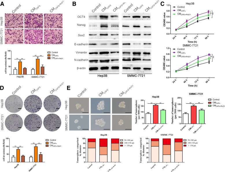
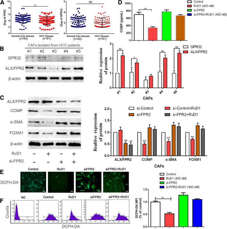
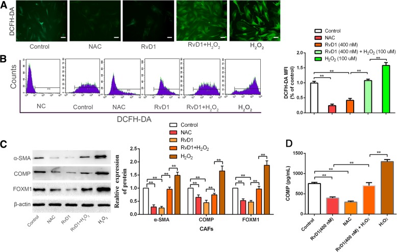
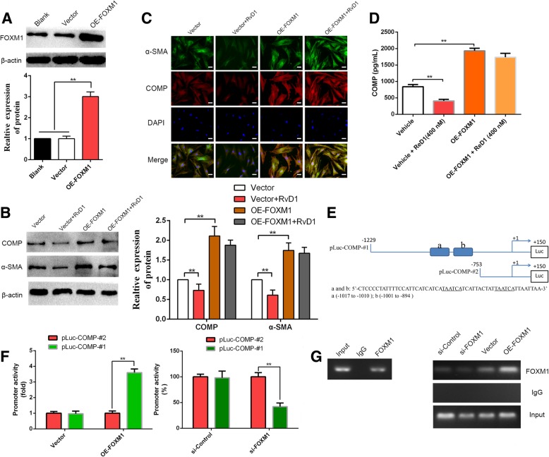
Background: Cancer stem cells (CSCs) require stromal signals for maintaining pluripotency and self-renewal capacities to confer tumor metastasis. Resolvin D1 (RvD1), an endogenous anti-inflammatory lipid mediator, has recently been identified to display anti-cancer effects by acting on stroma cells. Our previous study reveals that hepatic stellate cells (HSCs)-derived cartilage oligomeric matrix protein (COMP) contributes to hepatocellular carcinoma (HCC) progression. However, whether RvD1 inhibits paracrine of cancer-associated fibroblasts (CAFs)-derived COMP to prevent epithelial-mesenchymal transition (EMT) and cancer stemness in HCC remains to be elucidated.

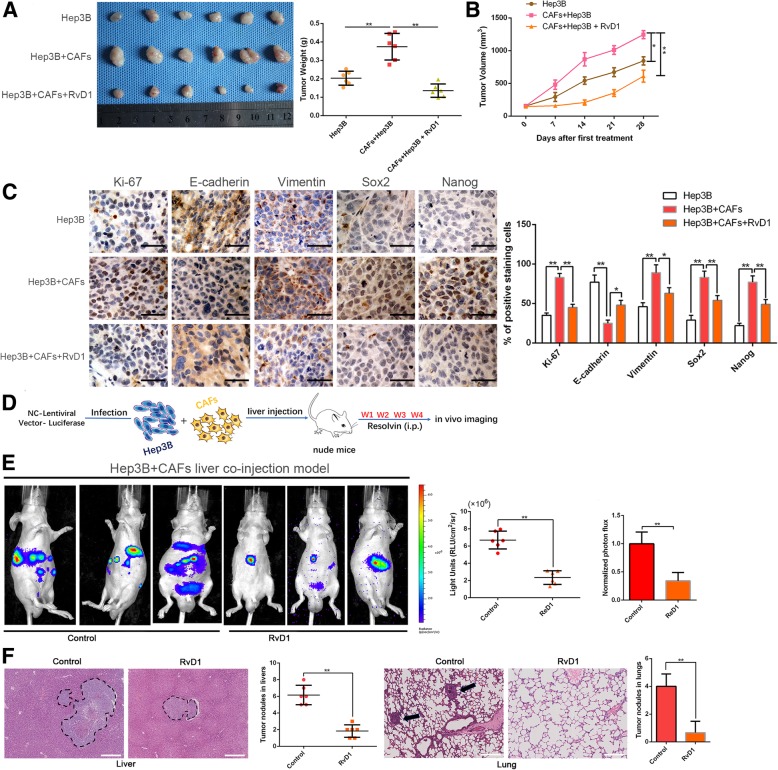
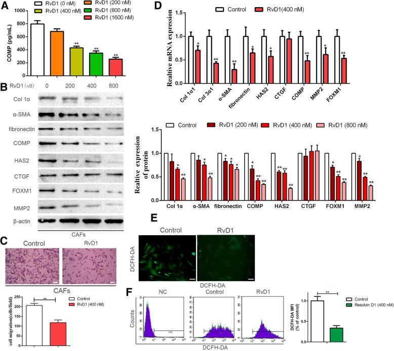
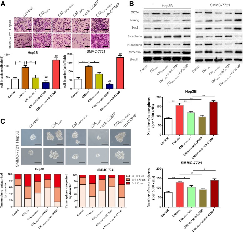
Methods: CAFs were isolated from HCC tissues. Direct and indirect co-culture models were established to analyze the interactions between HCC cells and CAFs in the presence of RvD1 in vitro. The transwell and tumor sphere formation assays were used to determine invasion and stemness of HCC cells. The subcutaneous tumor formation and orthotopic liver tumor models were established by co-implantation of CAFs and HCC cells to evaluate the role of RvD1 in vivo. To characterize the mechanism of RvD1 inhibited paracrine of COMP in CAFs, various signaling molecules were analyzed by ELISA, western blotting, reactive oxygen species (ROS) detection, immunofluorescence staining, dual luciferase reporter assay and chromatin immunoprecipitation assay.

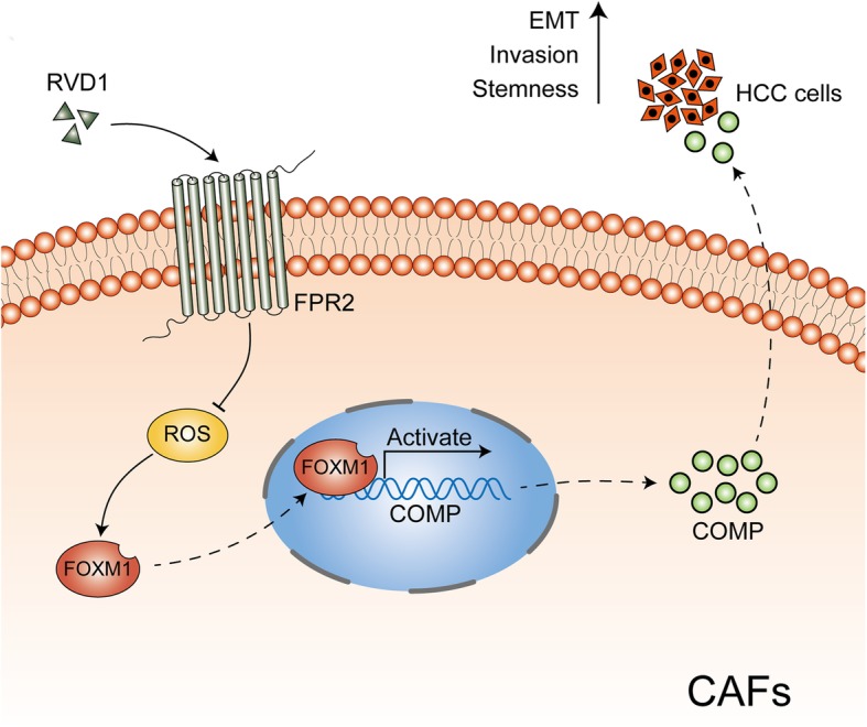
Results: Our data revealed that RvD1 treatment can impede the CAFs-induced cancer stem-like properties and the EMT of HCC cells under co-culture conditions. In vivo studies indicated that RvD1 intervention repressed the promoting effects of CAFs on tumor growth and metastasis of HCC. Furthermore, RvD1 inhibited CAF-induced EMT and stemness features of HCC cells by suppressing the secretion of COMP. Mechanistically, formyl peptide receptor 2 (FPR2) receptor mediated the suppressive effects of RvD1 on COMP and forkhead box M1 (FOXM1) expression in CAFs. Notably, RvD1 impaired CAF-derived COMP in a paracrine manner by targeting FPR2/ROS/FOXM1 signaling to ultimately abrogate FOXM1 recruitment to the COMP promoter.

Conclusion: Our results indicated that RvD1 impaired paracrine of CAFs-derived COMP by targeting FPR2/ROS/FOXM1 signaling to repress EMT and cancer stemness in HCC. Thus, RvD1 may be a potential agent to promote treatment outcomes in HCC.

Keywords: COMP; Cancer stemness; Cancer-associated fibroblasts; FOXM1; Hepatocellular carcinoma; ROS; Resolvin D1.


Conflict of interest statement

Ethics approval and consent to participate

All procedures performed in studies involving human participants were in accordance with the ethical standards of the Research Ethics Committee of The First Affiliated Hospital of Xi’an Jiaotong University and with the 1964 Helsinki declaration and its later amendments. ALL written informed consent to participate in the study was obtained from HCC patients for samples to be collected from them.

Consent for publication

Not applicable.

Competing interests

The authors declare that they have no competing interests.

Publisher’s Note

Springer Nature remains neutral with regard to jurisdictional claims in published maps and institutional affiliations.

Figures

Fig. 1
Fig. 1
RvD1 impedes the CAFs-induced cancer stemness and EMT of HCC cells. (a) Hep3B and SMMC-7721 cells were incubated with CM from CAFs (CMCAFs) or CM from CAFs pre-treated with RvD1(400 nM) (CMCAFs + RvD1) for 24 h, The cells were then seeded in a matrigel-coated invasion chamber for 24 h. The invasive cells were quantified by counting the number of cells in 5 random fields at × 100 magnification. Scale bars = 50 μm. n = three independent experiments, **P < 0.01 by ANOVA. (b) Hep3B and SMMC-7721 cells were treated with CMCAFs and CMCAFs + RvD1 for 48 h, and western blotting analysis was performed to test the expression of stemness markers (OCT4, Nanog, Sox2), and epithelial-mesenchymal transition markers (E-cadherin, N-cadherin, vimentin). (c) Hep3B and SMMC-7721 cells were incubated with CMCAFs and CMCAFs + RvD1 for 24, 48, 72 and 96 h, and cell viability was evaluated by MTT assay. n = three independent experiments, *P < 0.05 or **P < 0.01 by ANOVA. (d) Effects of CMCAFs and CMCAFs + RvD1 on the colony-forming ability of Hep3B and SMMC-7721 cells were assessed by colony formation assay. Images are representative of three independent experiments, and the colony number was counted and plotted. Scale bar = 1 cm. n = three independent experiments, **P < 0.01 by ANOVA. (E) Tumorsphere formation assay of Hep3B and SMMC-7721 cells inculated with CMCAFs or CMCAFs + RvD1. The number and size of tumorspheres were counted, measured and plotted. Magnification is × 200, and scale bars = 50 μm. n = three independent experiments, **P < 0.01 by ANOVA
Fig. 2
Fig. 2
RvD1 inhibits the CAFs-induced tumorgenicity of Hep3B cells along with reversing the mesenchymal and stem-like phenotypes. (a) Representative images of subcutaneous xenografts in nude mice implanted with Hep3B in the presence or absence of CAFs and treated with RvD1 (n = 6). (b) Xenografts weight (mg) and tumor sizes were monitored and undergone quantification analysis. n = 6, **P < 0.01 by ANOVA for tumor weight; *P < 0.05 or **P < 0.01 by repeated-measures ANOVA for tumor sizes. (c) Immunohistochemistry staining and the semi-quantification data of Ki-67, E-cadherin, Vimentin, Sox2 and Nanog in xenograft tissues from different groups. Magnification is × 400, the scale bar represents 20 μm. n = 6, *P < 0.05 or **P < 0.01 by ANOVA. (d) Schematic of establishing the orthotopic liver tumor model in nude mice using Luciferase-expressing Hep3B cells admixed with CAFs co-injection and administrated with RvD1 (n = 6 per group). (e) Representative BLI images of orthotopic liver tumor and metastasis in vivo were shown, and Luciferase signaling was analyzed. n = 6, **P < 0.01 by t test. (f) Representative hematoxylin and eosin–stained sections, and quantification analysis of tumor nodules in livers and metastasis nodules in lungs were shown. The scale bar represents 365 μm. n = 6, **P < 0.01 by t test
Fig. 3
Fig. 3
RvD1 abolishes secretion of COMP, down-regulates ROS level and suppresses FOXM1 expression in CAFs. (a) CAFs were treated with RvD1 (0, 200, 400 and 800 nM) for 24 h, then serum-starved for an additional 48 h, and the CAFs culture media was used to detect the secretion of COMP by Elisa assay. n = three independent experiments, **P < 0.01 by ANOVA. (b) CAFs were administrated with RvD1 (0, 200,400 and 800 nM) for 48 h, western blotting was used to detect the expression of Col 1α1, Col 3α1, α-SMA, fibronectin, HAS2, CTGF, COMP, MMP2 and FOXM1. n = three independent experiments, **P < 0.01 by ANOVA. (c) The migration capacity of CAFs in response to RvD1 (400 nM) was detected by Transwell-migration assay. Magnification is × 100, and scale bars = 50 μm. n = three independent experiments, **P < 0.01 by t test. (d) CAFs were administrated with RvD1(0, 400 nM) for 24 h, the expression of Col 1α1, Col 3α1, α-SMA, fibronectin, HAS2, CTGF, COMP, MMP2 and FOXM1 at mRNA level were assessed by qRT-PCR. n = three independent experiments, **P < 0.01 by t test. (e and f) CAFs were treated with RvD1, and the intracellular ROS level was determined by immunofluorescence analysis and flow cytometry using DCF-DA probe. Magnification is × 400, and scale bars = 20 μm. n = three independent experiments, **P < 0.01 versus control by t test
Fig. 4
Fig. 4
RvD1 suppresses the CAFs-derived COMP mediated cancer stemness of HCC cells. (a) Hep3B and SMMC-7721 cells were incubated with CMCAFs, CMCAFs + RvD1, CMCAFs + anti-COMP and CMCAFs + RvD1 + rh-COMP for 24 h, then the invasive ability of HCC cells were evaluated by the Matrigel-invasion assay. Magnification is × 100, and scale bars = 50 μm. n = three independent experiments, **P < 0.01 by ANOVA. (b) The expression of CSC and EMT markers after CMCAFs, CMCAFs + RvD1, CMCAFs + anti-COMP and CMCAFs + RvD1 + rh-COMP treatments were determined by western blotting. (c) Representative images of the tumorsphere formation assay after CMCAFs, CMCAFs + RvD1, CMCAFs + anti-COMP and CMCAFs + RvD1 + rh-COMP treatments in Hep3B and SMMC-7721 cells. Magnification is × 200, and scale bars = 50 μm. The number of tumorspheres was counted and plotted, and the percentage of tumorspheres with diameters of 50–100 μm, 100–150 μm or > 150 μm was calculated and plotted. Magnification, × 200.The scale bar represents 50 μm. n = three independent experiments, * P < 0.05 or ** P < 0.01versus control by ANOVA
Fig. 5
Fig. 5
RvD1 suppressed the expression of COMP, FOXM1 and ROS level mediated by ALX/FPR2. (a) The expression of ALX/FPR2, one of RvD1receptor, in HCC tissues was significantly lower than that in normal liver tissues, while the expression of GPR32 was no significant difference. Data from a database named R2: Genomics Analysis and Visualization Platform (http://r2.amc.nl). **P < 0.01by t test. (b) The expression of ALX/FPR2 and GPR32 in CAFs isolated from five HCC patients were determined by western blotting. * P < 0.05 or ** P < 0.01 by t test. (c) CAFs were transfected with siRNA targeting ALX/FPR2 (si-FPR2) or negative control (si-NC), and 24 h later, 400 nM RvD1 or vehicle were utilized to treat these cells for 48 h. Then ALX/FPR2, α-SMA, COMP and FOXM1 were detected by western blotting. n = three independent experiments, ** P < 0.01 by ANOVA. (d) CAFs were transfected with siRNA targeting ALX/FPR2 (si-FPR2) or negative control (si-NC), and 24 h later, 400 nM RvD1 or vehicle were utilized to treat these cells for 24 h, then serum-starved for an additional 48 h, and the CAFs culture media was used to detect the secretion of COMP by Elisa assay. n = three independent experiments, ** P < 0.01 by ANOVA. (e-f) CAFs were transfected with si-FPR2 then treated with RvD1, and the intracellular ROS level was determined by immunofluorescence analysis and flow cytometry analysis using DCF-DA probe. Magnification is × 400, and scale bars = 20 μm. n = three independent experiments, ** P < 0.01 by ANOVA
Fig. 6
Fig. 6
Manipulation of ROS level influenced the efficacy of RvD1 on the expression of COMP and FOXM1. (a and b) CAFs were treated with RvD1, NAC, RvD1 + H2O2, and H2O2, and the intracellular ROS level was determined by immunofluorescence analysis and flow cytometry analysis using DCF-DA probe. Magnification is × 400, and scale bars = 20 μm. n = three independent experiments, ** P < 0.01 by ANOVA. (c) CAFs were treated with RvD1, NAC, RvD1 + H2O2, and H2O2 for 48 h, the expression of α-SMA, COMP and FOXM1 were detected by western blotting. n = three independent experiments, ** P < 0.01 by ANOVA. (d) CAFs were treated with RvD1, NAC, RvD1 + H2O2, and H2O2, and the serection of COMP was detected by Elisa assay. n = three independent experiments, ** P < 0.01 by ANOVA
Fig. 7
Fig. 7
RvD1-mediated inhibits COMP expression through abrogation of FOXM1 recruitment to its promoter. (a) pcDNA/FOXM1 could significantly increase FOXM1 expression in CAFs at protein level. n = three independent experiments, ** P < 0.01 by ANOVA. (b-c) pcDNA/ Control and pcDNA/ FOXM1 were transfected into CAFs then treated with RvD1, the expression of α-SMA and COMP were examined by western blotting and double immunofluorescence analysis. Magnification is × 400, and scale bars = 20 μm. n = three independent experiments, ** P < 0.01 by ANOVA. (d) pcDNA/ Control and pcDNA/ FOXM1 were transfected into CAFs then treated with RvD1, the serection of COMP was measured by Elisa assay. n = three independent experiments, ** P < 0.01 by ANOVA. (e) The sequences and positions of putative FOXM1-binding elements on the COMP promoter. (f) CAFs were co-transfected with the COMP promoter–luciferase construct pLuc–COMP#1 or pLuc–COMP#2, and 50 nmol/L siFOXM1 or control siRNA, and pcDNA3.1-FOXM1 or pcDNA3.1-vector. Promoter activity was measured by a dual luciferase assay kit. n = three independent experiments, ** P < 0.01 by ANOVA. (g) Chromatins were extracted from CAFs, CAFs-siControl, CAFs-siFOXM1, CAFs-vector and CAFs-OE-FOXM1, and the binding of FOXM1 to the COMP promoter was detected by the ChIP assay
Fig. 8
Fig. 8
Schematic of the findings of the present study. CAFs-derived COMP induces EMT and stem cell-like phenotypes in HCC cells. However, RvD1, an endogenous proresolving and anti-inflammatory lipid mediator, binds to its receptor-FPR2 on the surface of CAFs, represses ROS mediated FOXM1 binding to the COMP promoter, and then inhibits COMP secretion to block this process

Similar articles

Cited by

References

    1. Kulik L, El-Serag HB. Epidemiology and Management of Hepatocellular Carcinoma. Gastroenterology. 2018. - PMC - PubMed
    1. Chen W, Zheng R, Baade PD, Zhang S, Zeng H, Bray F, Jemal A, Yu XQ, He J. Cancer statistics in China, 2015. CA Cancer J Clin. 2016;66:115–132. doi: 10.3322/caac.21338. - DOI - PubMed
    1. Donadon M, Solbiati L, Dawson L, Barry A, Sapisochin G, Greig PD, Shiina S, Fontana A, Torzilli G. Hepatocellular carcinoma: the role of interventional oncology. Liver Cancer. 2016;6:34–43. doi: 10.1159/000449346. - DOI - PMC - PubMed
    1. Tu K, Li J, Verma VK, Liu C, Billadeau DD, Lamprecht G, Xiang X, Guo L, Dhanasekaran R, Roberts LR, et al. Vasodilator-stimulated phosphoprotein promotes activation of hepatic stellate cells by regulating Rab11-dependent plasma membrane targeting of transforming growth factor beta receptors. Hepatology. 2015;61:361–374. doi: 10.1002/hep.27251. - DOI - PMC - PubMed
    1. Dou C, Liu Z, Tu K, Zhang H, Chen C, Yaqoob U, Wang Y, Wen J, van Deursen J, Sicard D, et al. P300 acetyltransferase mediates stiffness-induced activation of hepatic stellate cells into tumor-promoting Myofibroblasts. Gastroenterology. 2018;154:2209–2221 e2214. doi: 10.1053/j.gastro.2018.02.015. - DOI - PMC - PubMed

MeSH terms