. 2019 Apr 18;50(1):27.
doi: 10.1186/s13567-019-0644-0.

Newcastle disease virus selectively infects dividing cells and promotes viral proliferation

Affiliations

Newcastle disease virus selectively infects dividing cells and promotes viral proliferation

Zhili Chu et al. Vet Res. .

Abstract

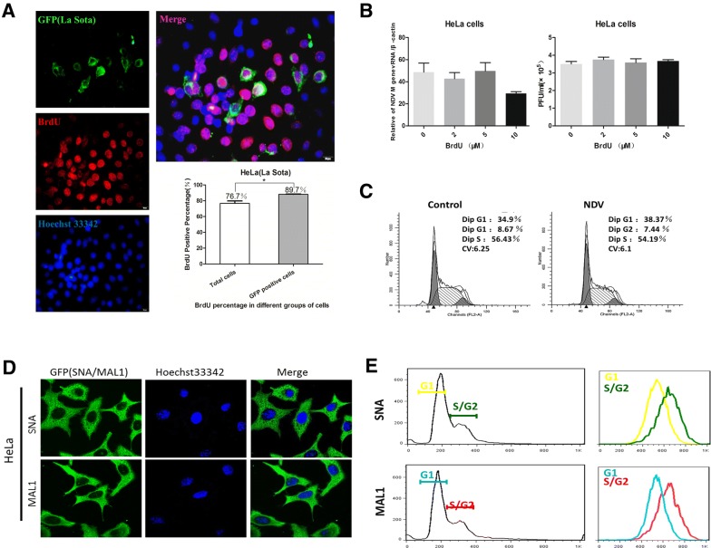
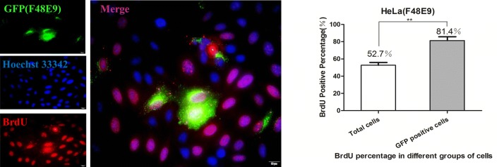
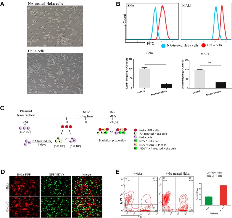
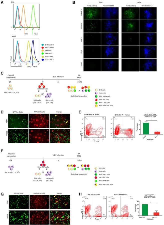
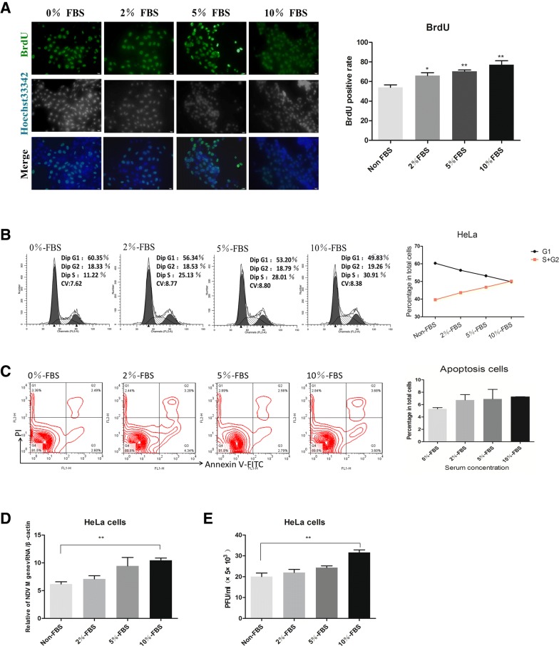
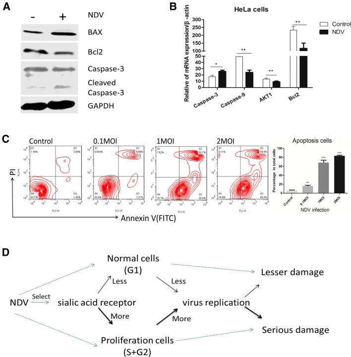
Newcastle disease virus (NDV) can select cells to infect, but the mechanism of its cell selectivity has not been comprehensively investigated. Here, we use HeLa cells to establish that NDV can selectively infect cells at the single-cell level. We labeled proliferating cells with 5'-bromo-2-deoxyuridine (BrdU) and examined the colocalization of BrdU with NDV in cells to clarify the relationships between NDV infection and cell proliferation. Receptors at the plasma membrane mediate NDV entry into host cells. We labeled sialic acid receptor isoforms, compared their densities between different cell types and measured the sialic acid receptor densities in different cell phases. Our results suggest that NDV displays host tropism to HeLa cells compared to BHK cells and that the differences in the receptor isoform expression patterns between cell types contribute to the selection of HeLa by NDV. At the single-cell level, the dynamics of receptor expression changes during different cell phases contributing to the selection of cells in S/G2 phase for NDV infection. Furthermore, cell proliferation benefits viral replication, and enhanced virus replication leads to increased damage to cells. The elucidation of the mechanisms underlying host cell selection by NDV may help in the screening and characterizing of additional candidate oncolytic virus strains.


Figures

Figure 1
Figure 1
A lentogenic NDV strain (La Sota) selectively infects proliferating cells. A Immunofluorescence analysis and quantification of NDV and BrdU staining in HeLa cells infected with the lentogenic NDV La Sota strain (0.1 MOI) and labeled with BrdU (5 μM) at 12 hpi. Scale bar, 20 μm. Data are presented as the mean ± SD of triplicate samples from three independent experiments. *P < 0.05 by t-test. B qRT-PCR analysis of NDV M gene expression normalized to β-actin expression in HeLa cells infected with different viral titers and treated with different concentrations of BrdU (0, 2, 5 and 10 μM). Data are presented as the mean ± SD of triplicate samples from four independent experiments. C Flow cytometry for cell cycle analysis of HeLa cells infected with NDV (0.1 MOI). D Fluorescein-labeled SNA and MAL1 were used to detect Sia expression on the surface of HeLa cells. E FACS analysis of HeLa cells colabeled with PI and either SNA or MAL1.
Figure 2
Figure 2
A virulent NDV strain (F48E9) selectively infects proliferating cells. Immunofluorescence analysis and quantification of NDV and BrdU staining in HeLa cells infected with the virulent NDV strain F48E9 (0.01 MOI) and labeled with BrdU (5 μM) at 12 hpi. Scale bar, 20 μm. Data are presented as the mean ± SD of triplicate samples from three independent experiments. **P < 0.01 by t-test.
Figure 3
Figure 3
NDV selectively infects HeLa cells but not neuraminidase-treated HeLa cells. A Morphologic observation of untreated and neuraminidase-treated HeLa cells. B The presence of sialic acid was detected in neuraminidase-treated HeLa cells by FACS analysis; cells treated without neuraminidase were used as controls. Data are presented as the mean ± SD of triplicate samples from three independent experiments. ***P < 0.001 by t-test. C Schematic of the coculture assay. D Fluorescence microscopy images of NDV (GFP) and RFP expression in cocultured cells. Scale bars represent 50 μm. E Flow cytometry analysis of RFP and GFP expression in HeLa-RFP/HeLa and HeLa-RFP/neuraminidase-treated HeLa cells. Numbers indicate the percentage of cells. n = 3 per group of four analyses. Data are presented as the mean ± SD of triplicate samples from three independent experiments. **P < 0.01 by t-test.
Figure 4
Figure 4
NDV selectively infects HeLa cells. A The presence of sialic acid was detected in HeLa and BHK cells by FACS analysis; unstained cells were used as a blank control. B Fluorescence microscopy images of NDV (GFP+) cells treated with different viral titers of NDV. Scale bars represent 50 μm. C Schematic of the coculture assay. D Fluorescence microscopy images of NDV (GFP+) and RFP in cocultured cells; the arrow indicates an NDV-infected BHK-RFP cell. Scale bars represent 50 μm. E Flow cytometry was used to analyze RFP and GFP expression in BHK-RFP/HeLa and BHK-RFP/BHK cell cocultures. Numbers reflect the percentage of cells. Data are presented as the mean ± SD of triplicate samples from three independent experiments. **P < 0.01 by t-test. F Schematic of the coculture assay. G Fluorescence microscopy images of NDV (GFP) and RFP in cocultured cells; the arrow indicates an NDV-infected HeLa-RFP cell. Scale bars represent 50 μm. H Flow cytometry was used to analyze RFP and GFP expression in HeLa-RFP/HeLa and HeLa-RFP/BHK cell cocultures. Numbers reflect the percentage of cells. Data are presented as the mean ± SD of triplicate samples from three independent experiments. *P < 0.05 by t-test.
Figure 5
Figure 5
Proliferating cells enhance NDV replication. HeLa cells were cultured with different concentrations of FBS for 24 h and then stained with BrdU (A), after which cell cycle distribution (B) and apoptosis (C) were analyzed by flow cytometry. Cells were pretreated with different concentrations of serum for 24 h and then infected with NDV. At 24 hpi, viruses were quantified by qRT-PCR based on M gene expression (D) and titration with plaque assay (E). Data are presented as the mean ± SD of triplicate samples from four independent experiments. **P < 0.01 by t-test.
Figure 6
Figure 6
Increased NDV replication leads to more cell apoptosis. A Western blot analysis of the expression of apoptosis-related proteins (BAX, Bcl2 and caspase-3) in HeLa cells at 48 hpi (0.1 MOI; La Sota). B qRT-PCR analysis of apoptosis-related gene expression in HeLa cells at 48 hpi (0.1 MOI; La Sota). C Flow cytometry analysis of cell apoptosis at 48 hpi (0.1, 1 or 2 MOI); data are pooled from replicates of at least three independent experiments. D Overall description of how NDV selectively infects proliferating cells to kill tumor cells. Data are presented as the mean ± SD of triplicate samples from a single experiment and are representative of three independent experiments. *P < 0.05, **P < 0.01 and ***P < 0.001 by t-test.

Similar articles

Cited by

References

    1. Cuadrado-Castano S, Sanchez-Aparicio MT, García-Sastre A, Villar E. The therapeutic effect of death: newcastle disease virus and its antitumor potential. Virus Res. 2015;209:56–66. doi: 10.1016/j.virusres.2015.07.001. - DOI - PMC - PubMed
    1. Elankumaran S, Rockemann D, Samal SK. Newcastle disease virus exerts oncolysis by both intrinsic and extrinsic caspase-dependent pathways of cell death. J Virol. 2006;80:7522–7534. doi: 10.1128/JVI.00241-06. - DOI - PMC - PubMed
    1. Bu X, Zhao Y, Zhang Z, Wang M, Li M, Yan Y. Recombinant Newcastle disease virus (rL-RVG) triggers autophagy and apoptosis in gastric carcinoma cells by inducing ER stress. Am J Cancer Res. 2016;6:924–936. - PMC - PubMed
    1. Ravindra PV, Tiwari AK, Ratta B, Chaturvedi U, Palia SK, Chauhan RS. Newcastle disease virus-induced cytopathic effect in infected cells is caused by apoptosis. Virus Res. 2009;141:13–20. doi: 10.1016/j.virusres.2008.12.008. - DOI - PubMed
    1. Ravindra PV, Tiwari AK, Sharma B, Chauhan RS. Newcastle disease virus as an oncolytic agent. Indian J Med Res. 2009;130:507–513. - PubMed

Grants and funding

LinkOut - more resources