Review
. 2019:134:289-315.
doi: 10.1016/bs.ctdb.2019.01.003. Epub 2019 Feb 12.

Rethinking sex determination of non-gonadal tissues

Affiliations
Review

Rethinking sex determination of non-gonadal tissues

Arthur P Arnold. Curr Top Dev Biol. 2019.

Abstract

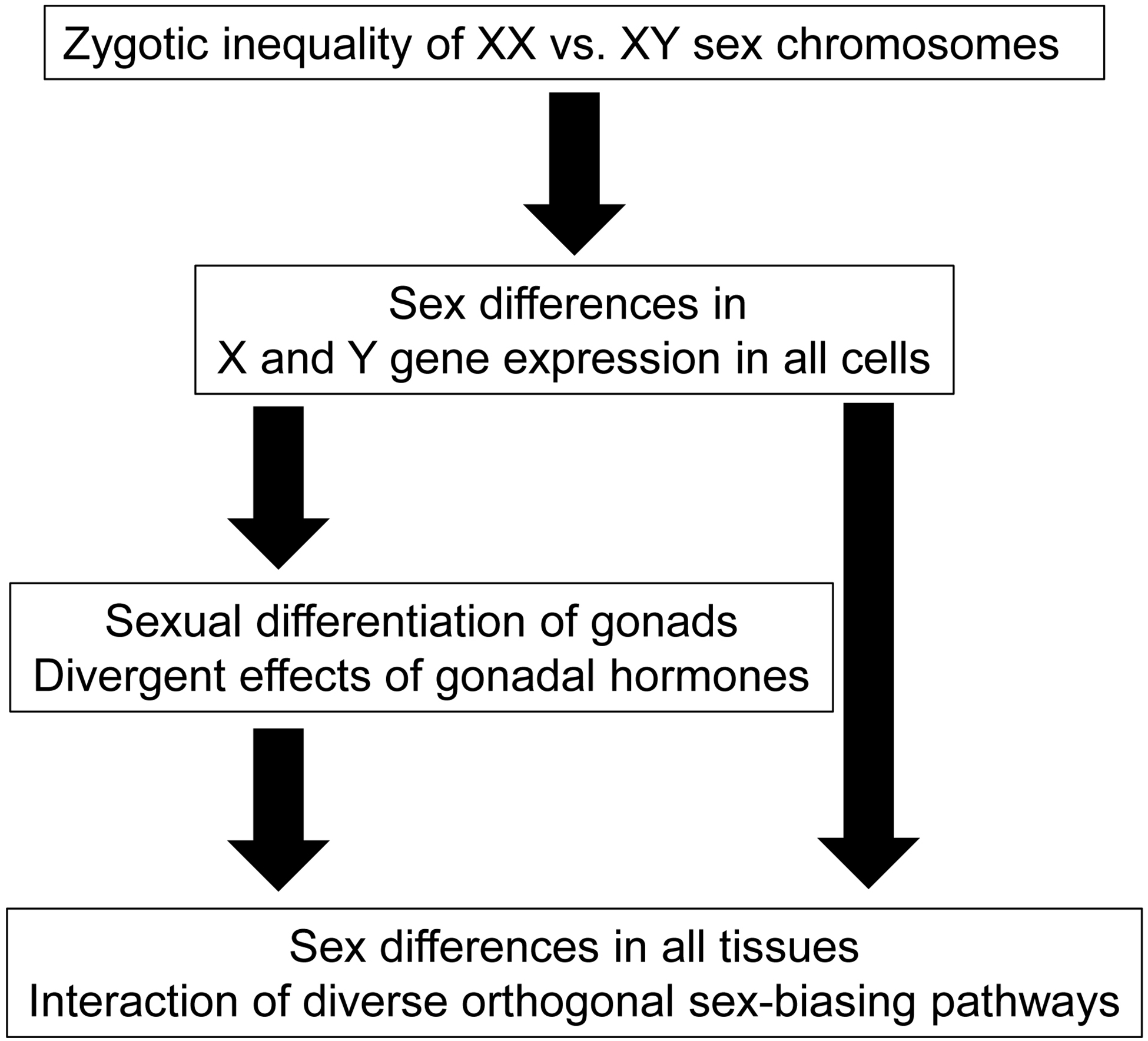
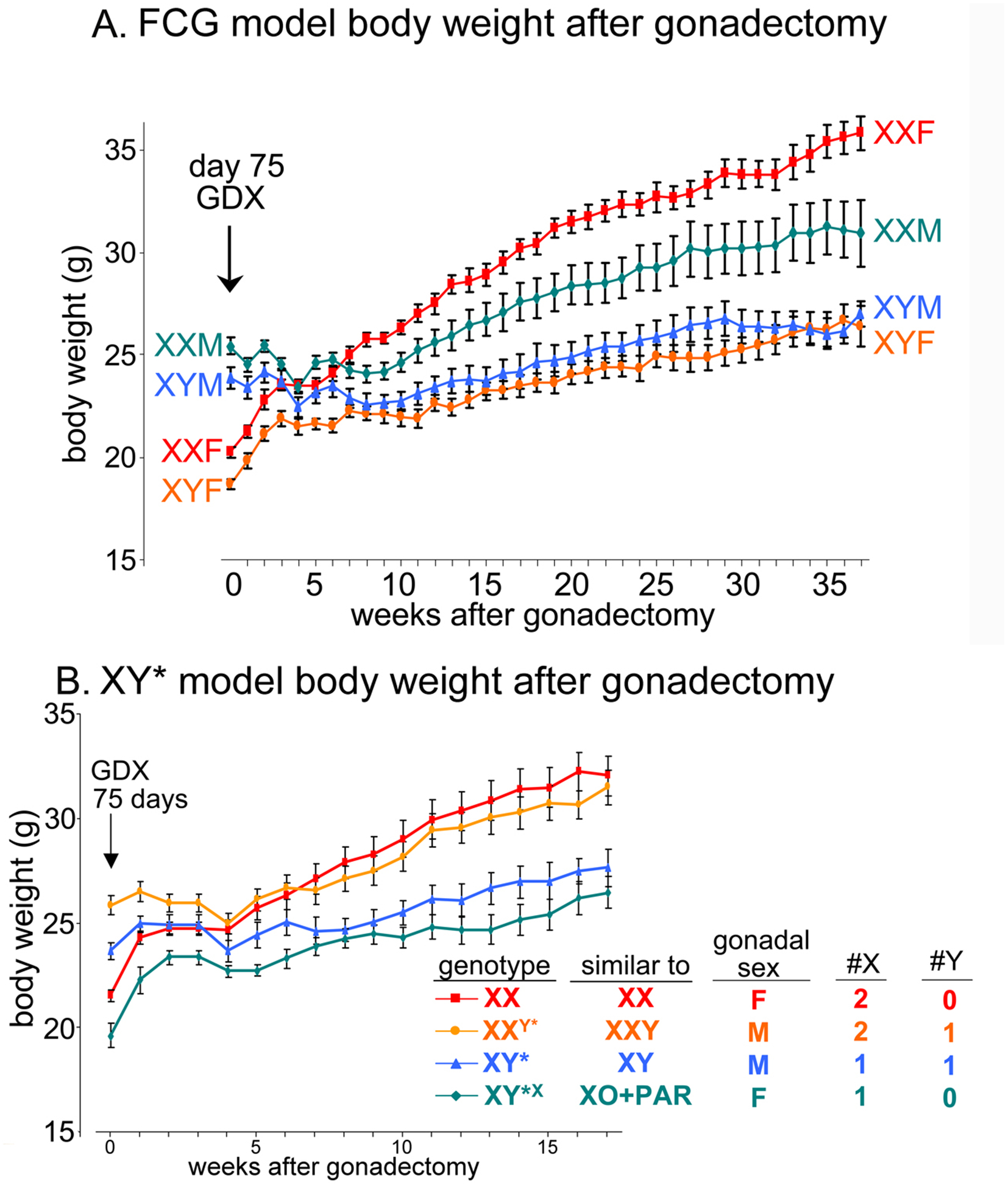
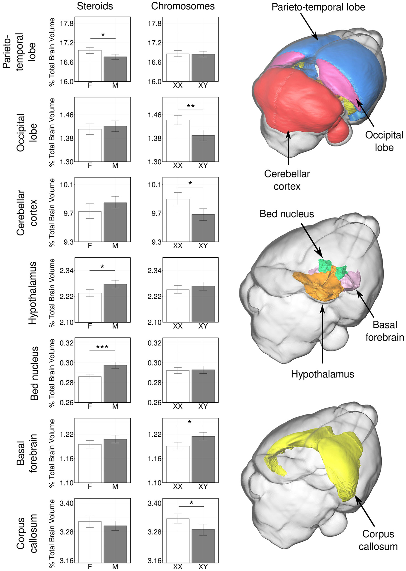
Evolution of genetic mechanisms of sex determination led to two processes causing sex differences in somatic phenotypes: gonadal differentiation and sex chromosome dosage inequality. In species with heteromorphic sex chromosomes, the sex of the individual is established at the time of formation of the zygote, leading to inherent sex differences in expression of sex chromosome genes beginning as soon as the embryonic transcriptome is activated. The inequality of sex chromosome gene expression causes sexual differentiation of the gonads and of non-gonadal tissues. The difference in gonad type in turn causes lifelong differences in gonadal hormones, which interact with unequal effects of X and Y genes acting within cells. Separating the effects of gonadal hormones and sex chromosomes has been possible using mouse models in which gonadal determination is separated from the sex chromosomes, allowing comparison of XX and XY mice with the same type of gonad. Sex differences caused by gonadal hormones and sex chromosomes affect basic physiology and disease mechanisms in most or all tissues.

Keywords: Cell-autonomous; Dosage compensation; Sex chromosomes; Sex determination; Sex difference; Sexual differentiation; X chromosome; Y chromosome.


Figures

Figure 1
Figure 1. The sex-chromosome-centric view of sexual differentiation.
All phenotypic sex differences stem from the difference in number and type of sex chromosomes present in the zygote. In the undifferentiated gonadal ridge, Sry expression in XY cells induces differentiation of testes. Ovaries differentiate in XX animals in the absence of Sry, because of the expression of autosomal or X-linked genes. Differences in gonadal hormone levels contribute to sex differences in tissue function. In parallel, sex differences are also caused in all non-gonadal tissues by differences in effects of X and Y genes. Hormonal and sex chromosome factors interact to promote or reduce sex differences in phenotypes.
Figure 2.
Figure 2.. Inherent bias in human X chromosome gene expression across tissues.
Analysis of over 5500 transcriptomes from 29 human tissues from GTEx consortium data shows the level of sex bias in expression of X genes reported to escape inactivation. Rows indicate different tissues, and columns represent X genes reported to escape X inactivation, mapped by chromosomal position. Red indicates expression higher in females, and blue indicates expression higher in males. Grey indicates expression too low to be analyzed. In the non-pseudoautosomal (non-PAR) region, about 23% of X genes overall showed higher expression in females, and in PAR1 region most genes were expressed higher in males. This general pattern can be attributed to escape from X inactivation in the non-PAR region in XX cells, and from some inactivation of PAR genes in XX cells but not XY cells. However, other sex-biasing factors (gonadal hormones, parental imprint) could contribute to sex differences as well, in either direction, and may account for colors that deviate from the pattern of adjacent genes (e.g., red squares in the PAR, and blue squares in the non-PAR). Reprinted from (Tukiainen et al., 2017) under terms of Creative Commons Attribution 4.0 International license.
Figure 3.
Figure 3.. The number of X chromosomes influences body weight in FCG and XY* mice.
A. In FCG mice, gonadal males at postnatal day 75 (week 0) weighed about 25% more than gonadal females. When gonads were removed on that day, the sex differences disappeared by 4 weeks after gonadectomy (GDX). After that, XX mice slowly gained much more body weight than XY mice, irrespective of previous gonadal type. B. In the XY* model, the same experiment replicates the larger body size of two groups with testes, relative to two groups with ovaries. After gonadectomy, the groups with two X chromosomes gained more weight than those with one X chromosome, illustrating that the body weight is affected by the number of X chromosomes. M = gonadal male, F = gonadal female. PAR = pseudoautosomal region. Reprinted from (Chen et al., 2012) under terms of the Creative Commons Attribution License.
Figure 4.
Figure 4.. Sex chromosome and hormonal effects on brain volumes from FCG mice.
Whole-brain MRI was used to measure volumetric variation in gonad-intact FCG mice. Among seven brain regions illustrated, some showed significant effects of gonadal hormones (parieto-temporal lobe of the cerebral cortex, the hypothalamus, and the bed nucleus of the stria terminalis). Others showed effects of sex chromosomes (basal forebrain, occipital lobe of the cerebral cortex, cerebellar cortex, corpus callosum). Asterisks indicate changes were significant at false discovery rate of 5% or less. Error bars represent 95% confidence intervals. Reprinted by permission of Springer Nature from (Corre et al., 2016).

Similar articles

Cited by

References

    1. Agate RJ, Grisham W, Wade J, Mann S, Wingfield J, Schanen C, Palotie A, Arnold AP (2003) Neural not gonadal origin of brain sex differences in a gynandromorphic finch. Proc Natl Acad Sci U S A 100:4873–4878. - PMC - PubMed
    1. Alsiraj Y, Thatcher SE, Charnigo R, Kuey C, Blalock E, Daugherty A, Cassis LA (2016) Female Mice with an XY Sex Chromosome Complement Develop Severe Angiotensin II-Induced Abdominal Aortic Aneurysms. Circulation. - PMC - PubMed
    1. Arnold AP (2011) The end of gonad-centric sex determination in mammals. Trends Genet 28:55–61. - PMC - PubMed
    1. Arnold AP (2014) Conceptual frameworks and mouse models for studying sex differences in physiology and disease: Why compensation changes the game. Exp Neurol 259:2–9. - PMC - PubMed
    1. Arnold AP (2017a) A general theory of sexual differentiation. J Neurosci Res 95:291–300. - PMC - PubMed

Substances