. 2019 Jun;212(2):417-430.
doi: 10.1534/genetics.119.302125. Epub 2019 Apr 18.

Overlapping Roles in Chromosome Segregation for Heterochromatin Protein 1 (Swi6) and DDK in Schizosaccharomyces pombe

Affiliations

Overlapping Roles in Chromosome Segregation for Heterochromatin Protein 1 (Swi6) and DDK in Schizosaccharomyces pombe

Kuo-Fang Shen et al. Genetics. 2019 Jun.

Abstract

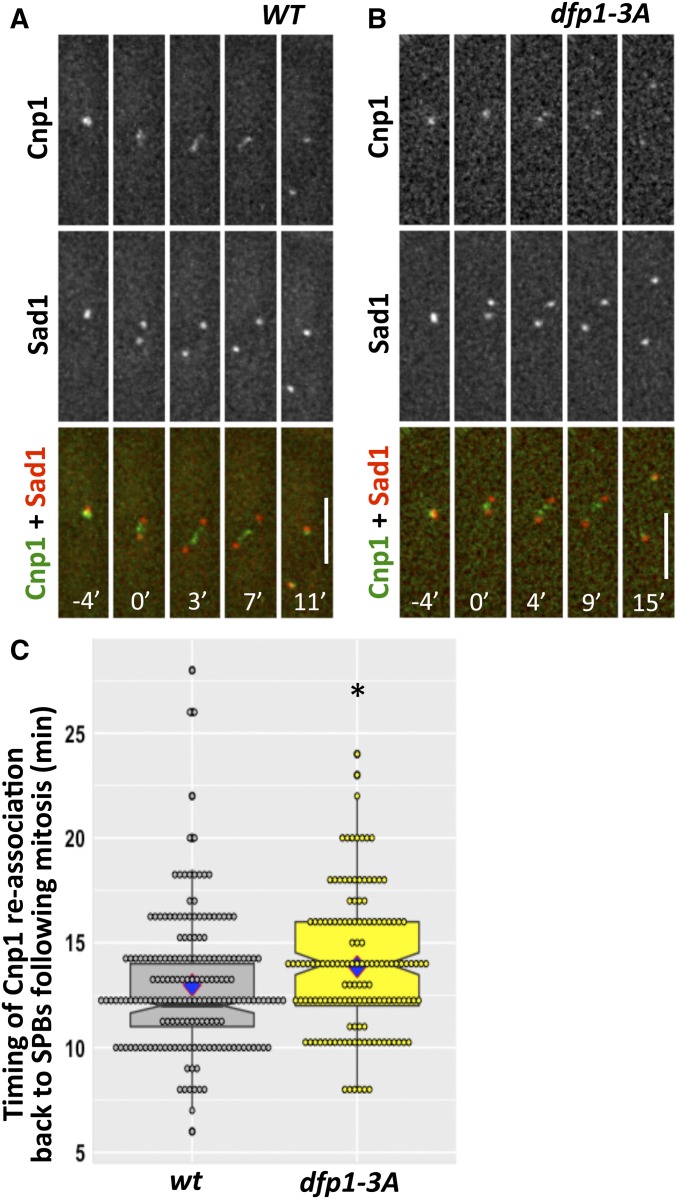
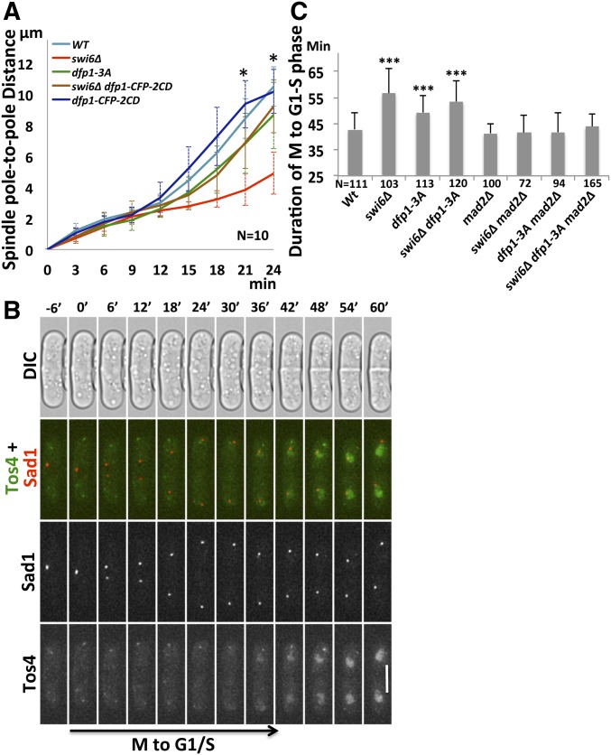
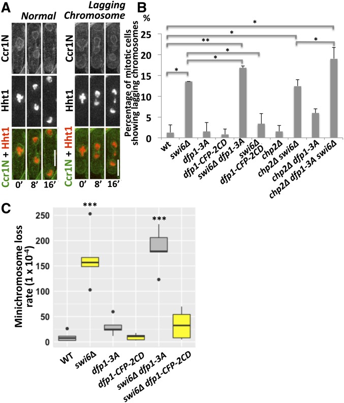
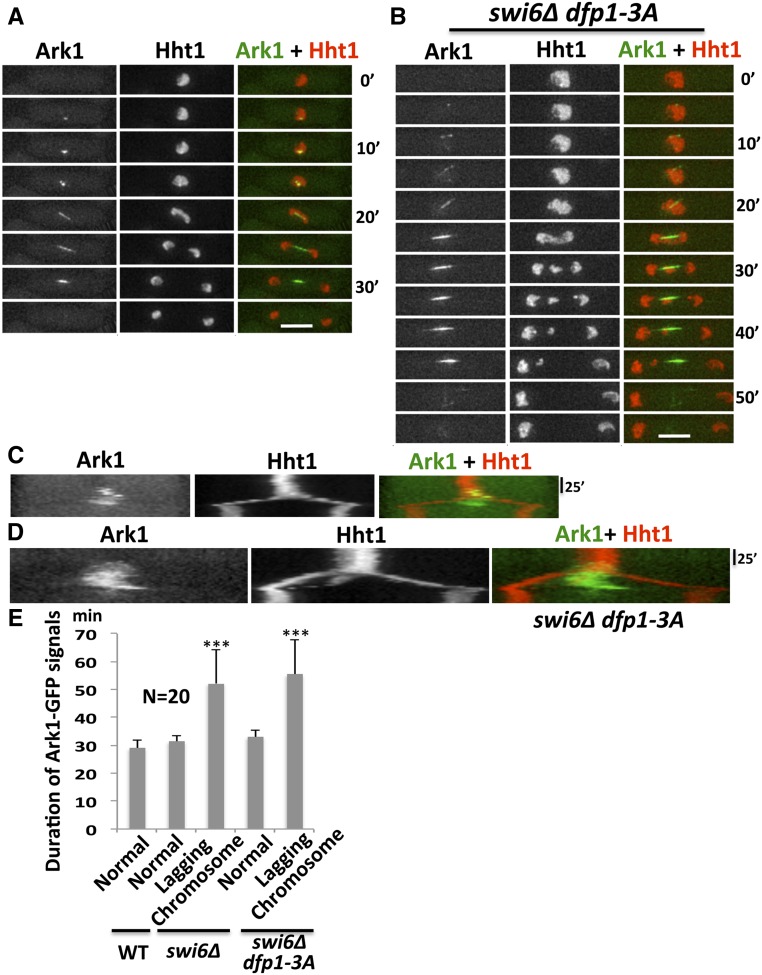
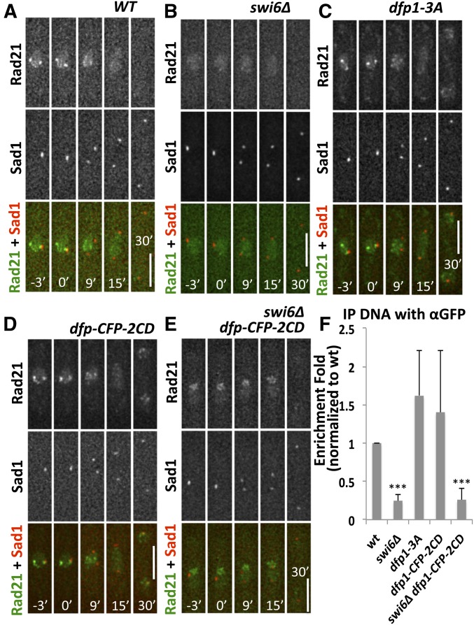
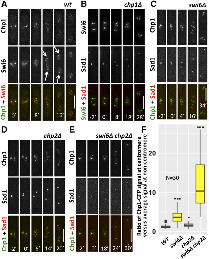
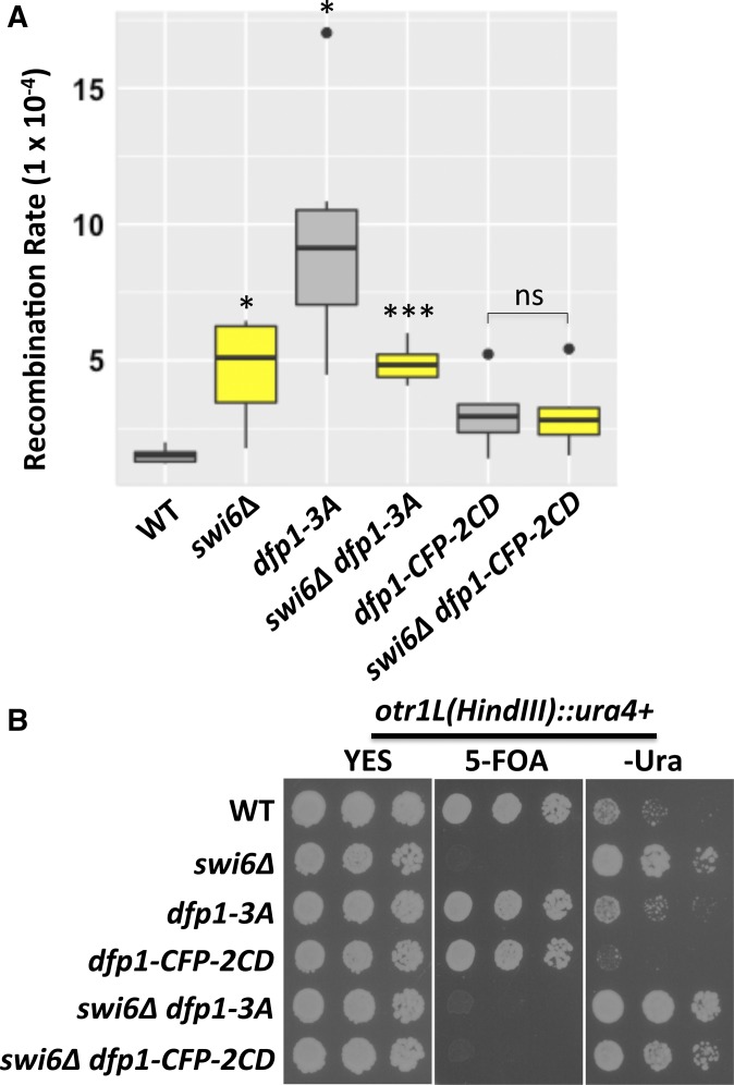
Fission yeast Swi6 is a human HP1 homolog that plays important roles in multiple cellular processes. In addition to its role in maintaining heterochromatin silencing, Swi6 is required for cohesin enrichment at the pericentromere. Loss of Swi6 leads to abnormal mitosis, including defects in the establishment of bioriented sister kinetochores and microtubule attachment. Swi6 interacts with Dfp1, a regulatory subunit of DBF4-dependent kinase (DDK), and failure to recruit Dfp1 to the pericentromere results in late DNA replication. Using the dfp1-3A mutant allele, which specifically disrupts Swi6-Dfp1 association, we investigated how interaction between Swi6 and Dfp1 affects chromosome dynamics. We find that disrupting the interaction between Swi6 and Dfp1 delays mitotic progression in a spindle assembly checkpoint-dependent manner. Artificially tethering Dfp1 back to the pericentromere is sufficient to restore normal spindle length and rescue segregation defects in swi6-deleted cells. However, Swi6 is necessary for centromeric localization of Rad21-GFP independent of DDK. Our data indicate that DDK contributes to mitotic chromosome segregation in pathways that partly overlap with, but can be separated from both, Swi6 and the other HP1 homolog, Chp2.

Keywords: Cdc7/Hsk1; Chp1 and SAC; Cnp1; fission yeast; heterochromatin; kinetochore; minichromosome; mitosis.


Figures

Figure 1
Figure 1
Disruption of Swi6 and Dfp1 interaction affects timing of reassociation of centromere to SPBs. We examined CFP-Cnp1 to mark the clustered centromeres, and Sad1-DsRed to mark the SPB, in live cells during mitosis in (A) wt (FY7554) and (B) dfp1-3A (FY7640). Time 0’ corresponds to the timepoint that precedes the first observed separation of the SPB and defines the initiation of mitosis. (C) Quantification of timing when CFP-Cnp1 locates back to the SPBs following mitosis. Significance was calculated with a two-tailed Student’s t-test with * P < 0.05. Bar, 5 µm. CFP, cyan fluorescent protein; SPB, spindle pole bodies; WT, wild-type.
Figure 2
Figure 2
Artificially tethering Dfp1 to the pericentromere via 2CD rescues mitotic spindle tension. (A) Spindle pole-to-pole distance was used as a marker for mitotic spindle tension, and was determined by the distance between Sad1-DsRed foci through mitosis in wt (FY8115), swi6∆ (FY8285), dfp1-3A (FY8051), dfp1-CFP-2CD (FY9117), and swi6dfp1-CFP-2CD (FY8269). (B) Duration of M-to-G1/S phase transition was determined by the timing between separation of duplicated SPBs (labeled by Sad1-DsRed) and the appearance of nuclear Tos4-GFP, an S phase marker, which coincides with the appearance of the septum, visualized by DIC. Time 0’ corresponds to the timepoint that precedes the first observed separation of the SPB and defines the initiation of mitosis. Representative image from wt (FY8222). (C) Quantification of timing from the assay in (B) for wt (FY8222), swi6∆ (FY8325), dfp1-3A (FY8254), mad2∆ (FY8393), mad2dfp1-3A (FY8394), and mad2swi6∆ (FY8390). The delay of M-to-G1/S phase transition in swi6∆ and dfp1-3A was abolished after deleting mad2. A two-tailed Student’s t-test was used to determine significance: * P < 0.05, *** P < 0.001. Error bars represent Standard Deviation (SD). Bar, 5 µm. CD, chromodomain; CFP, cyan fluorescent protein; SPB, spindle pole bodies; WT, wild-type.
Figure 3
Figure 3
DBF4-dependent kinase (DDK) contributes to proper chromosome segregation in the absence of Swi6. (A) We monitored mitosis in strains expressing ccr1N-GFP (D187 amino acids 1–275) (a membrane marker) and hht1-mRFP (a histone marker). Representative normal mitosis and lagging chromosome images are shown for wt (FY8023), swi6∆ (FY8214), dfp1-3A (FY8113), dfp1-CFP-2CD (FY8275), swi6dfp1-3A (FY8213), swi6dfp1-CFP-2CD (FY8375), chp2∆ (FY8410), chp2swi6∆ (FY8634), chp2dfp1-3A (FY8413), and chp2swi6dfp1-3A (FY8633). Time 0’ corresponds to the timepoint that precedes the first observed separation of the histone mass and defines the initiation of mitosis. (B) Quantification of mitotic cells showing lagging chromosomes in (A). (C) The frequency of chromosome loss was determined using a minichromosome with multiple genetic markers. Cells that survived on EMM +5-FOA +histidine +uracil +leucine +adenine +lysine plates have lost the right arm of the minichromosome, and were screened for leucine auxotrophy to determine whether the left arm was also missing. wt (FY4003), swi6∆ (FY5109), dfp1-3A (FY7824), dfp1-CFP-2CD (FY7719), swi6dfp1-3A (FY8027), swi6dfp1-CFP-2CD (FY7987), chp2∆ (FY8690), chp2dfp1-3A (FY8692), chp2swi6∆ (FY8700), and chp2swi6dfp1-3A (FY8702). Frequencies of minichromosome loss were calculated according to the Lea–Coulson method. A two-tailed Student’s t-test was used to determinate significance. P-values are reported as follows: * P < 0.05, ** P < 0.01, *** P < 0.001. Bar, 5 µm. WT, wild-type.
Figure 4
Figure 4
Prolonged nuclear Ark1-GFP correlates with increased lagging chromosomes. Localization of Ark1-GFP in relation to Hht1-mRFP (a histone marker) was observed in wt (FY8329), swi6∆ (FY8340), and swi6dfp1-3A (FY8376) during (A) representative normal mitosis and (B) lagging chromosome mitosis. (C and D) Kymograph analysis of (A and B), respectively shows prolonged duration of Ark1-GFP in strains with lagging chromosomes. (E) Duration of Ark1-GFP signals in (A and B). The difference in duration of Ark1-GFP signals in normal mitotic cells compared to lagging chromosome cells was significant using a two-tailed Student’s t-test: *** P < 0.001. Bar, 5 µm. RFP, red fluorescent protein; WT, wild-type.
Figure 5
Figure 5
Centromeric localization of Rad21 depends on Swi6. (A–E) The position of Rad21-GFP in relation to the centromere defined by an SPB marker, Sad1-DsRed, was observed in live cells. Time 0’ corresponds to the timepoint that precedes the first observed separation of the SPB and defines the initiation of mitosis. (A) WT (FY8115), (B) swi6∆ (FY8285), (C) dfp1-3A (FY8051), (D) dfp1-CFP-2CD (FY9117), and (E) swi6dfp1-CFP-2CD (FY8269). (F) Association of Rad21-GFP at the pericentromeric dh locus in strains FY8051, FY8115, FY8269, FY8285, and FY9117 was examined using chromatin IP-quantitative PCR, and is consistent with the visualization in (A–E). Significance was determined using a two-tailed Student’s t-test; *** P < 0.001. Bar, 5 µm. CD, chromodomain; CFP, cyan fluorescent protein; IP immunoprecipitation; SPB, spindle pole bodies; WT, wild-type.
Figure 6
Figure 6
Swi6 and Chp2 are required for the noncentromeric location of Chp1 in interphase. (A) Localization of Chp1-GFP and Swi6-chRFP was observed during mitosis in live WT cells (FY7973). Arrows indicate the position of Swi6 localized close to spindle poles. (B) Live-cell imaging of Swi6-GFP in relation to Sad1-DsRed was captured during mitosis in chp1∆ (FY7978). (C) Localization of Chp1-GFP and Sad1-DsRed in swi6∆ (FY7979). (D) Localization of Chp1-GFP and Sad1-DsRed in chp2∆ (FY8795). (E) Localization of Chp1-GFP and Sad1-DsRed in chp2swi6∆ (FY8726). Time 0’ corresponds to the timepoint that precedes the first observed separation of the SPB and defines the initiation of mitosis. (F) Chp1-GFP signals at centromere and noncentromere regions before mitosis were measured in (A and C–E), and the ratio of Chp1-GFP signals at centromere vs. noncentromere sites is presented. Bar, 5 µm. A two-tailed Student’s t-test was used to determine significance with * P < 0.05, *** P < 0.001. chRFP, mCherry red fluorescent protein; WT, wild-type.
Figure 7
Figure 7
Late replication timing correlates with increased gross chromosome rearrangement. (A) The frequency of chromosome rearrangement was determined using a minichromosome with multiple genetic markers (see Figure 3C). Cells that survived on EMM +5-FOA +histidine +uracil +leucine + adenine +lysine plates have lost the right arm of the minichromosome and maintain the left arm (Leu+). Recombination rates were calculated according to the Lea–Coulson method in the indicated strains; P-values are reported as follows: * P < 0.05, *** P < 0.001. (B) Pericentromere heterochromatin silencing was measured by expression of an ura4+ gene inserted at otr1L in wt (FY4293), swi6(FY4295), dfp1-3A (FY8144), dfp1-CFP-2CD (FY8221), swi6dfp1-3A (FY8146), and swi6dfp1-CFP-2CD (FY8191). Cells were spotted and grown on YES, YES+ 5-FOA, and EMM −Uracil plates at 32° for 3 days with 1:5 serial dilutions. Cells grow on EMM –Uracil plates, but not on YES + 5-FOA plates, when they lose heterochromatin silencing. CD, chromodomain; CFP, cyan fluorescent protein; WT, wild-type; YES, yeast extract with supplements.

Similar articles

Cited by

References

    1. Aasland R., Stewart A. F., 1995. The chromo shadow domain, a second chromo domain in heterochromatin-binding protein 1, HP1. Nucleic Acids Res. 23: 3168–3173. 10.1093/nar/23.16.3168 - DOI - PMC - PubMed
    1. Allshire R. C., Nimmo E. R., Ekwall K., Javerzat J. P., Cranston G., 1995. Mutations derepressing silent centromeric domains in fission yeast disrupt chromosome segregation. Genes Dev. 9: 218–233. 10.1101/gad.9.2.218 - DOI - PubMed
    1. Alper B. J., Lowe B. R., Partridge J. F., 2012. Centromeric heterochromatin assembly in fission yeast–balancing transcription, RNA interference and chromatin modification. Chromosome Res. 20: 521–534. 10.1007/s10577-012-9288-x - DOI - PMC - PubMed
    1. Bailis J. M., Bernard P., Antonelli R., Allshire R. C., Forsburg S. L., 2003. Hsk1-Dfp1 is required for heterochromatin-mediated cohesion at centromeres. Nat. Cell Biol. 5: 1111–1116. 10.1038/ncb1069 - DOI - PubMed
    1. Baker S. P., Phillips J., Anderson S., Qiu Q., Shabanowitz J., et al. , 2010. Histone H3 Thr 45 phosphorylation is a replication-associated post-translational modification in S. cerevisiae. Nat. Cell Biol. 12: 294–298. 10.1038/ncb2030 - DOI - PMC - PubMed

Publication types

MeSH terms