. 2019 Jun;180(2):757-766.
doi: 10.1104/pp.18.01377. Epub 2019 Apr 18.

A Mobile Auxin Signal Connects Temperature Sensing in Cotyledons with Growth Responses in Hypocotyls

Affiliations

A Mobile Auxin Signal Connects Temperature Sensing in Cotyledons with Growth Responses in Hypocotyls

Julia Bellstaedt et al. Plant Physiol. 2019 Jun.

Abstract

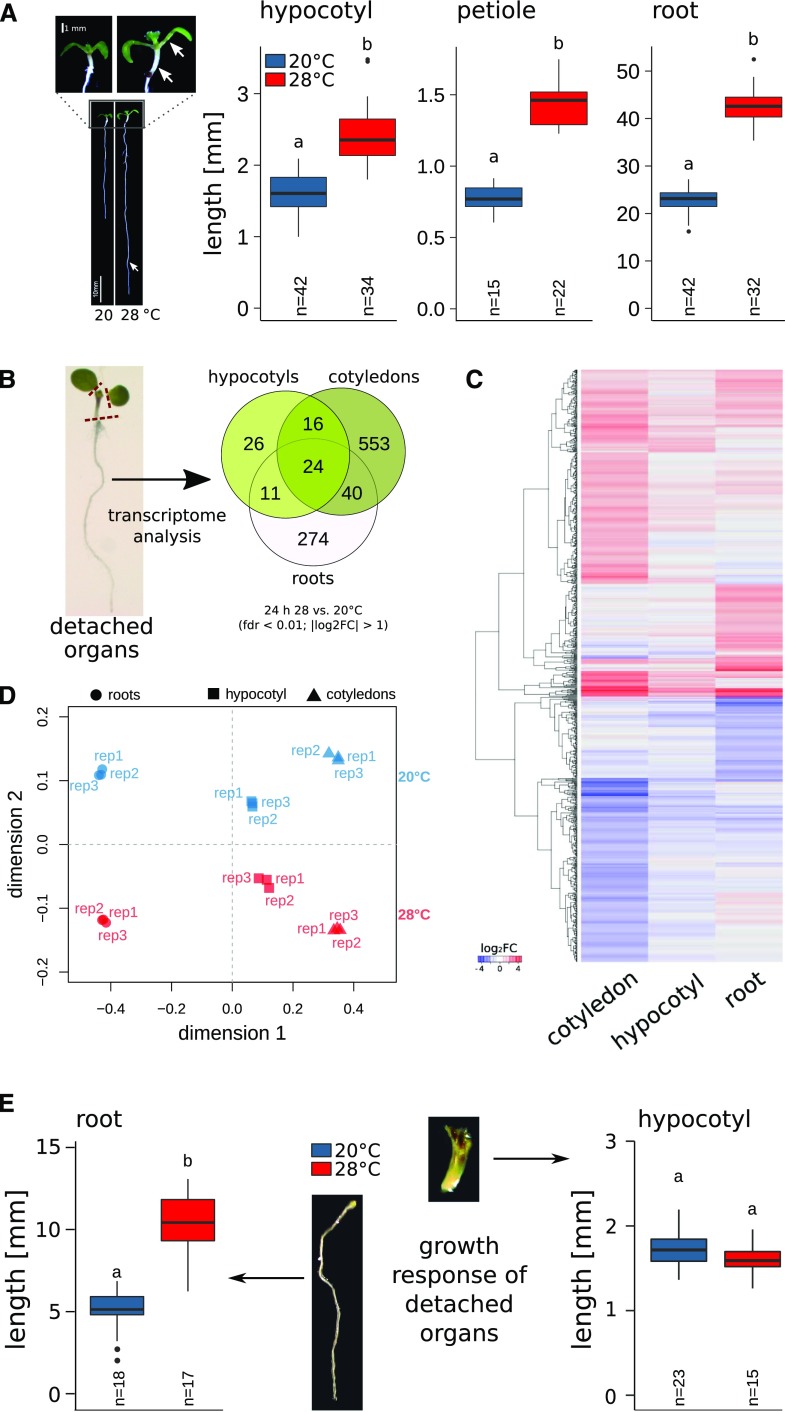
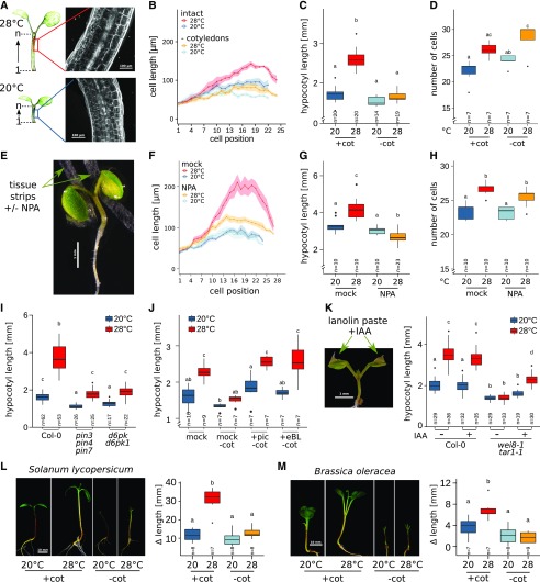
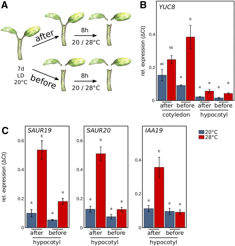
Plants have a remarkable capacity to adjust their growth and development to elevated ambient temperatures. Increased elongation growth of roots, hypocotyls, and petioles in warm temperatures are hallmarks of seedling thermomorphogenesis. In the last decade, significant progress has been made to identify the molecular signaling components regulating these growth responses. Increased ambient temperature utilizes diverse components of the light sensing and signal transduction network to trigger growth adjustments. However, it remains unknown whether temperature sensing and responses are universal processes that occur uniformly in all plant organs. Alternatively, temperature sensing may be confined to specific tissues or organs, which would require a systemic signal that mediates responses in distal parts of the plant. Here, we show that Arabidopsis (Arabidopsis thaliana) seedlings show organ-specific transcriptome responses to elevated temperatures and that thermomorphogenesis involves both autonomous and organ-interdependent temperature sensing and signaling. Seedling roots can sense and respond to temperature in a shoot-independent manner, whereas shoot temperature responses require both local and systemic processes. The induction of cell elongation in hypocotyls requires temperature sensing in cotyledons, followed by the generation of a mobile auxin signal. Subsequently, auxin travels to the hypocotyl, where it triggers local brassinosteroid-induced cell elongation in seedling stems, which depends upon a distinct, permissive temperature sensor in the hypocotyl.


Figures

Figure 1.
Figure 1.
Thermomorphogenesis in seedling organs is autonomous or interdependent. A, Temperature-induced elongation of hypocotyls, roots, and petioles (indicated by white arrows) in 8-d-old seedlings grown at 20°C or 28°C. B, Temperature-induced transcriptome responses in 6-d-old seedlings. Venn diagrams show overlap in differential gene expression after 24 h at 28°C versus control seedlings at 20°C (|log2 fold change [FC]| > 1, false discovery rate [fdr] < 0.01). C, Hierarchical clustering of DEGs using Euclidian distances of log2 FC data. D, MDS of DEGs (shown in C) based on the pairwise Pearson correlation (1 − cor) among all individual biological replicates. E, Elongation responses of detached seedling organs. Petioles and cotyledons or whole shoots were removed from 4-d-old seedlings grown at 20°C. Subsequently, detached organs were placed on growth medium and cultivated for an additional 4 d at 20°C or 28°C. Experiments were performed in long-day (16/8 h) conditions under 90 µmol m−2 s−1 white fluorescent light. Box plots show medians and interquartile ranges of total organ lengths; outliers (greater than 1.5× interquartile range) are shown as black dots. Different letters denote statistical differences at P < 0.05 as assessed by one-way ANOVA and Tukey’s honestly significant difference (HSD) posthoc test.
Figure 2.
Figure 2.
Hypocotyl cell elongation requires cotyledon-derived auxin. A, Lengths of individual cells in a consecutive hypocotyl cortex cell file were determined via confocal microscopy of propidium iodide-stained seedlings. B to D, Effects of cotyledon detachment on hypocotyl cell length (B), total hypocotyl length (C), and hypocotyl cell number in one consecutive cortex cell file (D). Seedlings were grown at 20°C for 4 d. Petioles and cotyledons were removed or seedlings were left intact prior to a shift to 28°C for an additional 3 d. E, Localized block of auxin transport by application of thin tissue strips soaked in medium with or without 100 µm NPA. F to H, Cell length (F), total hypocotyl length (G), and cell number (H) were determined, similar to A to D. I, Total hypocotyl lengths in 8-d-old auxin transport mutants. Seedlings grown at 20°C for 4 d were shifted to 28°C for an additional 4 d. J, Total hypocotyl length of intact seedlings (+cot) or with detached cotyledons (−cot) in the presence or absence of 1 µm picloram (pic) or 100 nm epi-brassinolide (eBL). Four-day-old seedlings grown at 20°C were transferred to medium containing the respective hormones and cultivated at 20°C or 28°C for an additional 3 d. K, Total hypocotyl lengths of 10-d-old seedlings with or without localized application of 1 mm IAA in lanolin paste to cotyledons. Seven-day-old seedlings grown at 20°C were treated with lanolin paste and transferred to 28°C for an additional 3 d. L and M, Temperature-induced changes in hypocotyl lengths of 11-d-old tomato (L) and cabbage (M) seedlings. Plants were initially grown for 8 d at 20°C. Cotyledons were detached or seedlings remained intact prior to the shift to 28°C for an additional 3 d. Δlength was calculated as the hypocotyl length on day 11 minus the length prior to shift. Experiments were performed in long-day conditions (16/8 h) under 30 µmol m−2 s−1 (E–H) or 90 µmol m−2 s−1 (A–D and I–M) white fluorescent light. Control plants in all experiments were treated similarly but were grown at 20°C for the whole time instead of shifting to 28°C. Bold lines in ribbon plots (B and F) show mean lengths of individual cells in a consecutive cortex cell file from the first cell after the root-shoot junction (1) upward to the shoot apex. The shadowed ribbon denotes the se. Box plots show medians and interquartile ranges; outliers (greater than 1.5× interquartile range) are shown as black dots. Different letters denote statistical differences at P < 0.05 as assessed by one-way ANOVA and Tukey’s HSD posthoc test.
Figure 3.
Figure 3.
Organ-specific expression analysis of auxin-related and growth-related genes. A, Seedlings cultivated for 7 d at 20°C were transferred to 20°C or 28°C at zeitgeber time 16 (ZT16) for 8 h prior to harvesting cotyledons and hypocotyls for expression analysis. Seedlings were either dissected after or before the temperature shift, including removal of the root. B, Reverse transcription quantitative PCR (RT-qPCR) expression analysis of the auxin biosynthesis gene YUC8 was assessed for cotyledons and hypocotyls. C, Genes relevant for hypocotyl elongation (SAUR19 and SAUR20) and an auxin response gene (IAA19) were assessed in hypocotyl samples. Seedlings were grown in long-day conditions (16/8 h) under 30 µmol m−2 s−1 white fluorescent light. RT-qPCR analyses were performed on three independent biological replicates. Bar plots show mean values, and error bars denote se. Different letters denote statistical differences at P < 0.05 as assessed by one-way ANOVA and Tukey’s HSD posthoc test.
Figure 4.
Figure 4.
BZR1-mediated hypocotyl thermomorphogenesis requires local permissive temperature sensing. A, PIF4 RT-qPCR expression analysis in cotyledons and hypocotyls dissected after the temperature treatment. The experimental setup was as described in Figure 3. B and C, Effects of cotyledon detachment on hypocotyl length in 35S:PIF4 (B), and bzr1-1D-OX (C) seedlings. Seedlings were initially grown in long-day conditions (16/8 h) under 90 µmol m−2 s−1 white fluorescent light at 20°C for 4 d. Petioles and cotyledons were removed or seedlings were left intact prior to a shift to 28°C for an additional 3 d. Control plants were treated similarly but grown at 20°C. Box plots show medians and interquartile ranges of total organ length; outliers (greater than 1.5× interquartile range) are shown as black dots. Different letters denote statistical differences at P < 0.05 as assessed by one-way ANOVA and Tukey’s HSD posthoc test. D, Model of spatial sensing and signaling specificities in seedling thermomorphogenesis.

References

    1. Adamowski M, Friml J (2015) PIN-dependent auxin transport: Action, regulation, and evolution. Plant Cell 27: 20–32 - PMC - PubMed
    1. Casal JJ, Balasubramanian S (2019) Thermomorphogenesis. Annu Rev Plant Biol 70: - PubMed
    1. Czechowski T, Stitt M, Altmann T, Udvardi MK, Scheible WR (2005) Genome-wide identification and testing of superior reference genes for transcript normalization in Arabidopsis. Plant Physiol 139: 5–17 - PMC - PubMed
    1. Delker C, Sonntag L, James GV, Janitza P, Ibañez C, Ziermann H, Peterson T, Denk K, Mull S, Ziegler J, et al. (2014) The DET1-COP1-HY5 pathway constitutes a multipurpose signaling module regulating plant photomorphogenesis and thermomorphogenesis. Cell Rep 9: 1983–1989 - PubMed
    1. Delker C, van Zanten M, Quint M (2017) Thermosensing enlightened. Trends Plant Sci 22: 185–187 - PubMed

Publication types

Substances