

Reconstitution of the lipid-linked oligosaccharide pathway for assembly of high-mannose N-glycans
- PMID: 31000718
- PMCID: PMC6472349
- DOI: 10.1038/s41467-019-09752-3
Reconstitution of the lipid-linked oligosaccharide pathway for assembly of high-mannose N-glycans
Abstract
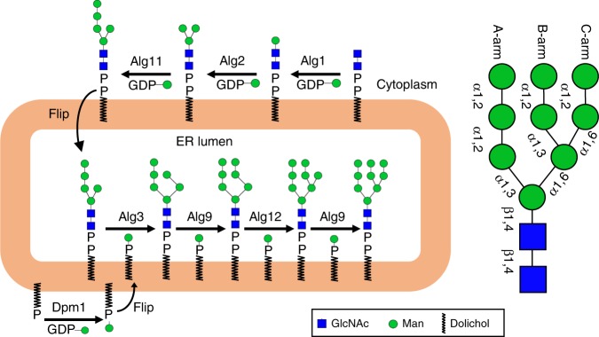
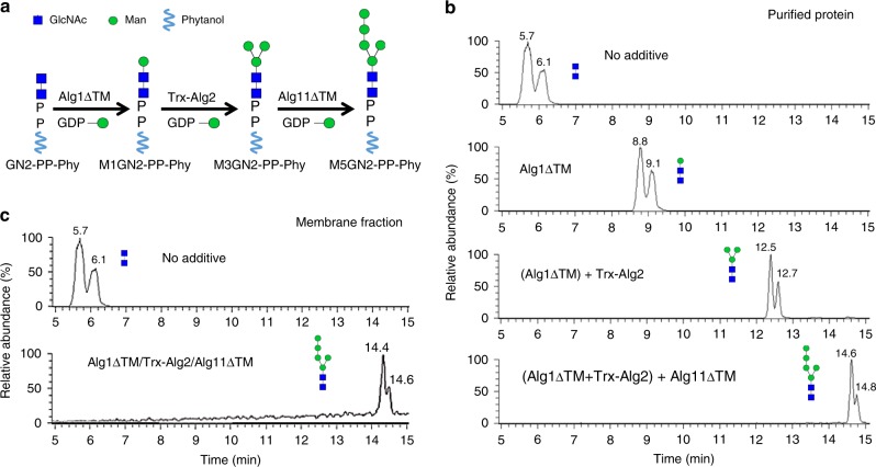
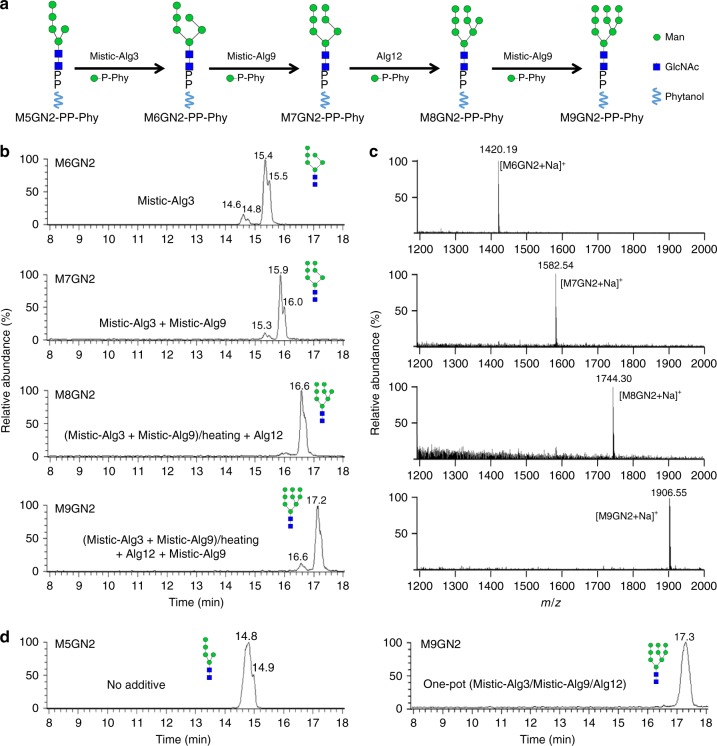
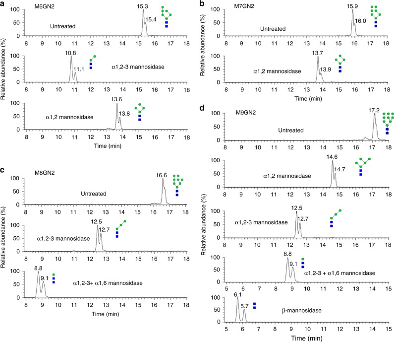
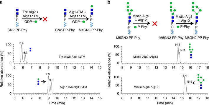
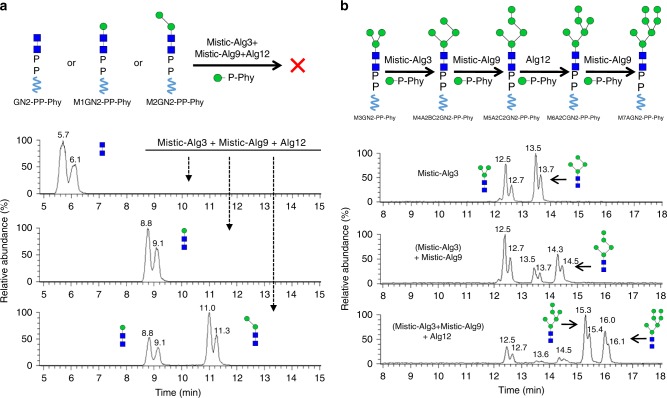
The asparagine (N)-linked Man9GlcNAc2 is required for glycoprotein folding and secretion. Understanding how its structure contributes to these functions has been stymied by our inability to produce this glycan as a homogenous structure of sufficient quantities for study. Here, we report the high yield chemoenzymatic synthesis of Man9GlcNAc2 and its biosynthetic intermediates by reconstituting the eukaryotic lipid-linked oligosaccharide (LLO) pathway. Endoplasmic reticulum mannosyltransferases (MTases) are expressed in E. coli and used for mannosylation of the dolichol mimic, phytanyl pyrophosphate GlcNAc2. These recombinant MTases recognize unique substrates and when combined, synthesize end products that precisely mimic those in vivo, demonstrating that ordered assembly of LLO is due to the strict enzyme substrate specificity. Indeed, non-physiological glycans are produced only when the luminal MTases are challenged with cytosolic substrates. Reconstitution of the LLO pathway to synthesize Man9GlcNAc2 in vitro provides an important tool for functional studies of the N-linked glycoprotein biosynthesis pathway.
Conflict of interest statement
The authors declare no competing interests.
Figures
Similar articles
-
Alg mannosyltransferases: From functional and structural analyses to the lipid-linked oligosaccharide pathway reconstitution.Biochim Biophys Acta Gen Subj. 2022 May;1866(5):130112. doi: 10.1016/j.bbagen.2022.130112. Epub 2022 Feb 22. Biochim Biophys Acta Gen Subj. 2022. PMID: 35217128 Review.
-
In vitro evidence for the dual function of Alg2 and Alg11: essential mannosyltransferases in N-linked glycoprotein biosynthesis.Biochemistry. 2006 Aug 8;45(31):9593-603. doi: 10.1021/bi060878o. Biochemistry. 2006. PMID: 16878994
-
Dolichol is not a necessary moiety for lipid-linked oligosaccharide substrates of the mannosyltransferases involved in in vitro N-linked-oligosaccharide assembly.Biochem J. 1995 Sep 15;310 ( Pt 3)(Pt 3):909-16. doi: 10.1042/bj3100909. Biochem J. 1995. PMID: 7575426 Free PMC article.
-
The accumulation of Man(6)GlcNAc(2)-PP-dolichol in the Saccharomyces cerevisiae Deltaalg9 mutant reveals a regulatory role for the Alg3p alpha1,3-Man middle-arm addition in downstream oligosaccharide-lipid and glycoprotein glycan processing.J Biol Chem. 2000 Feb 11;275(6):4267-77. doi: 10.1074/jbc.275.6.4267. J Biol Chem. 2000. PMID: 10660594
-
N-linked protein glycosylation in the ER.Biochim Biophys Acta. 2013 Nov;1833(11):2430-7. doi: 10.1016/j.bbamcr.2013.04.001. Epub 2013 Apr 10. Biochim Biophys Acta. 2013. PMID: 23583305 Review.
Cited by
-
Recent Progress in Chemo-Enzymatic Methods for the Synthesis of N-Glycans.Front Chem. 2020 Jun 16;8:513. doi: 10.3389/fchem.2020.00513. eCollection 2020. Front Chem. 2020. PMID: 32612979 Free PMC article. Review.
-
Integrating Biomaterials and Immunology to Improve Vaccines Against Infectious Diseases.ACS Biomater Sci Eng. 2020 Feb 10;6(2):759-778. doi: 10.1021/acsbiomaterials.9b01255. Epub 2020 Jan 12. ACS Biomater Sci Eng. 2020. PMID: 33313391 Free PMC article. Review.
-
Rft1 catalyzes lipid-linked oligosaccharide translocation across the ER membrane.Nat Commun. 2024 Jun 17;15(1):5157. doi: 10.1038/s41467-024-48999-3. Nat Commun. 2024. PMID: 38886340 Free PMC article.
-
Caenorhabditis elegans algn-2 Is Critical for Longevity Conferred by Enhanced Nonsense-Mediated mRNA Decay.iScience. 2020 Oct 20;23(11):101713. doi: 10.1016/j.isci.2020.101713. eCollection 2020 Nov 20. iScience. 2020. PMID: 33225240 Free PMC article.
-
Improving recombinant protein secretion in Aspergillus nidulans by targeting the N-glycosylation machinery.Metab Eng Commun. 2025 Jun 4;20:e00264. doi: 10.1016/j.mec.2025.e00264. eCollection 2025 Jun. Metab Eng Commun. 2025. PMID: 40575800 Free PMC article.
References
Publication types
MeSH terms
Substances
LinkOut - more resources
Full Text Sources
