

Proteins that physically interact with the phosphatase Cdc14 in Candida albicans have diverse roles in the cell cycle
- PMID: 31000734
- PMCID: PMC6472416
- DOI: 10.1038/s41598-019-42530-1
Proteins that physically interact with the phosphatase Cdc14 in Candida albicans have diverse roles in the cell cycle
Abstract
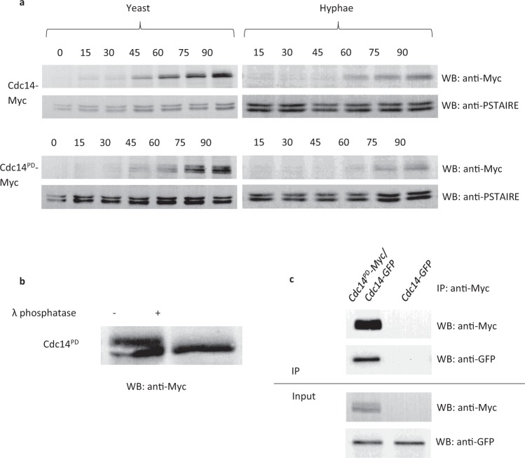
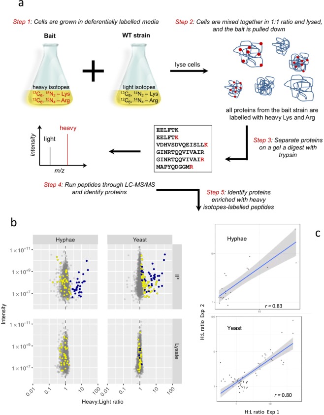
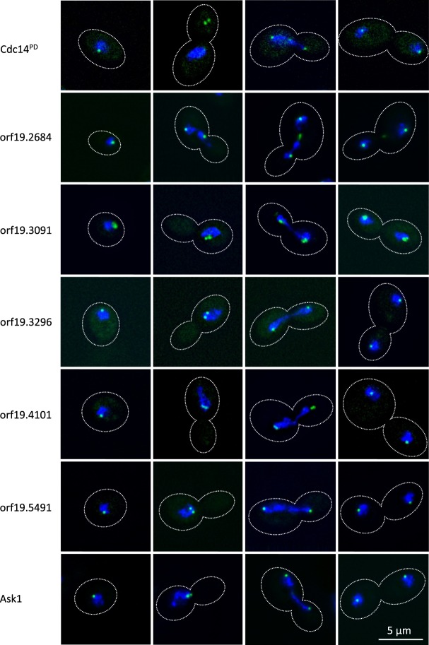
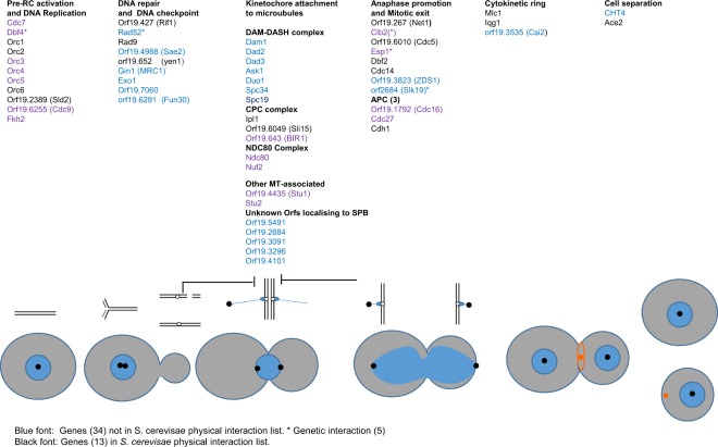
The chromosome complement of the human fungal pathogen Candida albicans is unusually unstable, suggesting that the process of nuclear division is error prone. The Cdc14 phosphatase plays a key role in organising the intricate choreography of mitosis and cell division. In order to understand the role of Cdc14 in C. albicans we used quantitative proteomics to identify proteins that physically interact with Cdc14. To distinguish genuine Cdc14-interactors from proteins that bound non-specifically to the affinity matrix, we used a substrate trapping mutant combined with mass spectrometry analysis using Stable Isotope Labelling with Amino Acids in Cell Culture (SILAC). The results identified 126 proteins that interact with Cdc14 of which 80% have not previously been identified as Cdc14 interactors in C. albicans or S. cerevisiae. In this set, 55 proteins are known from previous research in S. cerevisiae and S. pombe to play roles in the cell cycle, regulating the attachment of the mitotic spindle to kinetochores, mitotic exit, cytokinesis, licensing of DNA replication by re-activating pre-replication complexes, and DNA repair. Five Cdc14-interacting proteins with previously unknown functions localised to the Spindle Pole Bodies (SPBs). Thus, we have greatly increased the number of proteins that physically interact with Cdc14 in C. albicans.
Conflict of interest statement
The authors declare no competing interests.
Figures
Similar articles
-
Flp1, a fission yeast orthologue of the s. cerevisiae CDC14 gene, is not required for cyclin degradation or rum1p stabilisation at the end of mitosis.J Cell Sci. 2001 Jul;114(Pt 14):2649-64. doi: 10.1242/jcs.114.14.2649. J Cell Sci. 2001. PMID: 11683392
-
Dbf2-Mob1 drives relocalization of protein phosphatase Cdc14 to the cytoplasm during exit from mitosis.J Cell Biol. 2009 Feb 23;184(4):527-39. doi: 10.1083/jcb.200812022. Epub 2009 Feb 16. J Cell Biol. 2009. PMID: 19221193 Free PMC article.
-
Novel role for Cdc14 sequestration: Cdc14 dephosphorylates factors that promote DNA replication.Mol Cell Biol. 2007 Feb;27(3):842-53. doi: 10.1128/MCB.01069-06. Epub 2006 Nov 20. Mol Cell Biol. 2007. PMID: 17116692 Free PMC article.
-
The Multiple Roles of the Cdc14 Phosphatase in Cell Cycle Control.Int J Mol Sci. 2020 Jan 21;21(3):709. doi: 10.3390/ijms21030709. Int J Mol Sci. 2020. PMID: 31973188 Free PMC article. Review.
-
Closing mitosis: the functions of the Cdc14 phosphatase and its regulation.Annu Rev Genet. 2004;38:203-32. doi: 10.1146/annurev.genet.38.072902.093051. Annu Rev Genet. 2004. PMID: 15568976 Review.
Cited by
-
Characterization of a novel separase-interacting protein and candidate new securin, Eip1p, in the fungal pathogen Candida albicans.Mol Biol Cell. 2019 Sep 1;30(19):2469-2489. doi: 10.1091/mbc.E18-11-0696. Epub 2019 Aug 14. Mol Biol Cell. 2019. PMID: 31411946 Free PMC article.
-
A phylogenetically-restricted essential cell cycle progression factor in the human pathogen Candida albicans.Nat Commun. 2022 Jul 23;13(1):4256. doi: 10.1038/s41467-022-31980-3. Nat Commun. 2022. PMID: 35869076 Free PMC article.
-
Comprehensive Interactome Analysis for the Sole Adenylyl Cyclase Cyr1 of Candida albicans.Microbiol Spectr. 2022 Dec 21;10(6):e0393422. doi: 10.1128/spectrum.03934-22. Epub 2022 Oct 31. Microbiol Spectr. 2022. PMID: 36314909 Free PMC article.
-
Mass Spectrometry-Based Proteomics of Fungal Pathogenesis, Host-Fungal Interactions, and Antifungal Development.J Fungi (Basel). 2019 Jun 17;5(2):52. doi: 10.3390/jof5020052. J Fungi (Basel). 2019. PMID: 31212923 Free PMC article. Review.
-
Developmental Roles of the Hog1 Protein Phosphatases of the Maize Pathogen Cochliobolus heterostrophus.J Fungi (Basel). 2021 Jan 26;7(2):83. doi: 10.3390/jof7020083. J Fungi (Basel). 2021. PMID: 33530602 Free PMC article.
References
-
- Kullberg, B. J. & Filler, S. G. Candidemia in Candida and Candidiasis (ed. Calderone, R. A.) 327–340 (ASM Press, Washington DC 2002).
-
- Pfaller MA, Moet GJ, Messer SA, Jones RN, Castanheira M. Candida Bloodstream Infections: Comparison of Species Distributions and Antifungal Resistance Patterns in Community-Onset and Nosocomial Isolates in the SENTRY Antimicrobial Surveillance Program, 2008–2009. Antimicrob. Agents Chemother. 2011;55:561–566. doi: 10.1128/AAC.01079-10. - DOI - PMC - PubMed
-
- Runke, M. Skin and mucous infections in Candida and Candidiasis (ed. Calderone, R.) 307–325 (ASM Press, Washington 2002).
Publication types
MeSH terms
Substances
Grants and funding
- BB/J014443/1/RCUK | Biotechnology and Biological Sciences Research Council (BBSRC)/International
- BB/J002305/1/RCUK | Biotechnology and Biological Sciences Research Council (BBSRC)/International
- BB/M012166/1/RCUK | Biotechnology and Biological Sciences Research Council (BBSRC)/International
- BB/M012166/1/RCUK | Biotechnology and Biological Sciences Research Council (BBSRC)/International
LinkOut - more resources
Full Text Sources
Other Literature Sources
Molecular Biology Databases
