. 2019 May;33(5):714-723.
doi: 10.1038/s41433-019-0397-z. Epub 2019 Apr 19.

Long-term natural history of idiopathic epiretinal membranes with good visual acuity

Affiliations

Long-term natural history of idiopathic epiretinal membranes with good visual acuity

Kieu-Yen Luu et al. Eye (Lond). 2019 May.

Abstract

Background/objectives: To evaluate the long-term progression of idiopathic epiretinal membranes (iERMs) with good baseline visual acuity, and to identify predictors of visual decline.

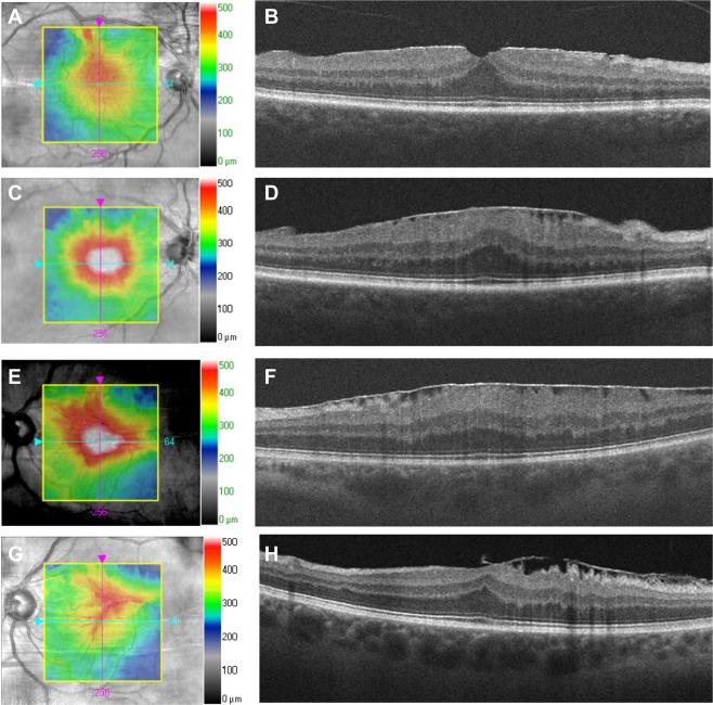
Design: Retrospective case series SUBJECTS METHODS: We reviewed records of 145 eyes with iERM and best-corrected visual acuity (BCVA) of 20/40 or greater at presentation, including BCVA, lens status, and central macular thickness (CMT) at yearly visits; as well as anatomic biomarkers including vitreomacular adhesion, pseudohole, lamellar hole, intraretinal cysts, disorganization of the inner retinal layers (DRIL), and disruption of outer retinal layers. Linear mixed effects and mixed-effects Cox proportional hazards models were used to identify clinical and anatomic predictors of vision change and time to surgery.

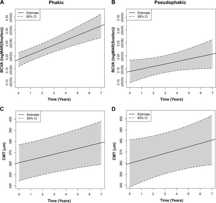
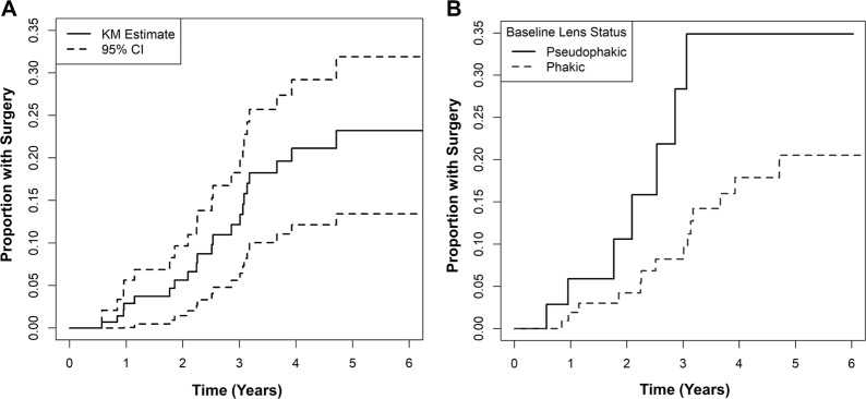
Results: At presentation, mean BCVA was 0.17 ± 0.10 logMAR units (Snellen 20/30) and mean CMT was 353.3 ± 75.4 μm. After a median follow-up of 3.7 years (range 1-7 years), BCVA declined slowly at 0.012 ± 0.003 logMAR units/year, with phakic eyes declining more rapidly than pseudophakic eyes (0.019 ± 0.003 vs. 0.010 ± 0.004 logMAR units/year). Metamorphopsia, phakic lens status, lamellar hole, and inner nuclear layer cysts were associated with faster visual decline. Cumulative rates of progression to surgery were 2.9, 5.6, 12.2, and 21.1% at years 1-4. Visual symptoms, metamorphopsia, greater CMT, and disruption of outer retinal layers were associated with greater hazard for surgery.

Conclusion: Eyes with iERM and visual acuity ≥ 20/40 experience slow visual decline, with 21% of eyes requiring surgery after 4 years. Clinical and anatomic predictors of vision loss may be distinct from factors associated with earlier surgical intervention.


Conflict of interest statement

Lawrence S. Morse: consultancy fees from Genentech. Glenn Yiu: grants from Alcon, Genentech, Iridex; and consultancy fees from Alimera, Allergan, Carl Zeiss Meditec, Iridex, and Southern California Desert Retina. The remaining authors declare that they have no conflict of interest.

Figures

Fig. 1
Fig. 1
Different patterns of long-term idiopathic ERM progression. Infrared images with topographic overlay (a, c, e, g) and OCT images (b, d, f, h) show two patients with different patterns of ERM progression. Patient RT (a-d) had a mild ERM and good visual acuity 20/20 at presentation (a-b), but progressed after 3 years with diffuse retinal thickening, inner retinal striae, and visual decline to 20/50 (c-d). Patient AM (e-h) had an idiopathic ERM with visual acuity of 20/30 at presentation (e-f), which spontaneously contracted with some restoration of foveal contour and visual improvement to 20/20 after 5 years (g-h)
Fig. 2
Fig. 2
Visual and anatomic progression of idiopathic ERM by lens status. Plots show estimates from linear mixed effects model for BCVA (a-b) and CMT (c-d) by time-varying lens status for phakic (a, c) and pseudophakic (b, d) eyes. The model fit for each plot is shown as a solid line, and the 95% CI is shown as dashed lines. Abbreviations: BCVA best-corrected visual acuity, CI confidence intervals, CMT central macular thickness, ERM epiretinal membranes
Fig. 3
Fig. 3
Progression to surgery for idiopathic ERM. Kaplan-Meier plot of time to surgery is shown for all eyes (a), and eyes categorized by baseline lens status (b). The estimate for all eyes is shown as a solid line, and the 95% CI is shown as dashed lines in (a). The estimate for pseudophakic eyes is shown as a solid line, and the estimate for phakic eyes is shown as a dashed line in (b). Abbreviations: CI confidence interval, ERM epiretinal membrane, KM Kaplan–Meier

References

    1. Hejsek L, Stepanov A, Dohnalova A, Rehakova T, Jiraskova N. The natural evolution of idiophatic epimacular membrane. Biomed Pap Med Fac Univ Palacky Olomouc Czech Repub. 2017;161:100–6. doi: 10.5507/bp.2016.065. - DOI - PubMed
    1. Bu SC, Kuijer R, Li XR, Hooymans JM, Los LI. Idiopathic epiretinal membrane. Retina. 2014;34:2317–35. doi: 10.1097/IAE.0000000000000349. - DOI - PubMed
    1. Konstantinidis L, Berguiga M, Beknazar E, Wolfensberger TJ. Anatomic and functional outcome after 23-gauge vitrectomy, peeling, and intravitreal triamcinolone for idiopathic macular epiretinal membrane. Retina. 2009;29:1119–27. doi: 10.1097/IAE.0b013e3181ac23da. - DOI - PubMed
    1. Moisseiev E, Kinori M, Moroz I, Priel E, Moisseiev J. 25-Gauge vitrectomy with epiretinal membrane and internal limiting membrane peeling in eyes with very good visual acuity. Curr Eye Res. 2016;41:1387–92. doi: 10.3109/02713683.2015.1114654. - DOI - PubMed
    1. Fraser-Bell S, Guzowski M, Rochtchina E, Wang JJ, Mitchell P. Five-year cumulative incidence and progression of epiretinal membranes: the Blue Mountains Eye Study. Ophthalmology. 2003;110:34–40. doi: 10.1016/S0161-6420(02)01443-4. - DOI - PubMed

Publication types