

Adhesive capsulitis: MRI correlation with clinical stages and proposal of MRI staging
- PMID: 31000937
- PMCID: PMC6467040
- DOI: 10.4103/ijri.IJRI_116_18
Adhesive capsulitis: MRI correlation with clinical stages and proposal of MRI staging
Abstract
Objective: The purpose of this study was to correlate the magnetic resonance imaging (MRI) findings of adhesive capsulitis with clinical stages and thereby propose a MR staging system.
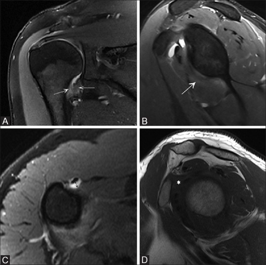
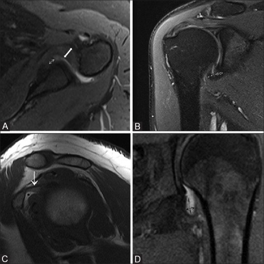
Materials and methods: This study consisted of 74 patients with clinically diagnosed adhesive capsulitis. The edema of the inferior glenohumeral ligament (IGHL), pericapsular edema, thickness of anterior band of IGHL, axillary pouch, thickness of coracohumeral ligament, and obliteration of fat in the subcoracoid triangle were evaluated by MRI.
Results: Thickening of the anterior band of IGHL showed most significant correlation with the clinical stages. The distribution of edema of IGHL and pericapsular edema also showed significant correlation with the clinical stages of adhesive capsulitis. Pericapsular edema and IGHL edema was not observed in stage IV.
Conclusion: MR is a useful tool for evaluation and prediction of clinical stage of adhesive capsulitis.
Keywords: Adhesive capsulitis; MRI staging; magnetic resonance imaging; shoulder.
Conflict of interest statement
There are no conflicts of interest.
Figures
Similar articles
-
Posterior capsule edema in adhesive capsulitis: comparison with established non-contrast MRI findings and multivariable analysis.Eur Radiol. 2024 Jan;34(1):260-269. doi: 10.1007/s00330-023-09966-6. Epub 2023 Aug 5. Eur Radiol. 2024. PMID: 37542655
-
Evaluation of Adhesive Capsulitis of the Shoulder With Fat-Suppressed T2-Weighted MRI: Association Between Clinical Features and MRI Findings.AJR Am J Roentgenol. 2016 Jul;207(1):135-41. doi: 10.2214/AJR.15.15200. Epub 2016 Apr 12. AJR Am J Roentgenol. 2016. PMID: 27070051
-
Anterior capsular abnormality: another important MRI finding for the diagnosis of adhesive capsulitis of the shoulder.Skeletal Radiol. 2019 Apr;48(4):543-552. doi: 10.1007/s00256-018-3064-8. Epub 2018 Sep 11. Skeletal Radiol. 2019. PMID: 30206678
-
Systematic review and meta-analysis of magnetic resonance imaging features for diagnosis of adhesive capsulitis of the shoulder.Eur Radiol. 2019 Feb;29(2):566-577. doi: 10.1007/s00330-018-5604-y. Epub 2018 Jul 5. Eur Radiol. 2019. PMID: 29978436
-
Adhesive capsulitis: review of imaging findings, pathophysiology, clinical presentation, and treatment options.Skeletal Radiol. 2019 Aug;48(8):1171-1184. doi: 10.1007/s00256-018-3139-6. Epub 2019 Jan 3. Skeletal Radiol. 2019. PMID: 30607455 Review.
Cited by
-
Optimal Cut-Off Value of the Coracohumeral Ligament Area as a Morphological Parameter to Confirm Frozen Shoulder.J Korean Med Sci. 2020 Apr 20;35(15):e99. doi: 10.3346/jkms.2020.35.e99. J Korean Med Sci. 2020. PMID: 32301291 Free PMC article.
-
Efficacy and safety of Tuina and intermediate frequency electrotherapy for frozen shoulder: MRI-based observation evidence.Am J Transl Res. 2023 Mar 15;15(3):1766-1778. eCollection 2023. Am J Transl Res. 2023. PMID: 37056812 Free PMC article.
-
Posterior capsule edema in adhesive capsulitis: comparison with established non-contrast MRI findings and multivariable analysis.Eur Radiol. 2024 Jan;34(1):260-269. doi: 10.1007/s00330-023-09966-6. Epub 2023 Aug 5. Eur Radiol. 2024. PMID: 37542655
-
The Use of a Suture Bridge Technique for Arthroscopic Rotator Cuff Repair in Patients Under 40 Years of Age Resulted in Successful Tendon Healing, Pain Relief, Improved Shoulder Function, and High Patient Satisfaction at a Minimum of 5-Year Follow-Up.Arthrosc Sports Med Rehabil. 2024 Sep 26;7(1):101009. doi: 10.1016/j.asmr.2024.101009. eCollection 2025 Feb. Arthrosc Sports Med Rehabil. 2024. PMID: 40041826 Free PMC article.
-
Can magnetic resonance imaging distinguish clinical stages of frozen shoulder? A state-of-the-art review.JSES Rev Rep Tech. 2024 May 15;4(3):365-370. doi: 10.1016/j.xrrt.2024.05.002. eCollection 2024 Aug. JSES Rev Rep Tech. 2024. PMID: 39157226 Free PMC article. Review.
References
-
- Codman EA. Boston, MA: Thomas Todd and Company; 1934. Tendinitis of the Short Rotators in the Shoulder: Rupture of the Supraspinatus Tendon and other Lesions in or about the Subacromial Bursa.
-
- Neviaser J. Adhesive capsulitis of the shoulder: Study of pathologic findings in periarthritis of the shoulder. J Bone Joint Surg. 1945;27:211–22.
-
- Ozaki J, Nakagawa Y, Sakurai G, Tamai S. Recalcitrant chronic adhesive capsulitis of the shoulder. Role of contracture of the coracohumeral ligament and rotator interval in pathogenesis and treatment. J Bone Joint Surg Am. 1989;71:1511–5. - PubMed
-
- Hannafin JA, Chiaia TA. Adhesive capsulitis: A treatment approach. Clin Orthop. 2000;372:95–109. - PubMed