Diffusion tensor imaging as an additional postoperative prognostic predictor factor in cervical myelopathy patients: An observational study
- PMID: 31000973
- PMCID: PMC6469325
- DOI: 10.4103/jcvjs.JCVJS_77_18
Diffusion tensor imaging as an additional postoperative prognostic predictor factor in cervical myelopathy patients: An observational study
Abstract
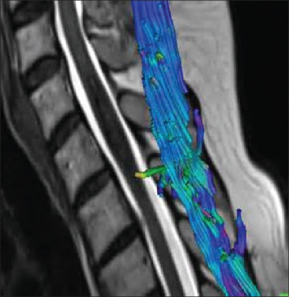
Introduction: Multiple investigation modalities have been invented for diagnosis and for planning management of degenerative cervical myelopathy, which include magnetic resonance imaging (MRI), computed tomography scan, and plain X-rays. Diffusion tensor imaging (DTI) of the spinal cord is a special variety of MRI where diffusion of water molecules across and along the tracts is mapped. The changes in anisotropy at the stenotic level can be a postoperative prognostic factor. The aim of this study was to establish postoperative prognostic predictive value of DTI in cases of degenerative cervical myelopathy.
Materials and methods: The study included 30 indoor patients in a tertiary care hospital diagnosed with degenerative compressive cervical myelopathy based on both clinical and radiological parameters with complete clinical data including follow-up. All patients with medical neurological diseases, cases who underwent repeat surgery, cases who developed surgical site infection, and those patients who were lost to follow-up were excluded from the study. The patients underwent operative decompression through either anterior or posterior approach with or without fixation with titanium implants as per indication. All patients underwent pre- and postoperative DTI. The fractional anisotropy (FA) and apparent diffusion coefficient (ADC) were noted in both pre- and postoperative imaging. Epidemiological data such as age and sex were noted. Pre- and postoperative modified Japanese Orthopedic Association (mJOA) scores were calculated.
Results: There was a significant improvement in FA values postoperatively. Preoperatively, both FA and ADC values showed a significant correlation with preoperative Neurological status of the patient while postoperatively only FA values were found to be significantly correlated. The regression equations for determining postoperative mJOA score based on preoperative FA and ADC values revealed mJOA = 9.77 + 12.1 (FA), mJOA = 14.2 + 2408.4 (ADC), and mJOA = 9.54 + 11.2 (FA) +1575.5 (ADC). This means that postoperative mJOA score, i.e., postoperative clinical status improvement can be determined using DTI variables which are an objective preoperative data. However, relative strength of prediction for FA value is 66.7% and for ADC value is 28.7%.
Conclusion: DTI tractography of the spinal cord will be a helpful objective prognostic factor for patients in whom surgery is planned. However, a study with larger subject size is required to increase the accuracy of determination of regression coefficient.
Keywords: Diffusion tensor imaging; myelopathy; prognostic.
Conflict of interest statement
There are no conflicts of interest.
Figures


References
-
- Payne EE, Spillane JD. The cervical spine; an anatomico-pathological study of 70 specimens (using a special technique) with particular reference to the problem of cervical spondylosis. Brain. 1957;80:571–96. - PubMed
-
- Bernhardt M, Hynes RA, Blume HW, White AA., 3rd Cervical spondylotic myelopathy. J Bone Joint Surg Am. 1993;75:119–28. - PubMed
-
- Nurick S. The pathogenesis of the spinal cord disorder associated with cervical spondylosis. Brain. 1972;95:87–100. - PubMed
-
- Rothman HA, Simeone FA. The Spine. 6th ed. Philadelphia: Saunders; 1992.