Cranially migrated lumbar intervertebral disc herniations: A multicenter analysis with long-term outcome
- PMID: 31000983
- PMCID: PMC6469315
- DOI: 10.4103/jcvjs.JCVJS_15_19
Cranially migrated lumbar intervertebral disc herniations: A multicenter analysis with long-term outcome
Abstract
Objective: Risk factors of cranial migration were investigated in patients with lumbar disc herniation (LDH) that migrated in the cranial direction and the long-term outcomes are discussed in this study.
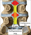
Materials and methods: Patients who underwent surgery for LDH at four different centers between 2012 and 2017 were studied. Extraligamentous discs were located in the lateral part of the posterior longitudinal ligament (PLL) within the spinal canal of the axial plane, and subligamentous discs were located under the PLL. The extent of cranial migration was calculated as a percentage of the height of the migrated corpus. Based on the extent of cranial migration, partial hemilaminectomy or hemilaminectomy was performed at different rates in each patient and the amount of laminectomy performed was recorded. During surgery, all free fragments were attempted to be removed. The appropriate technique was decided intraoperatively, and the surgery was performed on an individual patient basis.
Results: Of 1289 patients who underwent surgery for LDH, 654 (50.73%) had caudal migration, 576 (44.68%) had migration at the level of the disc, and 59 (4.57%) had cranial migration. Analysis of 59 patients with cranial migration according to the localization of the disc fragment revealed that 31 had extraligamentous and 28 had subligamentous fragments (P = 0.024).
Conclusions: Extraligamentous intervertebral disc fragments migrate more cranially than subligamentous intervertebral fragments. The anatomy of the PLL that varies along the corpus is the main reason for the weakness of the resistance of the disc material to the dorsolateral region, direction of discrete force vectors, and orientation of the disc fragment due to torsional vertebral movements.
Keywords: Caudal; cranial; direction; intervertebral; lumbar disc.
Conflict of interest statement
There are no conflicts of interest.
Figures


Similar articles
-
The Use of Tubular Retractors for Translaminar Discectomy for Cranially and Caudally Extruded Discs.Indian J Orthop. 2018 May-Jun;52(3):328-333. doi: 10.4103/ortho.IJOrtho_364_16. Indian J Orthop. 2018. PMID: 29887637 Free PMC article.
-
Are there typical localisations of lumbar disc herniations? A prospective study.Acta Neurochir (Wien). 1992;117(3-4):143-8. doi: 10.1007/BF01400611. Acta Neurochir (Wien). 1992. PMID: 1414514
-
The usefulness of magnetic resonance imaging for sequestered lumbar disc herniation treated with endoscopic surgery.J Xray Sci Technol. 2012;20(3):373-81. doi: 10.3233/XST-2012-0336. J Xray Sci Technol. 2012. PMID: 22948358
-
Clinicopathologic Features of Thoracolumbar Interdural Disc Herniations: A Retrospective Case Series with a Systematic Literature Review.World Neurosurg. 2020 Jul;139:e391-e398. doi: 10.1016/j.wneu.2020.04.015. Epub 2020 Apr 16. World Neurosurg. 2020. PMID: 32305597
-
Spontaneous regression of sequestrated lumbar disc herniations: Literature review.Clin Neurol Neurosurg. 2014 May;120:136-41. doi: 10.1016/j.clineuro.2014.02.013. Epub 2014 Feb 25. Clin Neurol Neurosurg. 2014. PMID: 24630494 Review.
Cited by
-
Posterior longitudinal ligament suturation after lumbar discectomy provides postoperative a large intradural area: First report.J Craniovertebr Junction Spine. 2023 Apr-Jun;14(2):181-186. doi: 10.4103/jcvjs.jcvjs_10_23. Epub 2023 Jun 13. J Craniovertebr Junction Spine. 2023. PMID: 37448510 Free PMC article.
References
-
- Davis CH., Jr . Extradural spinal cord and nerve root compression from benign lesions of the lumbar area. In: Youmans JR, editor. Neurological Surgery. Vol. II. Philadelphia: W. B. Saunders; 1973. pp. 1165–85.
-
- Vucetic N, Astrand P, Güntner P, Svensson O. Diagnosis and prognosis in lumbar disc herniation. Clin Orthop Relat Res. 1999;361:116–22. - PubMed
-
- Fardon DF, Williams AL, Dohring EJ, Murtagh FR, Gabriel Rothman SL, Sze GK, et al. Lumbar disc nomenclature: Version 2.0: Recommendations of the combined task forces of the North American Spine Society, the American Society of Spine Radiology and the American Society of Neuroradiology. Spine J. 2014;14:2525–45. - PubMed
-
- Modic MT. Magnetic Resonance Ýmaging of the Spine. New York: Year Book Medical; 1989. Degenerative disorders of the spine; pp. 83–95.
-
- Loupasis GA, Stamos K, Katonis PG, Sapkas G, Korres DS, Hartofilakidis G, et al. Seven- to 20-year outcome of lumbar discectomy. Spine (Phila Pa 1976) 1999;24:2313–7. - PubMed

