

Dorsal Spinal Intradural Intramedullary Epidermoid Cyst: A Rare Case Report and Review of Literature
- PMID: 31001035
- PMCID: PMC6454963
- DOI: 10.4103/jnrp.jnrp_304_18
Dorsal Spinal Intradural Intramedullary Epidermoid Cyst: A Rare Case Report and Review of Literature
Abstract
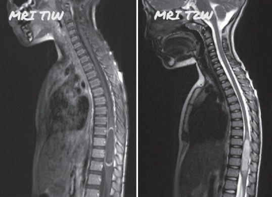
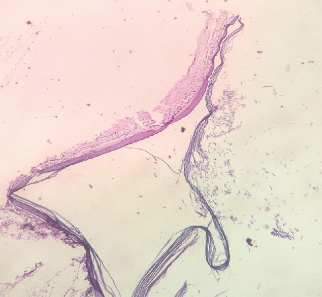
Epidermoid cysts are commonly seen intracranial lesions but their occurrence in the spine is rare. They account for <1% of all the benign tumors of the spine. These are benign epithelial-lined cysts filled with keratin. They are classified into two types: congenital or acquired. Congenital epidermoid cysts are more commonly associated with spinal dysraphic states such as syringomyelia, dermal sinus and spina bifida whereas the acquired cysts are associated with repeated lumbar punctures. Based on the location, they can be extradural, intradural, extramedullary, or intramedullary. Most of the epidermoids are intradural extramedullary. Intramedullary epidermoid cysts are very uncommon. We report a case of a 6-year-old female patient with dorsal epidermoid cyst with neurological deficits. Magnetic resonance imaging of the spine showed a well-defined lesion from D9 to D12 which was hypointense on T1W1 and heterogeneously hyperintense on T2W2. Surgery was performed to excise the lesion and to decompress the spinal cord. Histopathological examination of the excised lesion confirmed it as an epidermoid cyst.
Keywords: Epidermoid cyst; intradural; intramedullary lesion.
Conflict of interest statement
There are no conflicts of interest.
Figures
Similar articles
-
Intramedullary Epidermoid Cyst of the Conus Medullaris: A Case Report and Literature Review.Int Med Case Rep J. 2023 Sep 12;16:521-527. doi: 10.2147/IMCRJ.S430853. eCollection 2023. Int Med Case Rep J. 2023. PMID: 37720363 Free PMC article.
-
Intradural Extramedullary Epidermoid Cyst at the Conus Medullaris Level with Thoracic Syringomyelia: A Case Report.Acta Medica (Hradec Kralove). 2019;62(1):39-42. doi: 10.14712/18059694.2019.45. Acta Medica (Hradec Kralove). 2019. PMID: 30931896
-
A rare case of intradural and extramedullary epidermoid cyst after repetitive epidural anesthesia: case report and review of the literature.World J Surg Oncol. 2017 Jul 17;15(1):131. doi: 10.1186/s12957-017-1186-4. World J Surg Oncol. 2017. PMID: 28716031 Free PMC article. Review.
-
Giant intradural intramedullary epidermoid cyst Report of two cases with varied presentations.Asian J Neurosurg. 2014 Oct-Dec;9(4):244. doi: 10.4103/1793-5482.146653. Asian J Neurosurg. 2014. PMID: 25685236 Free PMC article.
-
Intramedullary epidermoid associated with an intramedullary spinal abscess secondary to a dermal sinus.Neurosurgery. 1992 Jan;30(1):118-21. doi: 10.1227/00006123-199201000-00022. Neurosurgery. 1992. PMID: 1738439 Review.
Cited by
-
Intramedullary Epidermoid Cyst of the Conus Medullaris: A Case Report and Literature Review.Int Med Case Rep J. 2023 Sep 12;16:521-527. doi: 10.2147/IMCRJ.S430853. eCollection 2023. Int Med Case Rep J. 2023. PMID: 37720363 Free PMC article.
-
Isolated thoracic intradural extramedullary epidermoid cyst: A technical note.Surg Neurol Int. 2024 May 24;15:170. doi: 10.25259/SNI_280_2024. eCollection 2024. Surg Neurol Int. 2024. PMID: 38840622 Free PMC article.
-
Spinal intramedullary epidermoid cysts: Three case presentations and literature review.Surg Neurol Int. 2020 Feb 7;11:17. doi: 10.25259/SNI_540_2019. eCollection 2020. Surg Neurol Int. 2020. PMID: 32123605 Free PMC article. Review.
-
Ossified spinal epidermoid cyst: A systematic review and case report.Heliyon. 2024 Aug 28;10(18):e37093. doi: 10.1016/j.heliyon.2024.e37093. eCollection 2024 Sep 30. Heliyon. 2024. PMID: 39315203 Free PMC article.
-
Filum terminale infected epidermoid cysts in pediatric age group; A case series.World Neurosurg X. 2024 Sep 21;24:100408. doi: 10.1016/j.wnsx.2024.100408. eCollection 2024 Oct. World Neurosurg X. 2024. PMID: 39391069 Free PMC article.
References
-
- Chandra PS, Manjari T, Devi BI, Chandramouli BA, Srikanth SG, Shankar SK, et al. Intramedullary spinal epidermoid cyst. Neurol India. 2000;48:75–7. - PubMed
-
- Roux A, Mercier C, Larbrisseau A, Dube LJ, Dupuis C, Del Carpio R, et al. Intramedullary epidermoid cysts of the spinal cord. Case report. J Neurosurg. 1992;76:528–33. - PubMed
-
- Amato VG, Assietti R, Arienta C. Intramedullary epidermoid cyst: Preoperative diagnosis and surgical management after MRI introduction. Case report and updating of the literature. J Neurosurg Sci. 2002;46:122–6. - PubMed
-
- Lunardi P, Missori P, Gagliardi FM, Fortuna A. Long-term results of the surgical treatment of spinal dermoid and epidermoid tumors. Neurosurgery. 1989;25:860–4. - PubMed
-
- Scarrow AM, Levy EI, Gerszten PC, Kulich SM, Chu CT, Welch WC, et al. Epidermoid cyst of the thoracic spine: Case history. Clin Neurol Neurosurg. 2001;103:220–2. - PubMed
Publication types
LinkOut - more resources
Full Text Sources