

Concurrent Spinal Epidural Tubercular and Pyogenic Abscess of Cervical Spine without Bony Involvement
- PMID: 31001041
- PMCID: PMC6454951
- DOI: 10.4103/jnrp.jnrp_318_18
Concurrent Spinal Epidural Tubercular and Pyogenic Abscess of Cervical Spine without Bony Involvement
Abstract
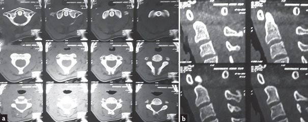
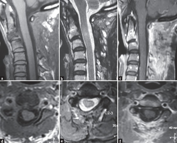
Spinal epidural abscess (SEA) presents with vertebral body involvement. SEA is mostly pyogenic in developed countries, but in developing countries, tuberculosis is more common cause. Young female presented with fever, neck, and right upper limb pain for 1 month followed by acute onset weakness in the right upper limb. Magnetic resonance imaging study of cervical spine showed contrast-enhancing lesion in C2-C3 epidural region. Surgery-aided by initiation of antibacterial and antitubercular treatment based on culture and histopathological study of pus helped us to get good clinical outcome. One should always keep in mind possibility of tubercular abscess while treating cases of spinal epidural lesions, though it is rare in the absence of osseous involvement. Pyogenic and tubercular abscess can present concurrently and sending pus for culture and sensitivity is must as it plays important role in identifying dual organisms.
Keywords: Abscess; cervical; concurrent; epidural; isolated; pyogenic; spinal; tuberculosis.
Conflict of interest statement
There are no conflicts of interest.
Figures


Similar articles
-
A pyogenic discitis at c3-c4 with associated ventral epidural abscess involving c1-c4 after intradiscal oxygen-ozone chemonucleolysis: a case report.Spine (Phila Pa 1976). 2009 Apr 15;34(8):E298-304. doi: 10.1097/BRS.0b013e318195a87e. Spine (Phila Pa 1976). 2009. PMID: 19365239
-
Tubercular spinal epidural abscess involving the dorsal-lumbar-sacral region without osseous involvement.J Infect Dev Ctries. 2011 Jul 27;5(7):544-9. doi: 10.3855/jidc.1494. J Infect Dev Ctries. 2011. PMID: 21795824
-
Hematogenous pyogenic spinal infections and their surgical management.Spine (Phila Pa 1976). 2000 Jul 1;25(13):1668-79. doi: 10.1097/00007632-200007010-00010. Spine (Phila Pa 1976). 2000. PMID: 10870142
-
Evaluation and Management of Pyogenic and Tubercular Spine Infections.Curr Rev Musculoskelet Med. 2018 Dec;11(4):643-652. doi: 10.1007/s12178-018-9523-y. Curr Rev Musculoskelet Med. 2018. PMID: 30280287 Free PMC article. Review.
-
Cervical Facet Joint Infection and Associated Epidural Abscess with Streptococcus intermedius from a Dental Infection Origin A Case Report and Review.Bull Hosp Jt Dis (2013). 2016 Sep;74(3):237-43. Bull Hosp Jt Dis (2013). 2016. PMID: 27620549 Review.
Cited by
-
Cervical Spinal Epidural Abscess: Diagnosis, Treatment, and Outcomes: A Case Series and a Literature Review.J Clin Med. 2023 Jul 5;12(13):4509. doi: 10.3390/jcm12134509. J Clin Med. 2023. PMID: 37445544 Free PMC article.
-
A Case of Five-Year Survival After Combined-Modality Treatment for Non-Small Cell Lung Cancer With Intraspinal Metastasis.Cureus. 2022 Jan 5;14(1):e20960. doi: 10.7759/cureus.20960. eCollection 2022 Jan. Cureus. 2022. PMID: 35154939 Free PMC article.
-
Spinal epidural abscess due to coinfection of bacteria and tuberculosis: A case report.World J Clin Cases. 2021 Jun 6;9(16):4072-4080. doi: 10.12998/wjcc.v9.i16.4072. World J Clin Cases. 2021. PMID: 34141768 Free PMC article.
-
Long-Segment Ventral Spinal Epidural Abscesses Caused by Mycobacterium Tuberculosis: A Report of 2 Cases and Review.JB JS Open Access. 2025 Jun 20;10(2):e24.00249. doi: 10.2106/JBJS.OA.24.00249. eCollection 2025 Apr-Jun. JB JS Open Access. 2025. PMID: 40547101 Free PMC article. Review.
References
-
- Chauhan A, Gupta BB. Spinal tuberculosis. Indian Acad Clin Med. 2007;8:110–4.
-
- Hsu LC, Leong JC. Tuberculosis of the lower cervical spine (C2 to C7). A report on 40 cases. J Bone Joint Surg Br. 1984;66:1–5. - PubMed
-
- Rajasekaran S, Shanmugasundaram TK, Prabhakar R, Dheenadhayalan J, Shetty AP, Shetty DK, et al. Tuberculous lesions of the lumbosacral region. A 15-year follow-up of patients treated by ambulant chemotherapy. Spine (Phila Pa 1976) 1998;23:1163–7. - PubMed
-
- Davis DP, Wold RM, Patel RJ, Tran AJ, Tokhi RN, Chan TC, et al. The clinical presentation and impact of diagnostic delays on emergency department patients with spinal epidural abscess. J Emerg Med. 2004;26:285–91. - PubMed
Publication types
LinkOut - more resources
Full Text Sources
Miscellaneous