. 2019 Apr 2:12:75.
doi: 10.3389/fnmol.2019.00075. eCollection 2019.

Transcription Factors Sp8 and Sp9 Regulate Medial Ganglionic Eminence-Derived Cortical Interneuron Migration

Affiliations

Transcription Factors Sp8 and Sp9 Regulate Medial Ganglionic Eminence-Derived Cortical Interneuron Migration

Guangxu Tao et al. Front Mol Neurosci. .

Abstract

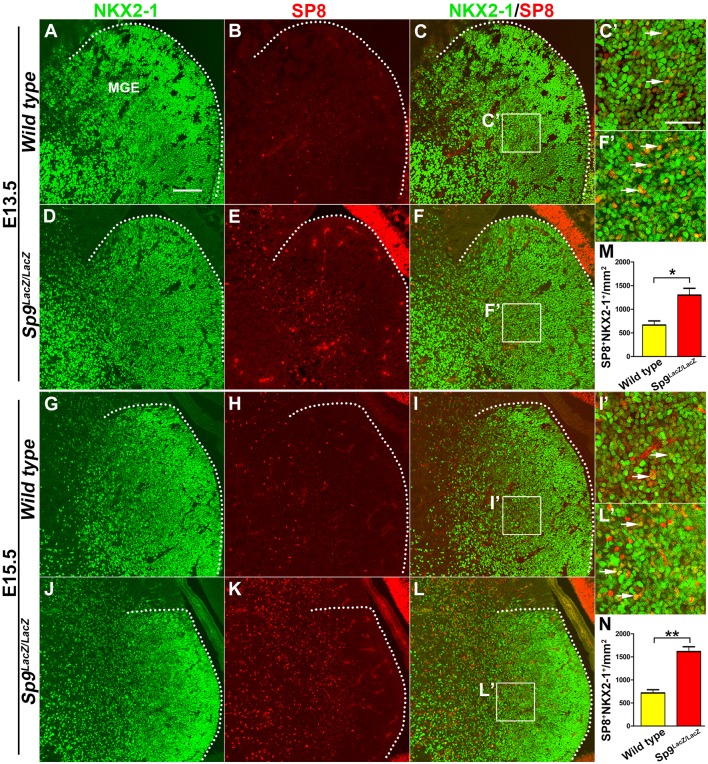
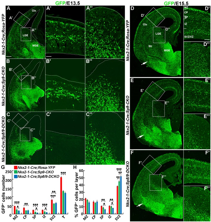
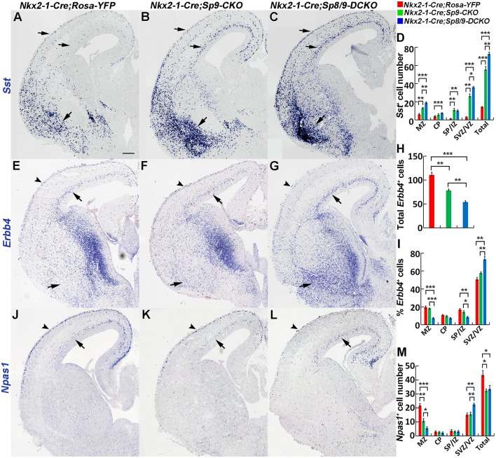
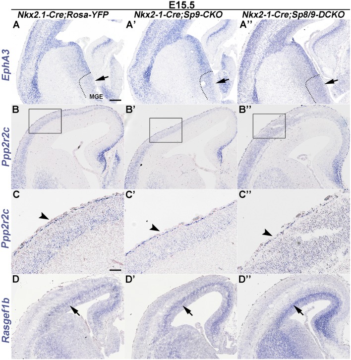
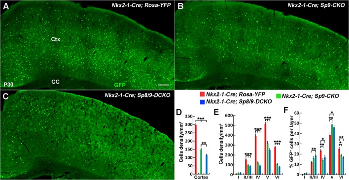
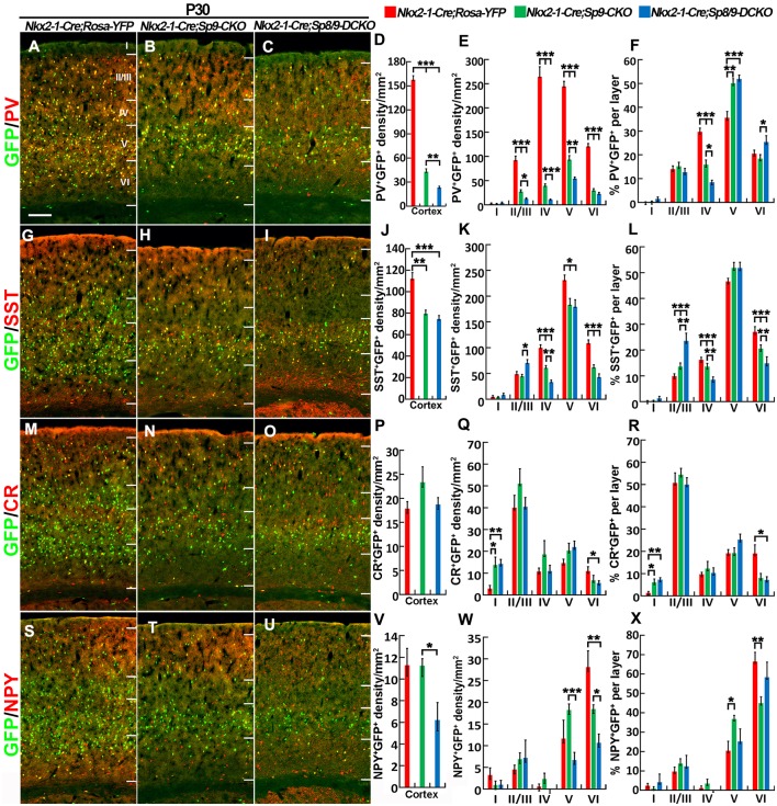
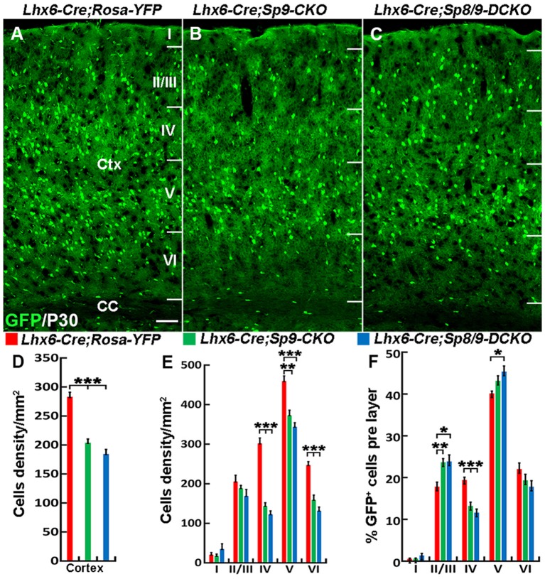
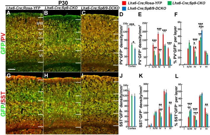
Cortical interneurons are derived from the subpallium and reach the developing cortex through long tangential migration. Mature cortical interneurons are characterized by remarkable morphological, molecular, and functional diversity. The calcium-binding protein parvalbumin (PV) and neuropeptide somatostatin (SST) identify most medial ganglionic eminence (MGE)-derived cortical interneurons. Previously, we demonstrated that Sp9 plays a curial transcriptional role in regulating MGE-derived cortical interneuron development. Here, we show that SP8 protein is weekly expressed in the MGE mantle zone of wild type mice but upregulated in Sp9 null mutants. PV+ cortical interneurons were severely lost in Sp8/Sp9 double conditional knockouts due to defects in tangential migration compared with Sp9 single mutants, suggesting that Sp8/9 coordinately regulate PV+ cortical interneuron development. We provide evidence that Sp8/Sp9 activity is required for normal MGE-derived cortical interneuron migration, at least in part, through regulating the expression of EphA3, Ppp2r2c, and Rasgef1b.

Keywords: Sp8; Sp9; cortical interneuron; medial ganglionic eminence; parvalbumin; somatostatin; tangential migration.


Figures

Figure 1
Figure 1
SP8 expression is upregulated in medial ganglionic eminence (MGE) of Sp9LacZ/LacZ null mutants. (A–L) NKX2-1/SP8 double-immunostained coronal sections at E13.5 and E15.5. (C′,F′,I′,L′) Higher magnification images of the boxed areas in (C,F,I,L). Upregulation of SP8 protein expression can be noted in the mutant MGE mantle zone; most of these SP8+ cells exhibit NKX2-1 coexpression. (M,N) Quantified data showed that the density of SP8+/NKX2-1+ cells was increased in Sp9LacZ/LacZ mutants compared with controls in E13.5 and E15.5. Scale bars: 100 μm in (A) for (A–L); 50 μm in (C’) for (C′,F′,I′,L′).
Figure 2
Figure 2
Tangential migration defects of MGE-derived cortical interneurons in Nkx2-1-Cre; Sp8/9-DCKO mice. (A–C) GFP immunostained E13.5 coronal hemisections. (A′–C″) Higher magnification images of the boxed areas in (A–C) show that MGE-derived GFP+ cortical interneurons migrate to the cortex much less efficiently in Nkx2-1-Cre; Sp8/9-DCKO (double mutant) mice than in controls and Nkx2-1-Cre; Sp9-CKO (single mutant) mice. Very few GFP+ cells can be noted in the lateral GE (LGE) subventricular zone (SVZ) of mutants (B″, C″). (D–F) GFP immunostained E15.5 coronal hemisections. (D′–F″) Higher magnification images of the boxed areas in (D–F). Note that more GFP+ cells ectopically accumulated in the ventral telencephalon in Nkx2-1-Cre; Sp8/9-DCKO mice than in controls and Nkx2-1-Cre; Sp9-CKO mice (arrows). (G,H) Quantification showing that mutant mice had fewer GFP+ cells in the cortex at E15.5. The percentage of GFP+ cells was reduced in the SP and the intermediate zone (IZ) and increased in the SVZ. Ctx, cortex. *P < 0.05; **P < 0.01; ***P < 0.001. Scale bars: 100 μm in (A) for (A–F); 25 μm in (A′) for (A′–C″); 50 μm in (D′) for (D′–F′′).
Figure 3
Figure 3
In situ RNA hybridization of Sst, Erbb4 and Npas1. (A–D) Sst mRNA expression was increased in the E15.5 neocortex (arrows) and ventral telencephalon (arrows) of mutants. More Sst+ cells were found in the ventral telencephalon of double mutants than single mutants and controls (arrows). (E–I) Abnormal migration of Erbb4+ cells (PV+ immature cortical interneurons) in mutant mice. Double mutants appeared to have relatively more Erbb4+ cells in the cortical SVZ (arrows) and in the ventral telencephalon (arrows) and relatively fewer Erbb4+ cells in the cortical marginal zone (MZ; arrowheads). (J–M) Decreased Npas1+ cells in the cortical MZ (arrowheads) and increased Npas1+ cells in the cortical SVZ (arrows) were observed in double mutants compared to single mutants and controls. *P < 0.05; **P < 0.01; ***P < 0.001. Scale bar: 200 μm in (A) for (A–C,E–G,J–L).
Figure 4
Figure 4
In situ RNA hybridization of Epha3, Ppp2r2c and Rasgef1b in coronal hemisections at E15.5. (A–A″) EphA3 was upregulated in the mutant MGE (arrows). (B–C″) Ppp2r2c expression was reduced in the mutant cortical MZ (arrowheads). (D–D″) Rasgef1b expression was increased in the mutant cortical SVZ (arrows). Note that dysregulation of these genes was more prominent in Sp8/9 double mutants than in Sp9 single mutants. Scale bars: 200 μm in (A) for (A–B″,D–D″); 100 μm in (C) for (C–C″).
Figure 5
Figure 5
The number of MGE-derived cortical interneurons was significantly reduced in double mutants compared with single mutants and controls at P30. (A–C) GFP+ cells in the cortex of Nkx2-1-Cre; Rosa-YFP (control), Nkx2-1-Cre; Sp9F/F; Rosa-YFP (single mutant) and Nkx2-1-Cre; Sp8F/F; Sp9F/F; Rosa-YFP (double mutant) mice. (D–F) Quantified data showed that the density of GFP+ cells in the somatosensory cortex was reduced in single mutants and further reduced in double mutants compared with controls. Ctx, cortex; CC, corpus callosum. *P < 0.05; **P < 0.01; ***P < 0.001. Scale bars: 200 μm in (A) for (A–C).
Figure 6
Figure 6
PV+ cortical interneurons are severely reduced in the Sp8/9 double mutant cortex at P30. (A–C,G–I,M–O,S–U) GFP and cortical interneuron markers [PV, SST, calretinin (CR) and NPY] in double-immunostained coronal sections of the somatosensory cortex. (D–F,J–L,P–R,V–X) Quantified data showed that the density of PV+/GFP+ cells was severely reduced in double mutants compared with single mutants and controls. The density of SST+/GFP+ and NPY+/GFP+ cells was also reduced in the mutant somatosensory cortex, whereas CR+/GFP+ cells were less affected. *P < 0.05; **P < 0.01; ***P < 0.001. Scale bars: 200 μm in (A) for (A–C,G–I,M–O,S–U).
Figure 7
Figure 7
The number of MGE-derived cortical interneurons is greatly reduced in the somatosensory cortex of Lhx6-Cre; Sp8F/F; Sp9F/F; Rosa-YFP mice compared with Lhx6-Cre; Rosa-YFP and Lhx6-Cre; Sp9F/F; Rosa-YFP mice at P30. (A–C) GFP+ cells in the somatosensory cortex. (D–F) Quantified data from the above experiments. The density of GFP+ cells was significantly reduced in mutants compared with controls. Ctx, cortex; CC, corpus callosum. *P < 0.05; **P < 0.01; ***P < 0.001. Scale bars: 200 μm in (A) for (A–C).
Figure 8
Figure 8
PV+ cortical interneurons are severely reduced in the Lhx6-Cre; Sp8F/F; Sp9F/F; Rosa-YFP somatosensory cortex. (A–C,G–I) GFP/PV and GFP/SST double-immunostained coronal sections. (D–F,J–L) The density of PV+/GFP+ cells was severely reduced in double mutants compared with single mutants and controls, whereas the density of SST+/GFP+ cells was less affected. *P < 0.05; **P < 0.01; ***P < 0.001. Scale bars: 200 μm in (A) for (A–C,G–I).

Similar articles

Cited by

References

    1. Antypa M., Faux C., Eichele G., Parnavelas J. G., Andrews W. D. (2011). Differential gene expression in migratory streams of cortical interneurons. Eur. J. Neurosci. 34, 1584–1594. 10.1111/j.1460-9568.2011.07896.x - DOI - PMC - PubMed
    1. Bell S. M., Schreiner C. M., Waclaw R. R., Campbell K., Potter S. S., Scott W. J. (2003). Sp8 is crucial for limb outgrowth and neuropore closure. Proc. Natl. Acad. Sci. U S A 100, 12195–12200. 10.1073/pnas.2134310100 - DOI - PMC - PubMed
    1. Cobos I., Long J. E., Thwin M. T., Rubenstein J. L. (2006). Cellular patterns of transcription factor expression in developing cortical interneurons. Cereb. Cortex 16, i82–i88. 10.1093/cercor/bhk003 - DOI - PubMed
    1. Colasante G., Sessa A., Crispi S., Calogero R., Mansouri A., Collombat P., et al. . (2009). Arx acts as a regional key selector gene in the ventral telencephalon mainly through its transcriptional repression activity. Dev. Biol. 334, 59–71. 10.1016/j.ydbio.2009.07.014 - DOI - PubMed
    1. Del Pino I., Rico B., Marin O. (2018). Neural circuit dysfunction in mouse models of neurodevelopmental disorders. Curr. Opin. Neurobiol. 48, 174–182. 10.1016/j.conb.2017.12.013 - DOI - PubMed

LinkOut - more resources