Review
. 2019 Mar 29:10:294.
doi: 10.3389/fphar.2019.00294. eCollection 2019.

Modulation of Allergic Inflammation in the Nasal Mucosa of Allergic Rhinitis Sufferers With Topical Pharmaceutical Agents

Affiliations
Review

Modulation of Allergic Inflammation in the Nasal Mucosa of Allergic Rhinitis Sufferers With Topical Pharmaceutical Agents

Annabelle M Watts et al. Front Pharmacol. .

Abstract

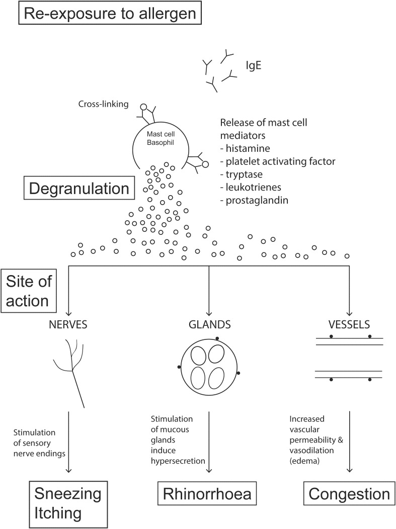
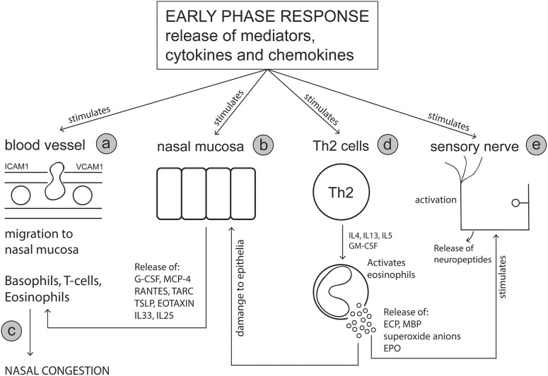
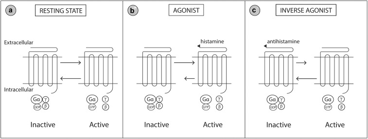
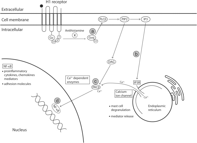
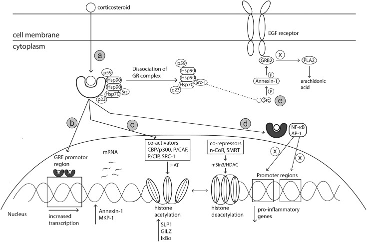
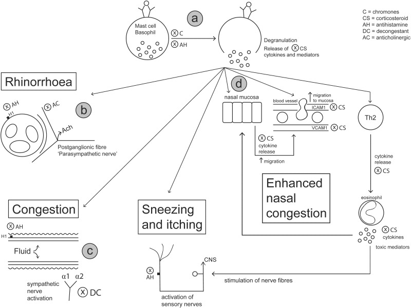
Allergic rhinitis (AR) is a chronic upper respiratory disease estimated to affect between 10 and 40% of the worldwide population. The mechanisms underlying AR are highly complex and involve multiple immune cells, mediators, and cytokines. As such, the development of a single drug to treat allergic inflammation and/or symptoms is confounded by the complexity of the disease pathophysiology. Complete avoidance of allergens that trigger AR symptoms is not possible and without a cure, the available therapeutic options are typically focused on achieving symptomatic relief. Topical therapies offer many advantages over oral therapies, such as delivering greater concentrations of drugs to the receptor sites at the source of the allergic inflammation and the reduced risk of systemic side effects. This review describes the complex pathophysiology of AR and identifies the mechanism(s) of action of topical treatments including antihistamines, steroids, anticholinergics, decongestants and chromones in relation to AR pathophysiology. Following the literature review a discussion on the future therapeutic strategies for AR treatment is provided.

Keywords: allergic rhinitis; anticholinergic; antihistamines; chromones; decongestants; intranasal; steroids.


Figures

FIGURE 1
FIGURE 1
The early phase response. Crosslinking of FCεR1-bound IgE antibodies on the mast cell surface in response to secondary allergen exposure stimulates the degranulation of mast cells. Degranulation induces the release of chemical mediators (primarily histamine) that stimulate sensory nerve endings, mucous glands and small vessels of the nasal mucosa to produce classic rhinitis symptoms: sneezing, nasal itching, rhinorrhoea and nasal congestion. The onset of action is typically within minutes of exposure and is sustained for 2–3 h forming the early-phase response.
FIGURE 2
FIGURE 2
The late phase response. Mediators and cytokines released during the early phase response act on various sites including nasal blood vessels, nasal epithelial cells, T cells and sensory nerves to initiate the symptoms of an allergic response. The late phase response is characterized by the involvement of key immune effector cells including basophils, T cells and eosinophils, which migrate to the nasal mucosa in response to early phase stimulus. The release of cytokines and mediators from these effector cells further perpetuates the allergic response and symptom manifestation. (a) Mast cell mediators act on adhesion molecules (e.g., ICAM-1 and VCAM-1) on blood vessel endothelial cells increasing vascular permeability thereby allowing effector cells such as eosinophils, T cells and basophils to migrate to the nasal mucosa. (b) Nasal mucosal cells are stimulated by mast cell products to secrete cell signaling molecules which further promote chemoattraction of effector cells to the nasal mucosa. (c) Nasal edema (congestion) is worsened by the influx of immune cells and their subsequent mediator release. (d) Cytokine release from T cells, activates and stimulates eosinophils to release toxic mediators. (e) Eosinophil derived mediators damage the nasal epithelium and leave nerve fibers exposed to histamine and other mediators promoting neurogenic inflammation. G-CSF, Granulocyte-colony stimulating factor; MCP-4, Monocyte chemotactic protein-4; RANTES, Regulated on activation normal T cell expressed and secreted; TARC, Thymus- and activation-regulated chemokine; TSLP, Thymic stromal lymphopoietin, GM-CSF, Granulocyte-macrophage colony-stimulating factor; ECP, Eosinophil cationic protein; MBP, Major basic protein.
FIGURE 3
FIGURE 3
Molecular model of the histamine 1 (H1) receptor. The H1 receptor is a G protein-coupled transmembrane receptor which acts as a ‘molecular switcher’ via interactions with their associated intracellular heterotrimeric G proteins (consisting of α, β, and γ subunits). G proteins regulate downstream intracellular signaling via their ability to catalyze the exchange of Gα bound GDP to GTP. The H1 receptor complex exists between two conformational states, active and inactive, which are directed by specific extracellular ligand binding to the G protein receptor. (a) When the active and inactive state are in equilibrium, the H1 receptor is in a resting state. (b) Histamine (an agonist) binds to and stabilizes the receptor in the active conformation which shifts the equilibrium toward the active state. (c) Antihistamines (an inverse agonist) binds to and stabilizes the receptor in the inactive conformation which shifts the equilibrium toward the inactive state. Gβ, Guanine nucleotide-binding protein beta; Gγ, Guanine nucleotide-binding protein gamma; Gα, Guanine nucleotide-binding protein alpha; GDP, Guanosine diphosphate; GTP, Guanosine triphosphate. Modified from Simons and Simons (2011).
FIGURE 4
FIGURE 4
Anti-inflammatory effects of antihistamines. Binding of antihistamines (an inverse agonist) to the transmembrane H1 receptor prevents the activation of intracellular signaling pathways that result in mast cell degranulation and NF-κB activation. Alternatively, when histamine (an agonist) binds to the H1 receptor, this signals the associated G protein subunit Gαq to activate the phospholipase C and phosphatidylinositol (PIP2) signaling pathways. (a) Gαq activates phospholipase C which cleaves phosphatidylinositol 4,5-bisphosphate (PIP2), a phospholipid constituent of the cell membrane, into diacyl glycerol (DAG) and inositol 1,4,5 triphosphate (IP3). (b) IP3 is then released into the cytoplasm where it binds to IP3 receptors situated in the endoplasmic reticulum (ER). The IP3 receptors are intracellular channels that facilitate calcium ion release. On binding with IP3, IP3 receptors are stimulated to release calcium ions from ER stores into the cytosol. Mast cell degranulation and subsequent mediator release is dependent on this flux in calcium ion availability in the cytosol. (c) Calcium ions and DAG (cleaved from PIP2) activate protein kinase C which is involved in activating the transcription factor NF-κB. (d) Activation of NF-κB results in increased transcription of proinflammatory genes. DAG, 1,2-diacyl-glycerol; PLCβ, phospholipase C β, PIP2, phosphatidylinositol 4,5-biphosphate; IP3, Inositol 1,4,5-triphosphate; IR, Inositol 1,4,5-triphosphate receptor type 1; PKCβ, protein kinase C beta; NADPH, nicotinamide adenine dinucleotide phosphate. Modified from Frolkis et al. (2010); Simons and Simons (2011), and Jewison et al. (2014).
FIGURE 5
FIGURE 5
Mechanism of action of corticosteroids. Corticosteroids act via various genomic and non-genomic pathways such as transactivation, transrepression, histone medication and Src kinase signaling, to reduce allergic inflammation. (a) Corticosteroids cross cell membranes and bind to a specific intracellular glucocorticoid receptor (GR). The complex of proteins bound to the receptor are released upon receptor-ligand binding, allowing the corticosteroid activated GR to translocate to the nucleus or interact with transcription factors in the cytoplasm. (b) Activated GR translocates to the nucleus and binds as a dimer to GRE located within the promotor region of specific anti-inflammatory genes. (c) Activated GR can modify chromatin structure to either enhance or prevent transcription of genes via interactions with coactivator and corepressor complexes which have inherent histone acetylation and histone deacetylation abilities, respectively. (d) Activated GR can bind directly with transcription factors including AP-1 and NF-κB to prevent binding to their respective promotor regions, thereby preventing the transcription of pro-inflammatory genes. (e) SRC, released upon dissociation of GC-GR complex, activates the annexin-1 protein. Annexin-1 then disrupts the signal transduction protein Grb2 which is linked with epidermal growth factor. Impairment of EGF reduces the production of leukotrienes and prostaglandins. Hsp90, heat shock protein 90; GRE, Glucocorticoid response elements; AP-1, activation protein 1; CBP/p300, CREB-binding protein; p/CAF, CBP/p300 associated factor; p/Cip, CBP/p300 co-integrator associated protein; SRC-1, steroid receptor coactivator 1; MKP-1, Mitogen-activated protein kinase 1; SLPI, secretory leukocyte protease inhibitor (SLPI); GILZ, Glucocorticoid-induced leucine zipper; IκBα, nuclear factor of kappa light polypeptide gene enhancer in B-cells inhibitor, alpha; N-CoR, nuclear receptor corepressor; SMRT, silencing mediator of retinoid and thyroid hormone receptor; HDAC, histone deacetylase; PLA2, phospholipase A2.
FIGURE 6
FIGURE 6
Intranasal medications prescribed for AR target different components of the allergic response to alleviate symptoms. (a) Antihistamines change the activity of histamine receptors to prevent the adverse effects of histamine on nerve endings, mucus glands and blood vessels. Stabilization of mast cells is provided by antihistamines and chromones, which prevent the degranulation of mast cells and downstream effects. (b) Anticholinergics prevent parasympathetic activation and secretion of mucus glands via antagonizing the action of acetylcholine on muscarinic receptors, thereby reducing the appearance of rhinorrhoea. (c) Decongestants activate adrenergic receptors which stimulate contraction of smooth muscles surrounding nasal vessels to prevent fluid leakage into tissues and reduce nasal congestion. (d) Corticosteroids act by modifying transcription of genes involved in allergic inflammation, thereby downregulating the production of cell signaling molecules and inhibiting the migration and activation of inflammatory cells. This action by corticosteroids limits the production of early phase symptoms (rhinorrhoea, sneezing and itching) and especially reduces nasal congestion associated with the late phase response.

References

    1. Abbinante-Nissen J. M., Simpson L. G., Leikauf G. D. (1995). Corticosteroids increase secretory leukocyte protease inhibitor transcript levels in airway epithelial cells. Am. J. Phys. 268(4 Pt 1), L601–L606. 10.1152/ajplung.1995.268.4.L601 - DOI - PubMed
    1. Abraham S. M., Lawrence T., Kleiman A., Warden P., Medghalchi M., Tuckermann J., et al. (2006). Antiinflammatory effects of dexamethasone are partly dependent on induction of dual specificity phosphatase 1. J. Exp. Med. 203 1883–1889. 10.1084/jem.20060336 - DOI - PMC - PubMed
    1. Adcock I. M. (2003). Glucocorticoids: new mechanisms and future agents. Curr. Allergy Asthma Rep. 3 249–257. 10.1007/s11882-003-0047-0 - DOI - PubMed
    1. Agache I., Akdis C. A. (2016). Endotypes of allergic diseases and asthma: an important step in building blocks for the future of precision medicine. Allergol. Int. 65 243–252. 10.1016/j.alit.2016.04.011 - DOI - PubMed
    1. Agache I., Rogozea L. (2018). Endotypes in allergic diseases. Curr. Opin. Allergy Clin. Immunol. 18 177–183. 10.1097/ACI.0000000000000434 - DOI - PubMed