. 2019 Apr 2:10:307.
doi: 10.3389/fneur.2019.00307. eCollection 2019.

Immediate Neutrophil-Variable-T Cell Receptor Host Response in Bacterial Meningitis

Affiliations

Immediate Neutrophil-Variable-T Cell Receptor Host Response in Bacterial Meningitis

Tina Fuchs et al. Front Neurol. .

Abstract

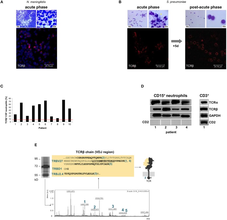
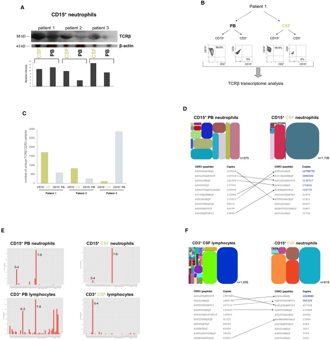
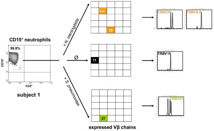
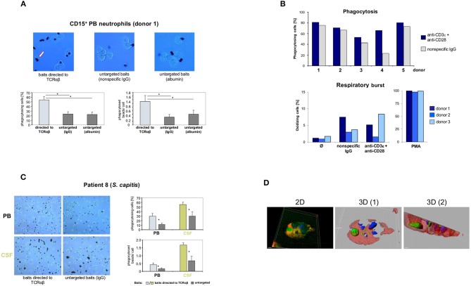
Bacterial meningitis is a life-threatening disease that evokes an intense neutrophil-dominated host response to microbes invading the subarachnoid space. Recent evidence indicates the existence of combinatorial V(D)J immune receptors in neutrophils that are based on the T cell receptor (TCR). Here, we investigated expression of the novel neutrophil TCRαβ-based V(D)J receptors in cerebrospinal fluid (CSF) from human patients with acute-phase bacterial meningitis using immunocytochemical, genetic immunoprofiling, cell biological, and mass spectrometric techniques. We find that the human neutrophil combinatorial V(D)J receptors are rapidly induced in CSF neutrophils during the first hours of bacterial meningitis. Immune receptor repertoire diversity is consistently increased in CSF neutrophils relative to circulating neutrophils and phagocytosis of baits directed to the variable immunoreceptor is enhanced in CSF neutrophils during acute-phase meningitis. Our results reveal that a flexible immune response involving neutrophil V(D)J receptors which enhance phagocytosis is immediately initiated at the site of acute bacterial infection.

Keywords: T cell receptors; V(D)J receptors; bacterial meningitis; neutrophil granulocytes; phagocytosis.


Figures

Figure 1
Figure 1
Rapid accumulation of T cell receptor αβ bearing neutrophils (TCRαβ+) in the cerebrospinal fluid (CSF) of patients with acute bacterial meningitis. CSF cytology images from two representative patients with acute bacterial meningitis caused by Neisseria meningitidis (A, patient 4) and Streptococcus pneumoniae, respectively (B, patient 5). The time between onset of specific symptoms and CSF collection was 9–15 h. The top row in each panel represents Giemsa stained CSF-cytospin preparations that demonstrate the massive accumulation of neutrophils (left). The presence of bacteria (asterisks) is shown at a higher magnification (right). Confocal immunofluorescence microscopy of CD15+-MACS purified CSF neutrophils (bottom) reveals consistent accumulation of TCRαβ expressing neutrophils (red) during the acute phase of the inflammatory response. Nuclei in panel A (blue) are counterstained with DRAQ5. CSF cytology in (B) is also shown after 5 days of antibiotic therapy revealing the presence of the TCRαβ in the neutrophil-driven acute phase and TCRαβ expression by T cells in the lymphocyte dominated post-acute phase. See also Figures S1, S2, S4 for CSF cytology images of additional patients. Isotype controls are shown in Figure S3. (C) Percentages of CSF neutrophils that stained positive for the TCRαβ in a cohort of 10 randomly selected patients with bacterial meningitis. Quantitative analysis of TCRαβ staining was performed in CD15+-purified CSF neutrophils that were collected during acute-phase bacterial meningitis. The red line represents the average percentage of TCRαβ+ neutrophils present in the circulation of healthy individuals. (D) RT-PCR demonstrates expression of the TCRα and the TCRβ constant chain genes in CSF CD15+-neutrophils from patients with acute-phase bacterial meningitis. Four patients (1–4) are representatively shown. CSF T cells (CD3+) from patient 1 are shown as positive control. Absence of expression of the lymphoid marker CD2 demonstrates that CD15+-MACS purified neutrophils were free of NK or T cells. GAPDH, loading control. (E) Direct mass-spectrometric identification of a rearranged Vβ-chain variant in CSF neutrophils from patient 2 (S. pneumoniae meningitis). Protein lysates from the patient's CSF CD15+-neutrophils were immunoprecipitated using an anti-TCRβ antibody and the predicted 58 kD band (boxed) was analyzed by MALDI-TOF mass spectrometry. Peaks 1–5 represent TCR Vβ-specific peptide fragments whose amino acid sequence identities with known TCR Vβ-chains are bolded. They are consistent with a TRBV27-TRBD1-TRBJ2.4 rearranged clonotype. For protein identification, peptide mass fingerprints were searched in the MASCOT database. See also Figure S7.
Figure 2
Figure 2
Induction of the variable TCRαβ in CSF neutrophils during the acute phase of bacterial meningitis. (A) The Western blot shows TCRαβ levels in CD15+ neutrophils from acute phase CSF (<15 h post-onset of symptoms) and simultaneously collected peripheral blood (PB) in patients with bacterial meningitis caused by S. aureus (patient 1), S. pneumoniae (patient 2) and E. coli, respectively (patient 3). A quantitative analysis of the TCRβ western blot signals in relation to β-actin are shown below. β-actin, protein loading control. The original blots this figure is based on are displayed in the Supplementary Material. (B) Experimental design of dual lineage NGS-based TCRβ transcriptome analyses in patients with acute bacterial meningitis (representatively shown for patient 1). Highly pure CD15+ neutrophils and CD3+ lymphocytes were obtained simultaneously from the patients' peripheral blood and CSF, respectively, and TCRβ transcriptomes were analyzed by ARM-PCR based high-throughput sequencing. The scattergrams document the purity of the isolated cell fractions. (C) Sequencing of the TCRβ transcriptomes of patients 1–3 reveals that the unique number of expressed TCRβ CDR3 variants is higher in CD15+ CSF neutrophils of patient 1 and 2 than in CD15+ PB neutrophils indicative of repertoire broadening at the site of inflammation. Shown are the unique TCRβ CDR3 variants. A unique CDR3 sequence is defined as a non-redundant fragment of amino acids which is derived from a stop-codon-free reading frame containing both translated conserved V and J motifs. An overview of the NGS results including effective reads, unique and total CDR3 numbers are shown in Table S2. (D) (top) Repertoire diversity tree plots visualize the relative abundance of the TCRβ CDR3 transcript variants that are expressed by CD15+ neutrophils in CSF and in peripheral blood (patient 1). Each spot represents a rearranged TCRβ transcript that encodes a unique CDR3β sequence. It is defined by a unique color and its area is proportional to the relative transcript frequency. The position of each spot within the plot area is defined according to its Vβ usage (x-axis: Vβ1 → Vβi) and Jβ usage (y-axis: Jβ1 → Jβi). Each plot has a distinct color code. Total numbers of identified non-redundant (“unique”) TCRβ CDR3 sequence variants are indicated for each diversity tree plot. CDR3, complementarity determining region 3. (bottom) Detailed list of the 10 most frequently expressed TCRβ CDR3 variants in each neutrophil population. Transcript copy numbers are indicated. TCRβ variants that are shared between PB and CSF neutrophils are connected by arrows. TCRβ variants that show an increase in CSF neutrophils are highlighted in blue. See also Figure S13. (E) Vβ gene usage of CD15+ neutrophils and CD3+ lymphocytes, respectively, in peripheral blood and CSF (patient 1). The 2D-plots demonstrate the relative usage of the Vβ genes for each cell compartment. This analysis is based on the number directly observed from the read count data. Note the markedly restricted Vβ gene usage in CSF cells relative to PB cells. X-axis: Vβ gene; y-axis: percentage of used Vβ genes. A normalized distribution of the Vβ gene usage is shown in Figure S15. (F) (top) Relative abundance of TCRβ CDR3 transcript variants expressed by CD15+ neutrophils and CD3+ lymphocytes in CSF (patient 2). Total numbers of identified non-redundant TCRβ CDR3 sequence variants are indicated for each leukocyte subpopulation. (bottom) Comparison of the 10 most frequently expressed TCRβ CDR3 variants reveals that neutrophils and lymphocytes in CSF share common repertoires (arrows), but also express a similar proportion of distinct TCRβ variants. Numbers designate transcript copy numbers. Note that two of the shared TCRβ CDR3 sequence variants exhibit higher expression rates in CSF neutrophils than in CSF lymphocytes (blue). See also Figure S16.
Figure 3
Figure 3
Exposure of neutrophils to bacterial pathogens that cause meningitis induces TCR Vβ repertoire changes in vitro. Shown are the TCR Vβ repertoires that are expressed by peripheral blood CD15+ neutrophils from a healthy subject after 9 h incubation with N. meningitidis (orange), S. pneumoniae (green) and in the absence of a bacterial pathogen, respectively (Ø, control). CD15+ neutrophils were obtained from a single blood draw. The detailed CDR3β length variants are shown in the right panel. The scattergrams (left) document the purity of the CD15+ neutrophils. Note that N. meningitidis and S. pneumoniae induce distinct TCR Vβ chain usage (center) and CDR3β repertoires (right). See also Figure S17.
Figure 4
Figure 4
Targeting of baits to the TCRαβ and TCRαβ activation via CD3/CD28 costimulation enhance neutrophil phagocytosis. (A) Representative unstained cytospin preparations (40x) of PB neutrophils from a healthy subject (donor 1) that were challenged with bead baits targeted to the TCRαβ or untargeted beads (controls) for 3 h. The arrow (left) highlights two phagocytosed beads. The quantitative analysis of neutrophil bead phagocytosis, defined as ingestion or binding of ≥1 bead, is shown in the bottom panel (percentage of phagocytosing cells, left; phagocytosed beads/cell, right). Note that targeting of beads to the TCRαβ significantly enhances PB neutrophil phagocytosis relative to untargeted beads. Purified PB CD15+ neutrophils were incubated with polystyrene bead baits (Ø 4.5 μm) coated with anti-TCRαβ antibodies and uptake of beads was recorded. Beads coated with equal amounts of non-specific IgG isotype antibodies, potentially binding to the Fcγ receptor, or albumin (irrelevant protein) served as controls (untargeted baits). See also Figures S18, S19 in the Supplementary material. (B) Anti-CD3/CD28-mediated TCRαβ activation enhances Fc receptor-independent phagocytosis of bacteria (top) but not respiratory burst in neutrophils (bottom). PB CD15+ neutrophils from five healthy individuals were infected for 10 min with non-opsonized FITC-labeled E. coli baits (cell/bacteria ratio 1:10) in the presence of anti-CD3ε/CD28 antibodies or non-specific IgG (top). Shown are the percentages of phagocytosing neutrophils. The bottom panel shows that CD3/CD28 costimulation under non-opsonizing conditions (three donors) has no effect on the neutrophil respiratory burst. Phagocytic and respiratory burst activities were assessed by flow cytometry. PMA, phorbol myristate acetate. (C) Increased phagocytosis of baits targeted to the TCRαβ in PB neutrophils and CSF neutrophils from a patient with Staphylococcus capitis meningitis (patient 8). PB and CSF neutrophils were simultaneously collected from the patient during the early phase of meningitis and subsequently challenged with beads targeted to the TCRαβ or untargeted beads (non-specific IgG) for 1.5 h. Representative cytospin preparations (20x, left) and the quantitative analysis of bead phagocytosis are shown (right). (D) 3D confocal immunofluorescence imaging identifies a CSF neutrophil from the same patient that has ingested a bead bait (green) targeted to the TCR. Two distinct cross-sections are shown (center, right). The 2D view is shown left. For a full 3D view see Movie S1. The bead bait was stained with FITC-labeled antibodies to mouse IgG, the cell membrane (red) and the nucleus (blue) were stained with Alexa 633-conjugated WGA and DAPI, respectively. All peripheral blood or CSF neutrophils were CD15+-MACS purified and incubated with bead baits at a cell density of 3 × 10⁶/ml and a cell/bead ratio of 1:1. Quantitation of phagocytosed beads was conducted by bright field microscopy of at least 12 randomly selected fields of vision. Error bars represent mean ± SD. *p < 0.001, Dunnett post-hoc test (A), Student's pair test (C).

Similar articles

Cited by

References

    1. Kim KS. Pathogenesis of bacterial meningitis: from bacteraemia to neuronal injury. Nat Rev Neurosci. (2003) 4:376–85. 10.1038/nrn1103 - DOI - PubMed
    1. Edmond K, Clark A, Korczak VS, Sanderson C, Griffiths UK, Rudan I. Global and regional risk of disabling sequelae from bacterial meningitis: a systematic review and meta-analysis. Lancet Infect Dis. (2010) 10:317–28. 10.1016/S1473-3099(10)70048-7 - DOI - PubMed
    1. Stephens DS, Greenwood B, Brandtzaeg P. Epidemic meningitis, meningococcaemia, and Neisseria meningitidis. Lancet. (2007) 369:2196–210. 10.1016/S0140-6736(07)61016-2 - DOI - PubMed
    1. van de Beek D, Drake JM, Tunkel AR. Nosocomial bacterial meningitis. N Engl J Med. (2010) 362:146–54. 10.1056/NEJMra0804573 - DOI - PubMed
    1. Oordt-Speets AM, Bolijn R, van Hoorn RC, Bhavsar A, Kyaw MH. Global etiology of bacterial meningitis: a systematic review and meta-analysis. PLoS ONE. (2018) 13:e0198772. 10.1371/journal.pone.0198772 - DOI - PMC - PubMed

LinkOut - more resources