Evaluation of in vitro Assays to Assess the Modulation of Dendritic Cells Functions by Therapeutic Antibodies and Aggregates
- PMID: 31001248
- PMCID: PMC6455063
- DOI: 10.3389/fimmu.2019.00601
Evaluation of in vitro Assays to Assess the Modulation of Dendritic Cells Functions by Therapeutic Antibodies and Aggregates
Abstract
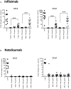
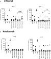
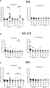
Therapeutic antibodies have the potential to induce immunogenicity leading to the development of anti-drug antibodies (ADA) that consequently may result in reduced serum drug concentrations, a loss of efficacy or potential hypersensitivity reactions. Among other factors, aggregated antibodies have been suggested to promote immunogenicity, thus enhancing ADA production. Dendritic cells (DC) are the most efficient antigen-presenting cell population and are crucial for the initiation of T cell responses and the subsequent generation of an adaptive immune response. This work focuses on the development of predictive in vitro assays that can monitor DC maturation, in order to determine whether drug products have direct DC stimulatory capabilities. To this end, four independent laboratories aligned a common protocol to differentiate human monocyte-derived DC (moDC) that were treated with either native or aggregated preparations of infliximab, natalizumab, adalimumab, or rituximab. These drug products were subjected to different forms of physical stress, heat and shear, resulting in aggregation and the formation of subvisible particles. Each partner developed and optimized assays to monitor diverse end-points of moDC maturation: measuring the upregulation of DC activation markers via flow cytometry, analyzing cytokine, and chemokine production via mRNA and protein quantification and identifying cell signaling pathways via quantification of protein phosphorylation. These study results indicated that infliximab, with the highest propensity to form aggregates when heat-stressed, induced a marked activation of moDC as measured by an increase in CD83 and CD86 surface expression, IL-1β, IL-6, IL-8, IL-12, TNFα, CCL3, and CCL4 transcript upregulation and release of respective proteins, and phosphorylation of the intracellular signaling proteins Syk, ERK1/2, and Akt. In contrast, natalizumab, which does not aggregate under these stress conditions, induced no DC activation in any assay system, whereas adalimumab or rituximab aggregates induced only slight parameter variation. Importantly, the data generated in the different assay systems by each partner site correlated and supported the use of these assays to monitor drug-intrinsic propensities to drive maturation of DC. This moDC assay is also a valuable tool as an in vitro model to assess the intracellular mechanisms that drive DC activation by aggregated therapeutic proteins.
Keywords: aggregates; anti-drug antibodies; dendritic cells; immunogenicity; in vitro assays; intracellular signaling.
Figures


Similar articles
-
Effect of growth hormone and IgG aggregates on dendritic cells activation and T-cells polarization.Immunol Cell Biol. 2017 Mar;95(3):306-315. doi: 10.1038/icb.2016.100. Epub 2016 Oct 7. Immunol Cell Biol. 2017. PMID: 27713394
-
The FcγRIIa-Syk Axis Controls Human Dendritic Cell Activation and T Cell Response Induced by Infliximab Aggregates.J Immunol. 2020 Nov 1;205(9):2351-2361. doi: 10.4049/jimmunol.1901381. Epub 2020 Sep 28. J Immunol. 2020. PMID: 32989091
-
Aggregation of human recombinant monoclonal antibodies influences the capacity of dendritic cells to stimulate adaptive T-cell responses in vitro.PLoS One. 2014 Jan 21;9(1):e86322. doi: 10.1371/journal.pone.0086322. eCollection 2014. PLoS One. 2014. PMID: 24466023 Free PMC article.
-
Immunogenicity of Bioproducts: Cellular Models to Evaluate the Impact of Therapeutic Antibody Aggregates.Front Immunol. 2020 May 5;11:725. doi: 10.3389/fimmu.2020.00725. eCollection 2020. Front Immunol. 2020. PMID: 32431697 Free PMC article. Review.
-
Immunogenicity of therapeutic proteins: influence of aggregation.J Immunotoxicol. 2014 Apr-Jun;11(2):99-109. doi: 10.3109/1547691X.2013.821564. Epub 2013 Aug 6. J Immunotoxicol. 2014. PMID: 23919460 Free PMC article. Review.
Cited by
-
Challenges of non-clinical safety testing for biologics: A Report of the 9th BioSafe European Annual General Membership Meeting.MAbs. 2021 Jan-Dec;13(1):1938796. doi: 10.1080/19420862.2021.1938796. MAbs. 2021. PMID: 34241561 Free PMC article.
-
Generation and characterization of chicken monocyte-derived dendritic cells.Front Immunol. 2025 Feb 4;16:1517697. doi: 10.3389/fimmu.2025.1517697. eCollection 2025. Front Immunol. 2025. PMID: 39967657 Free PMC article.
-
A High Threshold of Biotherapeutic Aggregate Numbers is Needed to Induce an Immunogenic Response In Vitro, In Vivo, and in the Clinic.Pharm Res. 2024 Apr;41(4):651-672. doi: 10.1007/s11095-024-03678-2. Epub 2024 Mar 22. Pharm Res. 2024. PMID: 38519817
-
A Semi-Mechanistic Mathematical Model of Immune Tolerance Induction to Support Preclinical Studies of Human Monoclonal Antibodies in Rats.Pharmaceutics. 2025 Jun 27;17(7):845. doi: 10.3390/pharmaceutics17070845. Pharmaceutics. 2025. PMID: 40733054 Free PMC article.
-
HLAII peptide presentation of infliximab increases when complexed with TNF.Front Immunol. 2022 Sep 13;13:932252. doi: 10.3389/fimmu.2022.932252. eCollection 2022. Front Immunol. 2022. PMID: 36177046 Free PMC article.
References
-
- Baudouin V, Crusiaux A, Haddad E, Schandene L, Goldman M, Loirat C, et al. . Anaphylactic shock caused by immunoglobulin e sensitization after retreatment with the chimeric anti – interleukin-2 receptor monoclonal antibody. Transplantation. (2003) 76:459–63. 10.1097/01.TP.0000073809.65502.8F - DOI - PubMed
MeSH terms
Substances
LinkOut - more resources
Full Text Sources
Research Materials
Miscellaneous

